| The Annals of Clinical and Analytical Medicine
525
The Annals of Clinical and Analytical Medicine
Case Report
Cervical spondyloarthropathy due to the dialysis-related amyloidosis
Cervical destructive spondyloarthropathy due to the
dialysis-related amyloidosis: imaging findings
DOI: 10.4328/ACAM.6068 Received: 05.11.2018 Accepted: 08.12.2018 Publihed Online: 10.12.2018 Printed: 01.07.2019 Ann Clin Anal Med 2019;10(4): 525-8 Corresponding Author: Hale Turnaoglu, Department of Radiology, Baskent University, Faculty of Medicine, 06490 Bahcelievler, Ankara, Turkey.
GSM: +905323106920 F.: +90 3122237333 E-Mail: [email protected] ORCID ID: https://orcid.org/0000-0002-0781-0036
Abstract
Dialysis-related amyloidosis that occurs secondary to the deposition of amyloid fibrils containing beta-2-microglobulin, is a type of amyloidosis affecting patients undergoing long-term hemodialysis. It involves the osteoarticular system predominantly. Destructive spondyloarthropathy, is a type of dialysis-related spondyloarthropa-thy, which frequently involves the cervical spine, have been reported only sporadically. We describe a case of a destructive spondyloarthropaspondyloarthropa-thy, in a 43-year-old long-term hemodialysis patient, presenting with myelopathy with particular interest to cervical computed tomography and magnetic resonance imaging findings. Keywords
Dialysis-Related Amyloidosis; Destructive Spondyloarthropathy; Myelopathy; Magnetic Resonance Imaging; Computed Tomography
Hale Turnaoglu1, Kemal Murat Haberal1, Oğuzcan Ünal1, Ozlem Isiksacan Ozen2, Ahmet Muhtesem Agildere1 1Department of Radiology, 2Department of Pathology, Baskent University Faculty of Medicine, Ankara, Turkey
| The Annals of Clinical and Analytical Medicine
Cervical spondyloarthropathy due to the dialysis-related amyloidosis
526
Introduction
Dialysis-related amyloidosis that consists of the deposition of amyloid fibrils containing beta-2-microglobulin, is a type of am-yloidosis affecting patients undergoing long-term hemodialysis. It affects the osteoarticular system predominantly. The most common manifestations are arthropathy of the axial skeleton, knees, shoulders, hips and carpal tunnel syndrome [1]. Dialysis-related spondyloarthropathy has been divided into three types: destructive spondyloarthropathy (DSA), amyloid deposition in spinal ligaments, and pseudotumor of the craniocervical junc-tion (amiloidoma). DSA, which frequently affects the cervical spine, have been reported only sporadically. We describe a case of a DSA, in a long-term hemodialysis patient, presenting with myelopathy with particular interest to cervical computed tomography (CT) and magnetic resonance imaging (MRI) find-ings.
Case Report
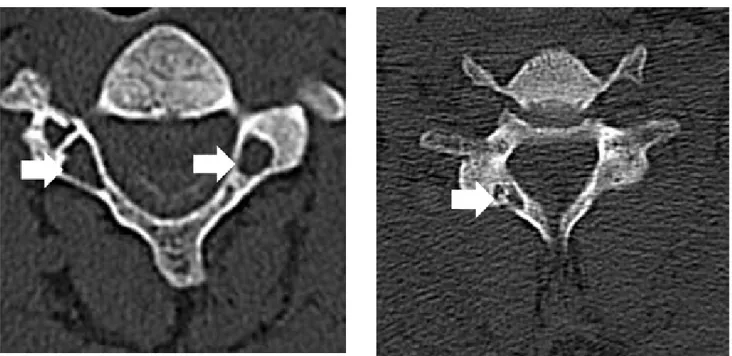
A 43-year-old man who had chronic renal failure secondary to unknown etiology presented with loss of strength in the legs and disturbances while walking. He had been receiving hemo-dialysis for 29 years. Cervical vertebral computed tomography (CT) scans showed narrowing in the cervical 2 (C2) - cervical 3 (C3) intervertebral space, and osteolytic areas with peripheral sclerosis in the laminae and pedicules of the various vertebrae (Figure 1). Magnetic resonance imaging (MRI) showed thicken-ing and decreased signal intensity on both T1and T2-weighted images, in the posterior longitudinal ligament and ligamentum flavum. Scattered increased signal intensity of the spinal cord was seen secondary to compression of thickened ligaments and narrowing of the spinal canal (Figure 2). No contrast was given because of the patient’s poor glomerular filtration rate. The
Figure 1. Axial cervical computed tomography scans showing the areas of osteolysis with peripheral sclerosis (a) in the C3 vertebral pedicles, and (b) C6 vertebral right lamina (white arrows).
Figure 2. Sagittal T1 (a) and T2-weighted turbo spin echo (b) images. Thickened posterior longitudinal ligament (white thick arrows) and ligamentum flavum (black arrows) with decreased intensity, indentation to the spinal cord (white thin arrows) and narrowing in the spinal canal. Scattered increased signal intensities, marked at the C1 level (grey arrow).
| The Annals of Clinical and Analytical Medicine
Cervical spondyloarthropathy due to the dialysis-related amyloidosis
527
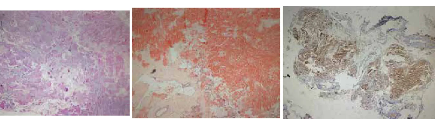
patient was operated. Histopathological examination revealed amyloid deposits and the presence of intense beta-2-micro-globulin fibrils (Figure 3). Informed consent was obtained from the patient for using his data.
Discussion
DSA is characterized by erosions of the anterosuperior and/or anteroinferior aspects of the vertebral body, severe narrowing of the intervertebral disk space and erosions and cysts of ad-jacent vertebral plates, with absence of significant osteophyte formation, radiographically. In advanced stages of the disease, subluxation, listhesis, or vertebral body collapse may occur [2]. CT is the best modality for detecting osseous erosion or small areas of osteolysis in cortical bone. CT can demonstrate the distribution and extent of the destructive changes [2]. In the case presented here, areas of osteolysis with peripheral sclero-sis in the cervical vertebral bodies, laminae, and pedicles were demonstrated by CT (Figure 1).
MRI shows the extent and distribution of osseous, articular, spi-nal cord and soft-tissue involvement, adding to the informa-tion obtained from radiographic and CT images. MRI may show amyloid deposits in the intervertebral disk, in the synovium of apophyseal joints, and in the ligaments. Although bone lesions show decreased signal intensity on T1-weighted images in most patients, T2-weighted images show various signal intensity patterns that range from hypointense to hyperintense. The vari-ability in signal intensity is probably caused by the combination of amyloid deposits and fluid collection within the subchondral lesions. Identification of an intraosseous lesion with low signal intensity on both T1- and T2-weighted images is helpful in the diagnosis of amyloidosis. After the gadolinium-based contrast material injection, the bone lesions usually show moderate en-hancement [3]. Compression of the spinal cord and myelopathy caused by extradural deposition and thickening of ligaments may occur [3]. MRI is well suited, as in the case presented here, for assessing the compression of the spinal cord and myelopa-thy, caused by the thickening of ligamentum flavum and pos-terior longitudinal ligament (Figure 2). The diseases of the dif-ferential diagnosis usually includes spondylodiscitis, metastatic malignancy, multiple myeloma, secondary hyperparathyroidism (renal osteodystrophy), ossification of the posterior longitudi-nal ligament, and cervical spondylosis. In some cases, it can be difficult to differentiate changes secondary to dialysis-related amyloidosis from spondylodiscitis. In spondylodiscitis, involved structures show decreased signal intensity on T1-weighted MR
images and in most cases increased signal intensity on T2-weighted and STIR images. Low signals present in T2-T2-weighted images helps the exclusion of an infection [3]. Brown tumors of hyperparathyroidism are sometimes difficult to differentiate from amyloid cysts in dialysis patients. The location of the bone lesions is helpful [4]. Also, cysts tend to increase in number and size associated with the duration of dialysis [3]. In the meta-static disease, lesions are more diffuse and less circumscribed compared with dialysis-related amyloidosis. Multiple myelomas can be differentiated from dialysis-related amyloidosis by urine and serum protein electrophoresis. A bone scan may be used for detecting other locations, and magnetic resonance imaging has been recommended to assist with the diagnosis [5]. Ossification of the posterior longitudinal ligament can be recognized by the presence of calcifications on the plain radiography and/or CT [6]. In the cervical spondylosis, features related to the cervical spine and intervertebral space, such as osteophyte formation and intervertebral space narrowing are evident.
The gold standard of the diagnosis is the histological identifi-cation of beta-2-microglobulin, a major constituent of amyloid fibrils, in the material which is obtained by surgery. The pathol-ogy diagnosis is made with hematoxylin-eosin and Congo red. Under polarized light, these areas exhibit characteristic apple-green birefringence [1] (Figure 3).
In the treatment, medical therapy is limited to symptomatic approaches to reduce pain and inflammation. In the patients suffering from cervical pain may be referred for surgical evalu-ation. For relieving of the pain, surgical procedures, such as circumferential reconstructive surgery involving pedicle screw fixation, anterior strut bone grafting, posterior and/or anterior decompression, posterior nerve root decompression or spinous process wiring may perform, due to the severity and involve-ment of the disease [8]. The best treatinvolve-ment of hemodialysis-related amyloidosis is renal transplantation. Renal transplanta-tion can provide a very rapid symptomatic relief and prevents the progression of the disease. However, the effect of trans-plantation on existent amyloid depositions is controversial [7].
Conclusion
In the long-term dialysis patients, imaging diagnosis is neces-sary for the evaluation of dialysis-related amyloidosis before serious complications arise. The changes of the vertebral body, ligaments, facet joints and intervertebral spaces in dialysis-re-lated amyloidosis and the complications due to these changes, can recognize and identify by CT and MRI.
Figure 3. Photomicrographs of surgical specimen of the ligamentum flavum. (a) Histological finding with hematoxylin and eosin staining shows amorphous and eosi-nophilic material (H&E x100). (b) Amyloid deposits are seen with congo red staining (Congo red x100). (c) Also, β2-M positive staining of the material is seen (Immuno-peroxidase x100).
| The Annals of Clinical and Analytical Medicine
Cervical spondyloarthropathy due to the dialysis-related amyloidosis
528
Scientific Responsibility Statement
The authors declare that they are responsible for the article’s scientific content including study design, data collection, analy-sis and interpretation, writing, some of the main line, or all of the preparation and scientific review of the contents and ap-proval of the final version of the article.
Animal and human rights statement
All procedures performed in this study were in accordance with the ethical standards of the institutional and/or national re-search committee and with the 1964 Helsinki declaration and its later amendments or comparable ethical standards. No ani-mal or human studies were carried out by the authors for this article.
Conflict of interest
None of the authors received any type of financial support that could be considered potential conflict of interest regarding the manuscript or its submission.
References
1. Moslavac S, Dzidic I, Kejla Z, Tomas D. Hemodialysis associated amyloidosis with cervical spinal cord compression and incomplete tetraplegia: a case report. Spinal Cord. 2007; 45(12): 799-801.
2. Oruckaptan H, Karli Oguz K, Isikay I, Ruacan S. Amyloidoma of the temporal bone and upper cervical spine; presentation of a rare clinical entity with a brief literature review. Turk Neurosurg. 2009; 19(2): 159-62.
3. Kiss E, Keusch G, Zanetti M, Jung T, Schwarz A, Schocke M, et al. Dialysis-related amyloidosis revisited. AJR Am J Roentgenol. 2005; 185(6): 1460-7.
4. Yamamoto S, Gejyo F. Historical background and clinical treatment of dialysis-related amyloidosis. Biochim Biophys Acta. 2005; 1753(1): 4-10.
5. Schiffl H. Impact of advanced dialysis technology on the prevalence of dialy-sis-related amyloidosis in long-term maintenance dialysis patients. Hemodial Int. 2014; 18(1): 136-41.
6. Choi BW, Song KJ, Chang H. Ossification of the posterior longitudinal ligament: a review of literature. Asian Spine J. 2011; 5(4): 267-76.
7. Mourad G, Argilés A. Renal transplantation relieves the symptoms but does not reverse beta 2-microglobulin amyloidosis. J Am Soc Nephrol. 1996; 7(5): 798-804.
8.Abumi K, Ito M, Kaneda K. Surgical treatment of cervical destructive spondylo-arthropathy (DSA). Spine. 2000; 25(22): 2899-905.
How to cite this article:
Turnaoglu H, Haberal KM, Ünal O, Ozen ÖI, Agildere AM. Cervical destructive spondyloarthropathy due to the dialysis-related amyloidosis: imaging findings. Ann Clin Anal Med 2019;10(4): 525-7.