Pathological findings of experimental Aeromonas hydrophila infection
in Nile tilapia (Oreochromis niloticus)
*Banu YARDIMCI1, Yılmaz AYDIN2
1 Department of Preclinical Science, Faculty of Veterinary Medicine, University of Ondokuz Mayis, Samsun; 2Department of Pathology, Faculty of Veterinary Medicine, University of Ankara, Ankara, Turkey.
Summary: In this study, clinical findings with macroscopic and microscopic pathologic changes of the tissues and organs
after necropsy examination at the 1st, 2nd, 3rd, 5th and 7th days following intraperitoneally injection of Nile tilapia (Oreochromis
niloticus) with Aeromonas hydrophila was aimed to be observed. Clinically; weakness, anorexia, swimming closer to surface,
darkness in color with hyperemia and lysis of the fins were observed. In the macroscopic examination the liver was seen to be yellowish brown and crispy with haemorrhagic and greyish white foci on the surface. The gall bladder was tightly full with emerald green bile. The kidney and hearth had haemorrhagic foci whilst intestine lumina observed to be filled with yellow coloured mucoid liquid. The degenerative changes, cytoplasmic fat vacuols and lymphocyte infiltration in lever with focal necrosis of hepatocytes and pancreatic cells were observed histologically. The skin, spleen, gill and eyes did not reveal any significant symptoms. The haemorrhagies and intensive lymphocyte infiltrations in liver, kidney and hearth both in macroscopic and microscopic levels revealed a visceral haemorrhagic septicemia. In this sense; clinical, macroscopic and microscopic findings observed during the present study complied with the Aeromonas hydrophila infections recorded in the other fresh water fishes.
Key words: Aeromonas hydrophila, bacterial haemorrhagic septicemia, Nile tilapia (Oreochromis niloticus), pathology
Nil tilapyalarında (Oreochromis niloticus) deneysel Aeromonas hydrophila enfeksiyonundaki patolojik bulgular
Özet: Bu çalışmada; intraperitoneal olarak Aeromonas hydrophila ile enfekte edilen Nil tilapyalarında (Oreochromis
niloticus), enjeksiyonu izleyen 1., 2., 3., 5. ve 7. günlerdeki klinik bulgular ile ötenaziyi takiben yapılan nekropsiler sonrasında doku
ve organlarda gözlenen patolojik bulguların, makroskobik ve mikroskobik düzeyde incelenerek değerlendirilmesi amaçlandı. Klinik olarak halsizlik, iştahızlık, yüzeye yakın yüzme, renkte koyulaşma ile yüzgeç diplerinde hiperemi ve erime gibi bulgular gözlendi. Nekropside karaciğerin sarımsı kahverenkli olduğu ve kıvamının gevrekleşip, üzerinde kanama ve boz beyaz odaklar gözlendi. Safra kesesinin zümrüt yeşili renkli safra sıvısıyla gergin bir şekilde dolu olduğu dikkati çekti. Böbrekte ve kalpte kanama odakları gözlenirken, bağırsak lümenlerinin sarımtırak renkli, mukuslu sıvı ile dolu olduğu izlendi. Histopatolojik olarak karaciğerde dejeneratif değişiklikler, sitoplazmalarında yağ vakuolleri ve lenfosit infiltrasyonu ile hepatosit ve pankreas hücrelerinde fokal nekroz gözlendi. Böbrekte, parankim dejenerasyonu, tubul epitellerinde nekroz ve fokal lenfosit infiltrasyonu görüldü. Deri, dalak, solungaç ve gözlerde belirgin bir bulgu gözlenmedi. Karaciğer, böbrek ve kalpte gerek makroskobik gerekse de mikroskobik düzeyde hemorajilerin ve lenfosit infiltrasyonunun yoğun olarak gözlenmesi visseral bir hemorajik septisemi tablosu çizmektedir. Bu bağlamda mevcut çalışma süresince gözlenen klinik, makroskobik ve mikroskobik bulgular genel itibariyle diğer tatlı su balıklarında şekillenen A. hydrophila enfeksiyonlarındaki temel bulgularla benzerlik gösterdi.
Anahtar sözcükler: Aeromonas hydrophila, bakteriyel hemorajik septisemi, Nil tilapyası (Oreochromis niloticus), patoloji.
* This research has been summarized from the same entitled PhD thesis. Introduction
Aeromonas hydrophila is one of the most important
agents of the outbreaks in fresh water fish, in which skin ulcers, hemorrhage and necrosis of the visceral organs are the major symptoms. Synonyms are; Bacterial
hemorrhagic septicemia, Aeromonad septicemia, or Red Pest (24).
The incubation period of the disease depend on fish species and resistance, environmental conditions and the season. This period varies 2-4 days in natural infections and 8-48 hours in experimental infection models (5,14).
Clinically; common symptoms such as weakness, being close to the pool wall and floor, stagnation and anorexia with free mucus or intestine shaped faeces in the water can be observed. (1,3,4,6).
In the acute form of disease, a fatal septicemia may occur so rapidly that fish die before they have time to develop anything but a few gross signs of disease. When clinical signs of infection are present, affected fish may show exophthalmia, reddening of the skin, and an accumulation of fluid in the scale pockets (10). The abdomen may become distended as a result of an edema and the scales may bristle out from the skin to give a “washboard” appearance. The gills may hemorrhage and ulcers may develop on the dermis and motile aeromonads were isolated from the eyes, liver and kidneys of affected fish. (7,24). The condition at first affected one eye, progressed into the other eye, after which the orbits ruptured causing blindness and death. Similarly, Yambot and Inglis (28) described an acute mortality among Nile tilapia (Oreochromis niloticus) in which the most apparent clinical signs included an opaqueness in one or both eyes, accompanied by exophthalmia and eventual bursting of the orbit (28).
Systemic infections were characterized by diffuse necrosis in several internal organs and the presence of melanin-containing macrophages in the blood (27). Internally, the liver and kidneys are target organs of an acute septicemia. The liver may become pale or have a greenish coloration while the kidney may become swollen and friable. These organs are apparently attacked by bacterial toxins and lose their structural integrity (1,14).
Histopathologically, fish may exhibit epithelial hyperplasia in the foregut; leptomeningeal congestion in the brain, as well as a thrombosis and inflammation in the perisclerotic region and corneal epithelium of the eye (11). There also be a severe branchitis, as indicated by leukocytic infiltration and dilation of the central venous sinus (13). Chronic motile aeromonad infections manifest themselves primarily as ulcerous forms of disease, in which dermal lesions with focal hemorrhage and inflammation are apparent. Both the dermis and epidermis are eroded and the underlying musculature becomes severely necrotic (14).
In this study, clinical findings with macroscopic and microscopic pathologic changes of the tissues and organs after necropsy examination at the 1st, 2nd, 3rd, 5th and 7th
days following intraperitoneal injection of Nile tilapia
(Oreochromis niloticus) with Aeromonas hydrophila was
aimed to be observed.
Materials and Methods
In the study, mean 50-80 g in weight and clinically healthy 78 Nile tilapia (Oreochromis niloticus) were provided from Ankara University Agricultural Faculty Department of Fisheries and Aquaculture. 18 of them for determination of the LC50 value and the other 60 were
used in the experiments. The experiments were
performed in the Ankara University Agricultural Faculty Department of Fisheries and Aquaculture Experiment Unit. In the experiments 6 aquariums in 100 lt volume filled with dechlorized, minimally 48 hours rested and ventilated company’s water were used. During the study water levels were adjusted to 27 ± 1 oC, 8 mg/L soluble
oxygen and 7.38 pH. Aeromonas hydrophila strain used in the study was provided from Ministry of Health, Refik Saydam Hygiene Center, Refik Saydam National Culture Collection (RSKK 05049) and LC50 was defined as 109
CFU according to the Kärber method (2).
For orientation, 50 fishes were distributed in 5 aquariums ten by ten 15 days before assay. During this period, fish were fed in a ratio of %2 BW/day. Each fish was intraperitoneally injected with 0.1ml A. hydrophila suspension of (1x10⁸ CFU) sublethal concentration under
LC50 from the mid-point of the ventral fins. A control
group was formed to the sixth aquarium (2 for per group, totally 10) in which the fish was injected with 0.1ml PBS with the same method. In order to ease the necropy and tissue processing, groups were named as A for the first, B for the second, C for the third, D for the fifth and E for the seventh day respectively.
Fishes were euthanatized on 1st, 2nd, 3rd, 5th and 7th
days after recording the clinical findings. Following the necropsy, tissue samples from skin, gill, fin, muscle, liver, kidney, spleen, hearth, stomach, intestine, brain, eye and gonads were taken for histopathological examination, and reisolation and identification of the agent. The samples taken for histopathological examination were fixed in Bouin’s solution for 6 hours and then embedded in parafin following routine tissue processing. Tissue sections in 5-6 µm width were stained with haematoxylin-eosin and evaluated under light microscope.
Results
Clinical findings: 8-10 hours following the
injections all fishes were observed to be stagnant. By the first day post-injection, reluctance to eat, darkening in skin, and mild hyperemia of the fin bases were observed. By the second day, focal hyperemia of the skin over the pectoral fins and swimming closer to surface and aquarium wall was identified. By the third day, hyperemia of the fin bases were more obvious and fin rot especially on the tips of the pectorals were observed. By the fifth day darkening of the skin was more obvious than the hyperemia. By the seventh day, disappearance of eating, unstable swimming on the bottom of the aquarium, significant hyperemia on the base of the fins and severe fin rot was observed.
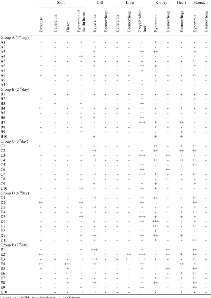
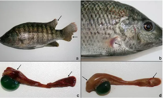
Macroscopic findings: Macroscopic findings of the
skin, gill, liver, kidney, hearth, and stomach were completely defined in Table 1 for each groups (Figure 1a-d).
Table 1. Distribution of macroscopical findings according to the organs. Tablo 1. Organlara göre makroskobik bulguların dağılımı.
Skin ____________________________________________ Gill ________________ Liver _____________________________ Kidney _________________ Heart _________ Stomach _______________
Darkness Hyperemia Fin rot Hyperemia of the
fin ba se s Hyperemia Haemorrhag e Hyperemia Haemorrhag e
Greyish white foci Hyperemia Haemorrhag
e Haemorrhag e Hyperemia Haemorrhag e Group A (1st day) A1 + - - - + - - - + + - - - - A2 + - - + ++ - - - ++ - - - A3 - - - - + - - - ++ ++ - - + - A4 - - - ++ + - - - A5 + - - - ++ - A6 + - - - + - - - ++ + - - + - A7 - - - - + - - - - - A8 - - - - + - - - ++ - A9 + - - + - - - + - A10 - - - - + - - - - - Group B (2 ndday) B1 + - - + - - - B2 + - - - + - - - + - B3 - + - + - - - - ++ - - - B4 ++ + - ++ - - - - ++ - - + - - B5 - - - ++ - - - + - B6 - - - + + - - - ++ - - - B7 - - - - +++ + - ++ - - B8 - + - - + - - - + + - - + - B9 - - - + - - - + - - B10 - - - - + - - - - + - + - - Group C (3rdday) C1 ++ - - + - - - - + ++ - + ++ - C2 - - - - ++ - - - + ++ - ++ ++ - C3 + - - - + - - + +++ - ++ - - - C4 + - - - ++ - - - + ++ - ++ ++ - C5 - - - ++ - - - ++ - C6 - - - - ++ - ++ - - - C7 + - - - ++ - - - +++ + - - ++ - C8 + - - - + - - - + - ++ - - - C9 - - - - + - - - + + - - + - C10 - - - ++ - - - - ++ + - - - - Group D (5 thday) D1 - + - - ++ - - - ++ ++ - - ++ - D2 ++ - - ++ - - - - ++ - - - ++ - D3 - - - - ++ - - + - - + - + - D4 - - - - ++ - - - ++ - ++ + ++ - D5 - - - ++ - - - - +++ + - + + - D6 - - - - + - - + ++ +++ - - + - D7 - - - + + +++ - - ++ - D8 + - - - - - - - + + - - - - D9 - - - + ++ - - - ++ ++ - + - - D10 - + - - - + - - ++ - Group E (7 thday) E1 + - - + +++ - - - + - ++ - ++ - E2 ++ - - - ++ +++ - ++ + ++ - E3 - - - ++ +++ - - +++ +++ + - - ++ - E4 ++ - +++ - ++ - - - ++ - - ++ + - E5 + - + - - - + - ++ - ++ - E6 + - ++ + ++ - - + + - - + ++ - E7 - - - - - - - + ++ + ++ + ++ - E8 - - + - ++ - - - + ++ - - ++ - E9 - - - + - - - + ++ - - - ++ - E10 + - - ++ ++ - - - ++ - + + - -
Figure 1. a. Darkness in skin with fin rot in caudal and dorsal fins (E4, arrows). b. Hyperemia at the bases of the pectoral fin (B4, arrow).
c. Large haemorrhagic foci on the parietal surface of the liver (E3, arrows).
d. Greyish white foci on liver (C7, arrows) and enlarged gall bladder filled with emerald-green secretion. Şekil 1. a. Enfeksiyona bağlı deri renginde koyulaşma ile kaudal (kuyruk) ve dorsal (sırt) yüzgeçte erime (E4, oklar).
b. Pektoral yüzgeç tabanında hiperemi (B4, ok).
c. Karaciğerin pariyetal yüzünde geniş kanama odakları (E3, oklar).
d. Karaciğerde boz beyaz odaklar (C7, oklar) ve safra kesesinde zümrüt yeşili renginde aşırı safra salgısı birikimi
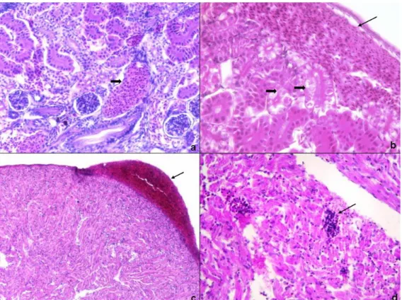
Figure 2. a. Haemorrhage in liver (E2; HE, 40x).
b. Degenerative changes in liver (thin arrow) and necrosis of pancreatic cells (thick arrow) (D4; HE, 400x). c. Diffuse lipidosis of liver (thick arrow) and hyperemia (thin arrow) (C5; HE, 100x).
d. Focal necrosis of hepatocytes (thin arrow) and lymphocyte infiltration (thick arrow) (D7; HE ,200x). Şekil 2. a. Karaciğerde kanama (E2; HE, 40x).
b. Karaciğerde dejeneratif değişiklikler (ince ok) ve pankreas hücrelerinde nekroz (kalın ok) (D4; HE, 400 x). c. Karaciğerde diffuz yağlanma(kalın ok) ve hiperemi (ince ok ) (A6; HE, 100x).
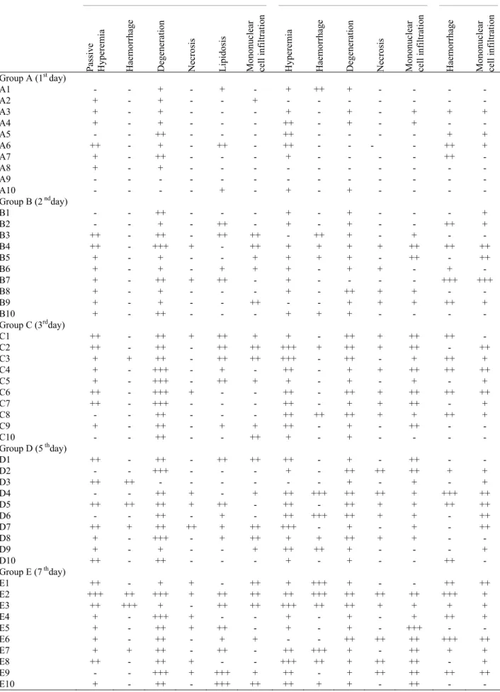
Table 2. Distribution of histopathological findings according to the organs. Tablo 2. Organlara göre mikroskobik bulguların dağılımı.
Liver _____________________________________________________________________ Kidney ________________________________________________________ Heart ______________________ Pa ssive Hyperemia Haemorrhag e Degeneration Necros is
Lipidosis Mononuclear cell inf
iltr atio n Hyperemia Haemorrhag e Degeneration Necros is
Mononuclear cell inf
iltr
atio
n
Haemorrhag
e
Mononuclear cell inf
iltr atio n Group A (1st day) A1 - - + - + - + ++ + - - - - A2 + - + - - + - - - A3 + - + - - - + - + - + + + A4 + - + - - - ++ - + - + - - A5 - - ++ - - - ++ - - - - + + A6 ++ - + - ++ - ++ - - - - ++ + A7 + - ++ - - - + - - - - ++ - A8 + - + - - - A9 - - - - - A10 - - - - + - + - + - - - - Group B (2 ndday) B1 - - ++ - - - + - + - - - + B2 - - + - ++ - + - + - - ++ + B3 ++ - ++ - ++ ++ - ++ + - + - - B4 ++ - +++ + - ++ + + + + ++ ++ ++ B5 + - + - - + + + + - ++ - ++ B6 + - + - + + + - + + - + - B7 + - ++ + ++ - + - - - - +++ +++ B8 + - + - - - + - ++ + + - - B9 + - + - - ++ - - + + + ++ + B10 + - ++ - - - + + + - - - - Group C (3rdday) C1 ++ - ++ + ++ + + - ++ + ++ ++ - C2 ++ - ++ - ++ ++ +++ + ++ + ++ - ++ C3 + + ++ - ++ ++ +++ - ++ - + ++ + C4 + - +++ - + - ++ - + + ++ ++ ++ C5 + - +++ - ++ + + - + - + - + C6 ++ - +++ + - - ++ - ++ + ++ ++ ++ C7 ++ - +++ - - - ++ - + + ++ - + C8 - - ++ - - - ++ ++ ++ + + ++ + C9 + - ++ - + + ++ - + - ++ - - C10 - - ++ - - ++ + - + - - - - Group D (5 thday) D1 ++ - ++ - ++ ++ ++ - + - ++ - - D2 - - +++ - - - + - ++ ++ ++ + + D3 ++ ++ - - - + - + - + D4 - - ++ + - + ++ +++ ++ ++ + +++ ++ D5 ++ ++ ++ + ++ - ++ - ++ + + ++ ++ D6 - - ++ - + - ++ +++ ++ + + - ++ D7 ++ + ++ ++ + ++ +++ - + - + - ++ D8 + - +++ - + ++ + + ++ + + - - D9 + - + - - + ++ ++ + - - - + D10 ++ - ++ - - - + - + - - ++ - Group E (7 thday) E1 ++ - + + - ++ + +++ + - - ++ ++ E2 +++ ++ +++ + ++ ++ ++ +++ ++ ++ ++ +++ + E3 ++ +++ + - ++ ++ +++ ++ ++ + + + + E4 + - +++ + - - + - + - + ++ + E5 + - ++ + ++ - + - + - +++ - - E6 + - ++ - + + - - ++ ++ ++ +++ ++ E7 + + ++ - ++ - ++ +++ + - ++ + + E8 ++ - ++ + - - +++ ++ + ++ ++ - + E9 - - +++ + +++ + ++ - + ++ ++ ++ ++ E10 + - ++ - +++ ++ ++ + + - ++ - -
Microscopic findings: Major histopathologic
findings of the disease were observed in liver, kidney and hearth tissue which were completely defined in Table 2 (Figure 2a-d,3a-d).There was also hyperemia at the fin-skin border and haemorrhage of the same region in group D and E. Lymphocyte infiltration was observed in fin-skin border of all groups which was more significant in group D and E. Branchial arters of gills were seen to be filled with erythrocytes in all groups. Hyperemia of brain vessels in all groups and focal lymphocyte infiltration in group C, D, and E was also seen.
Microbiologic findings: Tissue samples taken from
the organs that have lesions were cultured in blood agar in order to identify the bacteria. And then biochemical tests (β-hemolyse, esculin hydrolyse, H2S production,
glucose, maltose, sucrose, mannitol fermentation) were performed for the identification of the colonies that proliferated in the agar. As a result, reisolation and identification of the bacteria was achieved from the organs that have lesions.
Discussion and Conclusion
Although motile aeromonads appropriately receive much notoriety as pathogens of fish, it is important to note that these bacteria also compose part of the normal
intestinal microflora of healthy fish. Therefore, the presence of these bacteria, by itself, is not indicative of disease and, consequently, stress is often considered to be a contributing factor in outbreaks of disease caused by these bacteria. (16,17,19,26).
Abrupt temperature change, handling, crowding, inadequate feed and oxygen are known to be the predisposing factors which contribute to the infection of
A. hydrophila (20,24). Such stressors are most commonly
associated with environmental and physiological parameters that adversely fish under intensive culture. As one such example, Eissa et al. (8) have shown that the prevalence of motile aeromonad septicemia in cultured and wild Nile tilapia (Oreochromis niloticus) was 10% and 2.5% respectively. During this study water heat was adjusted to 27 ± 1 oC, fish were placed in ratio of
10lt/fish and fed with %2 BW/day. By that way, with providing the optimum environmental conditions, stress factors were eliminated and investigation of the bacterial activity and morbidity of the agent under sublethal bacteria concentration was performed.
Clinically, fish that have motile aeromonad septicemia are reported to be stagnant, be close to the wall and floor of the aquarium, anorectic and defecate luminal shaped faeces or free mucus (1,4,6). In this study
Figure 3. a. Severe hyperemia in kidney (thick arrow) and degenerative changes in tubule epithels (thin arrow) (C2; HE, 200x). b. Haemorrhage in kidney (thin arrow) and necrosis at tubule epithels (thick arrow) (E2; HE, 400x).
c. Pericardial haemorrhage (arrow) (C2; HE, 100x).
d. Lymphocyte infiltration between the cardiac muscle fibres (arrow) (B7; HE, 400x).
Şekil 3. a. Böbrekte şiddetli hiperemi (kalın ok) ve tubul epitellerinde dejeneratif değişiklikler (inceok) (C2; HE,200x). b. Böbrekte kanama (ince ok) ve tubul epitellerinde nekroz (kalın ok) (E2; HE,400x).
c. Kalpte perikardda kanama (ok) (C2; HE,100x).
all clinical findings except luminal shaped or free mucus defecation was observed. According to the present authors, the absence of this finding was due to the 7 day long acute infection model which was more significant in chronic infections.
In carps, when the clinical signs of infection are present, affected fish may show exopthalmia, reddening or darkening of the skin, and an accumulation of fluid in the scale pockets. The abdomen may become distended as a result of an edema and the scales may bristle out from the skin to give a “washboard” appearance (7,10,24). In a natural infection model from Nile tilapia
(Oreochromis niloticus) farm in Saudi Arabia, it was
reported that fish show ecchymosis, loss of scale and few ulcers in the trunk area, redness at the margins of the eye orbits. Corneal opacity with slight exophthalmia in the eyes of the morbid fish was also reported (1). In present study none of case in any groups showed corneal opacity or exophthalmus. Also there was neither any ulceration nor scale loss was observed. In the base of the pectoral fins from where it attaches to the trunk significant hyperemic areas, in direct proportion with the severity of the disease, was observed. Also starting from the 3rd day
post injection, fin rot (especially in the pectoral fins) was seen. Significant darkening in the skin was also identified which was more evident by the time pass. Huizinga et al. (14) reported that focal haemorrhage and dermal lesions accompanied by ulcerative form of the disease were observed in chronic motil aeromonad infection significantly and target organs in acute septicaemia were liver and kidneys. According to present authors, mildness of the skin lesions in this experiment was due to the visceral organ based acute infection model of this short duration follow up study.
In a prior study about pathological findings in carps experimentally infected with A. hydrophila (9), intraperitoneally injected fishes, showed pathological changes mostly in visceral organs, on the other hand, intramuscularly injected fishes had changes mostly on their skins and muscles. Furthermore, it is also observed in a study (21) about A. hydrophila infection in cultured carps (Carassus autarus) that bacterial virulence in fishes intraperitoneally injected were higher than the fishes intramuscularly injected. According to the present authors, the reason why the virulence was higher in intraperitoneal injection group than the intramuscular was the faster and higher morbidity rate of intraperitoneal injection which enables direct contact of the bacteria with visceral organs. In present study, following intraperitoneal injections, focusing of the lesions in the visceral organs such as kidney, liver, and hearth in both macroscopic and miscroscopic levels, verifies this theory. In a study by Afifi et al. (1), although a significant splenomegaly associated with phagocytic activity of the melano-macrophage centers of the spleen was reported,
interestingly it was not observed in any case of this study. Huizinga et al. (14) reported that no lesions were observed in hearth and spleen in the acute cases in which diffuse tissue necrosis of liver and kidneys were present. In present study, although lack of any finding about splenomegaly was found meaningful, linear haemorrhages on the ventricular and atrioventricular border in hearth showed disparity with the literature.
In this study, microscopically the most significant findings were observed in liver, kidney, and hearth. In liver, sinusoids were enlarged and Remak cords were dissociated, observation of focal necrosis in hepatocytes and pancreas cells, and sharp-shaped fat vacuoles in hepatocyte cytoplasmas were similar with literature data (23). The reason of the lipidosis and necrosis of the liver was reported to be associated with toxins and extracellular products such as hemolysin, protease, elastase produced by A. hydrophila (1,12,15,18,22,25).
In a study about histopathology of motil aeromonad septicemia in carps (Carassius auratus), haemosiderin deposition in the atrophic hepatocytes and interstitial tissue macrophages and diffuse hematoidin deposition in the spleen was reported (21). Also Ventura & Grizzle (13) reported that fish with only cutaneous infections may have several types of concealed lesions including increased amounts of lipofuscin and haemosiderin in the liver and spleen; however most visceral organs were not necrotic. In present study, pigments such as haemosiderin, lipofuscin, and hematoidine were not observed in the liver and spleen of any case. In the liver; detection of subcapsular, perivascular and peripacreatic lymphocyte infiltration was consistent with those in literature (1,7,9,13,24).
In the kidneys; subcapsular and interstitial haemorrhage, parenchyme degeneration and cytoplasmic vacuol formation, necrosis of the tubular epithelium with focal lymphocyte infiltration were considered to be associated with interstitial nephritis which were seemed to be parallel with previous studies (9,24).
Observation of diffuse haemorrhage and lymphocyte infiltration in liver, kidney and hearth in both macroscopic and microscopic level depicted a visceral haemorrhagic septicemia. As a result it was concluded that the macroscopic and microscopic findings obtained during the whole study was found to be fundamentally similar with the findings of A. hydrophila infections of the other fish species.
Acknowledgements
The authors thanks to Prof Dr. Selçuk SEÇER, Prof Dr. Hijran YAVUZCAN and Res.Asist. F. Sertel SEÇER on behalf of the Ankara University Agricultural Faculty Department of Fisheries and Aquaculture for supports in this study.
References
1. Afifi SH, Al-Thobiati S, Hazaa MS (2000):
Bacteriological and histopathological studies on Aeromonas hydrophila infection of Nile tilapia (Oreochromis niloticus) from fish farms in Saudi Arabia. Assiut Vet Med
J, 84, 195-205.
2. Arda M (1971): Hastalık Etkenlerinin Titrasyon ve Nötralizasyon Testlerinde Uygulanan Laboratuar Metodları, Ankara Üniversitesi Veteriner Fakültesi, Yayın 273, Tatbikat Kılavuzu 175, 105s.
3. Arda M, Seçer S, Sarıeyyüpoğlu M (2005) : İnfeksiyöz
hastalıklar. 69-105. In : M Arda, S Seçer and M
Sarıeyyüpoğlu (Eds), Balık Hastalıkları. Medisan 61, Ankara
4. Austin B, Austin DA (1987): Bacterial fish pathogens. 191-197. In: E Horwood (Ed), Disease in Farmed and Wild Fish. Chichester.
5. Bach R, Chen PK, Chapman CB (1978): Changes in the
spleen of the channel catfish Ictalurus punctatus rafinesque induced by Aeromonas hydrophila. J Fish Dis,
1, 205-207.
6. Baran I., Timur M, Aydın N, İstanbulluoğlu E, Aydıntuğ MK (1980): Çifteler-Sakaryabaşı balık üretim
ve araştırma istasyonunda alabalıklarda (Salmo gairdneri irideus) görülen bakteriyel hemorajik septisemi hastalığı üzerine incelemeler. Ankara Üniv Vet Fak Derg,
27,467-473.
7. Cipriano RC (2001): Aeromonas hydrophila and motile aeromonad septicemias of fish.
Erişim:[http://w3.kunsan.ac.kr/~psw/technote/main.cgi/aer omonas.pdf?down_num=1093572320&
board=pds&command=down_load&d=& filename = aeromonas.pdf]. Erişim tarihi: 11.07.2006
8. Eissa IAM, Badran AF, Moustafa M, Fetaih H (1994):
Contribution to hareketlie Aeromonas septicemie in some cultured and wild freshwater fish. Vet Med J Giza, 42,
63-69.
9. Erer H (1981): The investigation of the pathologic findings of experimental Aeromonas hydrophila infection (Bacterial Haemorrhagic Septicaernia) in carp. Doktora Tezi, Ankara Üniversitesi.
10. Faktorovich KA (1969): Histological changes in the liver,
kidneys, skin and brain of fish sick with red rot. 83-101. In:
KA Faktorovich (Ed), Infectious Diseases of Fish and Their Control, Division of Fisheries Research, Bureau of Sport Fisheries and Wildlife, Washington, DC.
11. Fuentes RJM, Perez HJA (1998): Isolation of Aeromonas
hydrophila in the rainbow trout (Oncorhynchus mykiss).
Vet Mexico, 29, 117-119.
12. Gado MSM (1998): Studies on the virulence of
Aeromonas hydrophila in Nile Tilapia (Oreochromis niloticus). Assiut Vet Med J, 40, 190-200.
13. Grizzle JM, Kirju Y (1993): Histopathology of gill, liver
and pancreas and serum enzyme levels of channel catfish infected with Aeromonas hydrophila complex. J Aquat
Anim Health, 5, 36-50.
14. Huizinga HW, Esch GW, Hazen TC (1979):
Histopathology of red-sore disease (Aeromonas hydrophila) in naturally and experimentally infected largemouth bass Micropterus salmoides (Lacépéde). J Fish Dis, 2 263-277.
15. Kanai K, Wakabayashi H (1984): Purification and some
properties of protease from Aeromonas hydrophila. B Jpn
Soc Sci Fish, 50, 1367-1374.
16. Kaper JB, Lochman H, Colwell RR, Joseph SW (1981):
Aeromonas hydrophila: Ecology and toxigenicity of isoloates from an estuary. J Appl Bacteriol, 50, 359-377.
17. Khalil AH, Mansour EH (1997): Toxicity of crude
extracellular products of Aeromonas hydrophila in tilapia, Tilapia nilotica. Lett Appl Microbiol, 25, 269-273.
18. Lallier R, Bernard F, Lalonda G (1984): Difference in
the extracellular products of two strains of Aeromonas hydrophila virulent and weakly virulent for fish. Can J
Microbiol, 30, 900-904.
19. Len PP (1987): Mesophilic spoilage of marine fish: bay
trout (Arripis trutta), bream (Acanthopagrus butcheri) and mullet (Aldrichetta forsteri). Food Technol Aust, 39,
277-282.
20. Leung KY, Yeab IV, Lam TJ, Sin YM (1994): Serum
resistance as a good indicator for virulence in Aeromonas hydrophila strains isolated from diseased fish in South East Asia. J Fish Dis, 18, 511-518.
21. Miyazaki T, Kaige N (1985): A histopathological study
on motile Aeromonad disease of crucian carp. Fish Pathol,
21, 181-185.
22. Nieto TP, Santos Y, Rodriguez LA, Ellis AE (1991): An
extracellular acetyl choline estrase produced by Aeromonas hydrophila is a major lethal toxin for fish.
Microb Pathogenesis, 11, 101-110.
23. Paperna I (1996): Bacterial infections. 10-28. In: I Paperna (Ed), Parasites, Infections and Diseases of Fishes, An update, CIFA Technical Paper, Rome, FAO.
24. Roberts RJ (2001): The bacteriology of teleosts. 315-321. In: RJ Roberts (Ed), Fish Pathology, WB Saunders, Philadelphia.
25. Rodriguez LA, Ellis AE, Nieto TP (1992): Effects of
acetylcholinesterase toxin of Aeromonas hydrophila on the central nervous system of fish. Microb Pathogenesis, 14,
411-415.
26. Trust TJ, Bull ML, Currie BR, Buckley JT (1974):
Obligate anaerobic bacteria in the gastrointestinal microflora of the grass carp (Ctenopharyngodon idella), goldfish (Carassius auratus), and rainbow trout (Salmo gairdneri). J Fish Res Board Can, 36, 1174-1179.
27. Ventura MT, Grizzle JM (1988): Lesions associated with
natural and experimental infections of Aeromonas hydrophila in channel catfish Ictalurus punctatus (Rafinesque). J Fish Dis, 11, 397-407.
28. Yambot AV, Inglis V (1994): Aeromonas hydrophila
isolated from Nile tilapia (Oreochromis niloticus L.) with “Eye Disease”. International Symposium on Aquatic
Animal Health, Seattle, WA (USA), 4th-8th September, University of California, School of Veterinary Medicine, Davis, CA: 103
Geliş tarihi: 19.10.2009 / Kabul tarihi: 22.01.2010
Address for correspondence
Yrd. Doç.Dr. Banu Yardımcı
Ondokuz Mayis University, Faculty of Veterinary Medicine Department of Preclinical Science
Atakum / Samsun, Turkey e-mail: [email protected]