187 Ankara Üniversitesi Tıp Fakültesi Mecmuası 2009, 62(4) CERRAHİ BİLİMLER / SURGICAL SCIENCES
Olgu Sunumu / Case Report
Sinonasal adenocarcinomas (SAs) are local aggressive tumors and occur in middle and advanced aged male patients.
A polypoid mass involving to the left nose and paranasal sinuses was determined in endoscopical examination of a 57-year-old female patient admitting with epistaxis. Punch biopsy revealed a low grade adenocarcinoma. The mass was totally excised and its histopathological features were similar to punch biopsy. Glandular epithelium was positive for EMA, CK7, CK19, LMWCK and S-100. The findings were concordant with a low grade adenocarcinoma originating from sinonasal mu-cosa. No recurrence was determined 20 months after total excision.
SAs are a heterogenous group of neoplasms characterizing by variable clinical behavior and dif-ferent epidemiological features. In the difdif-ferential diagnosis, many tumoral and tumor like lesions should be considered. The differential diagnosis is very important because of the risk of recur-rence.
Key Words : Adenocarcinoma, Sinonasal Region.
Sinonazal adenokarsinomalar (SA) lokal agresif tümörlerdir ve çoğunlukla orta ve ileri yaştaki er-keklerde görülür.
Burun kanaması şikayeti ile başvuran 57 yaşındaki kadın hastanın endoskopik muayenesinde sol burun boşluğu ve paranazal sinüsleri tutan polipoid kitle lezyonu saptandı. Punç biyopsi dü-şük dereceli adenokarsinoma ile uyumluydu. Kitle total olarak çıkarıldı ve histopatolojik özellikle-ri punç biyopsininki ile benzerdi. Glandüler epitel hücreleözellikle-ri EMA, CK7, CK19, LMWCK ve S-100 için pozitifdi. Bulgular, sinonasal mukozadan kaynaklanan düşük dereceli adenokarsinoma ile uyum-luydu. Total eksizyon sonrası 20. ayda hastada nüks gelişmedi.
SA değişken klinik davranış ve farklı epidemiyolojik özellikler ile karakterli heterojen bir grup ne-oplazmdır. Ayırıcı tanıda, bir çok tümor ve tümor benzeri lezyon hesaba katılmalıdır. Bu lezyonlar arasındaki ayırıcı tanı nüks riski nedeniyle önemlidir.
Anahtar Sözcükler: Adenokarsinoma, Sinonazal Bölge.
1 Gaziosmanpaşa Üniversitesi Tıp Fakültesi Tıbbi Patoloji Anabilim Dalı
Sinonasal Low Grade Adenocarcinoma; A case report and
the analysis of its histochemical and immunohistochemical
characteristics
Sinonazal Düşük Dereceli Adenokarsinoma; Bir olgu sunumu ve histokimyasal / immünohistokimyasal özelliklerin analizi
Reşit Doğan Köseoğlu, İbrahim Aladağ
Tumors of nasal and paranasal sinuses account for 0.4% of all human neo-plasms (1). Sinonasal adenocarcino-mas (SAs) are the third most frequent malignancies following squamous car-cinoma and lymphoma, respectively (1). Dulgerov et al (2) reported that SAs had a rate of 11.4% in all tumors of this region. If salivary gland type ad-enocarcinomas are excluded, SAs can be broadly classified into enteric and nonenteric subtypes based on their histopathological resemblance to in-testinal and submucosal seromucinous glands, respectively (3). The World Health Organization (WHO)
classifi-cation recognizes four major categories of primary SAs; low grade adenocarci-noma, papillary adenocarciadenocarci-noma, in-testinal type adenocarcinoma (ITAC) and polymorphous low grade adeno-carcinoma (4).
SAs are clinically local aggressive tumors and predominantly occur in middle and advanced aged male patients. The ethmoid sinus, maxillary sinus and na-sal cavity are the most common sites of origin (1). Nasal obstruction, epistaxis and rhinorrhoea are the most common symptoms. Probable effects of some factors such as wood and leather dust Received: 12.08.2009 • Accepted: 08.06.2010
Corresponding author Yrd. Doç. Dr. Reşit Doğan Köseoğlu
Gaziosmanpaşa University, School of Medicine, Department of Pathology, Tokat, 60100, Turkey
Phone : +90 356 212 95 00 Fax : +90 356 213 31 79 E-mail Address : [email protected]
188 Sinonasal Low Grade Adenocarcinoma; A case report and the analysis of its histochemical and immunohistochemical characteristics Ankara Üniversitesi Tıp Fakültesi Mecmuası 2009, 62(4)
are suggested for the development of SA (1,5). This occupational relation-ship is valid for especially ITAC and elderly men are chiefly involved and ethmoid sinus is the site of origin (1). Sporadic SAs often affect women and occur in the maxillary sinus (1). In here, we present a 57-year-old female
patient with low grade SA that showed tubulopapillary growth pattern and ar-gue its differential diagnosis, including immunohistochemical characteristics, in accompanying of the literature.
Case history
A 57-year-old female patient was admitted with intermittent epistaxis from left nose and nasal obstruction for last 10 days. The patient was a housewife and the medical history of the patient was unremarkable. General physical exam-ination was normal and there was no abnormal finding of hemogram and blood chemistry. In the endoscopical view of nose and paranasal sinuses, a polypoid mass originating from the
ostium of sphenoid sinus and at the posterior of lower concha was deter-mined. The polypoid lesion extended to nasopharynx.
Histopathological findings
Punch biopsy revealed a polypoid lesion consisting of very crowded glandular structures with back to back pattern. Most glands had papillary structures (Figure 1A). Although cytologically bland and uniform appearance was dominant, nucleomegaly and mild pleomorphism were noticed in some areas. Lining epithelium of the glands was columnar or cuboidal with rare intraepithelial cyst formations Mitosis was very rarely observed. The nuclei with inconspicuous nucleoli were ba-sally located (Fig. 1B). Elongated and distorted glandular structures resem-bling stromal invasion were noticed in the central of the lesion (Fig. 1C). On the basis of these findings, we di-agnosed the case as a low grade adeno-carcinoma originating from sinonasal
mucosa and favored total excision of the mass. The histopathological find-ings of resected polypoid mass were similar to those of previous punch bi-opsy. Polypoid lesion was covered with respiratory type epithelium. More ad-vanced cytological and architectural atypia were not determined according to previous punch biopsy.
Histochemical and immunohisto-chemical findings
Cystic dilated glands and microcysts were filled by deep blue staining material with Periodic acid-Schiff (PAS)-Alcian blue (AB) at pH 2.5 (Fig. 1D). This material was diastase resistant and showed a weak staining with mucicar-men. Histochemical findings pointed out acidic type mucin. The features of antibodies used in the current study and the immunohistochemical results are shown in Table 1. Glandular epi-thelium for EMA, CK7, CK19, LM-WCK and S-100 was focally positive (Fig. 2). Covering respiratory epitheli-um showed also focal staining for CK7 and CK19.
All these findings were concordant with a low grade adenocarcinoma originating from sinonasal mucosa. After the diag-nosis, the patient is closely followed-up with routine clinical, endoscopical and radiological examinations. No re-currence was determined 20 months after total excision.
Discussion
SAs are a heterogenous group of neoplasms characterized by variable clinical be-havior and different epidemiological features. SAs, especially the group of low grade adenocarcinoma have been poorly characterized from the histo-pathological point of view (5). Basic histopathological features of low grade SAs are cystically dilated glands and tubulopapillary structures lined by bland cuboidal and/or columnar epi-thelial cell with intraepiepi-thelial cyst for-mation. In our case, all these findings were present and a focus of
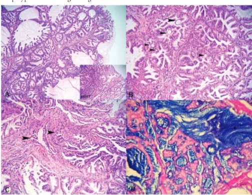
nuclear/ar-Figure 1 A: Polypoid tumoral lesion consisting of crowded tubulopapillary structures with back to back pattern (HE, X10). Inset shows a more crowded area of glands in the tumoral lesion.
Figure 1 B: Intraepithelial microcysts (arrow heads) in the lining epithelium of tubulopapillary structures (HE, X20).
Figure 1 C: Stromal invasion area consisting of distorted glandular structures (HE, X20). Figure 1 D: Intraluminal and intracystic material positive with alcian blue in pH2.5 (Periodic acid Schiff-alcian blue, X30).
189 Journal Of Ankara University Faculty of Medicine 2009, 62(4)
Reşit Doğan Köseoğlu, İbrahim Aladağ chitectural atypia favoring the stromal invasion was also noticed. Intralumi-nal spaces and intraepitelial microcysts were filled with positive material for diastase resistant PAS, AB at pH 2.5 and mucicarmine. Intraluminal mate-rial was acidic type mucin. Similar his-tochemical findings were reported by Skalova et al (4) and Franchi et al (6).
Sinonasal ITACs and low grade SAs have some similar histopathological features. The distinction between low grade SAs and ITACs may be easy in many cases. Evident nuclear pleomor-phism and architectural characteristics reminiscent of a colonic neoplasm are very helpful for this discrimination. But, especially low grade ITACs with
papillary configuration may be very similar to low grade SAs. However, distinction between these is very im-portant because ITACs have an aggres-sive behavior characterized by repeated local recurrences and poor outcome (7). However, low grade SAs have a more indolent clinical course (4). Im-munohistochemically, some distinc-tive characteristics may be suggested for the differential diagnosis. CK7 and CK20 antibodies may be useful for this aim. CK7+/CK20- immuno-phenotype is the immuno-phenotype of normal upper respiratory mucosa. Low grade SAs show also this type. CK7-/CK20+ immunopheno-type shows intestinal differentiation and this phenotype is seen in sinonasal ITACs (8). In our case, both of histo-pathological and immunohistochemi-cal findings favored to low grade SA. Immunophenotype of our case was CK7+/CK20-. In addition, other epi-thelial markers (LMWCK, EMA and CK19) and S-100 were also positive in the present case, while neuroendocrine markers, p53, c-myc, CA19-9, CD10 and CEA were negative. Some authors have reported that expressions of p53, c-myc and CA19-9 have been found in malignant tumors of salivary gland and other organs (especially gastroin-testinal tract) more commonly than their benign counterparts or lesions (9, 10). However, some myoepithelial markers (such as alpha-SMA, CD10) showed stronger staining in benign myoepithelial cells surrounding the malignant epithelial cells than those surrounding benign cells in salivary gland (11). Our case was negative for all of these antibodies.
The differential diagnosis of low grade SAs includes also columnar cell papilloma (Schneiderian papilloma) and poly-morphous low grade adenocarcinoma of minor salivary glands. Columnar cell papilloma has several histological features similar to those of low grade SAs, such as cytologically bland ap-pearance, epithelium with eosinophil-ic staining cytoplasm, intraepithelial microcysts containing mucin, regular simple papillary structures. The
dis-Table 1: The features of the antibodies and the results of immunohistochemical staining.
Antibodies Clone Dilution Result
CK7 Clone OV-TL 12/30 1/200 Focally positive
CK19 Clone BA17 1/50 Focally positive
CK20 Clone Ks20.8 1/50 Negative
LMWCK AE1 1/200 Focally positive
EMA GP1.4 1/1000 Diffusely positive
NSE E27 1/200 Negative
ChA Clone SP12 1/200 Negative
Synp Polyclonal 1/300 Negative
CEA COL-1 1/100 Negative
p53 D07 1/100 Negative
S-100 Polyclonal 1/200 Focally positive
SMA Polyclonal 1/100 Negative
CD10 56C6 1/60 Negative
CA19-9 121SLE 1/100 Negative
c-myc 9E10 1/60 Negative
Abbreviations: CK; cytokeratin, LMWCK; low molecular weight cytokeratin, EMA; epithelial membrane antigen, NSE; neuron specific enolase, ChA; chromogranin A, Synp; synaptophy-sin, CEA; carcino embryonic antigen, SMA; smooth muscle antigen.
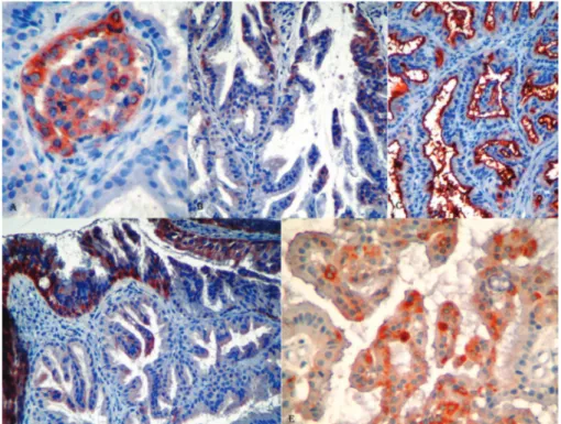
Figure 2: Focal cytoplasmic staining for LMWCK (A), CK 7 (B), CK 19 (D) and S-100 (E) on the lining epithelium of tubulopapillary structures. Pseudostratified epithelium covering the surface of polypoid tumoral lesion shows also cytoplasmic staining for CK 19 (D), (AEC, X40, X20, X20 and X40, respectively).
Diffuse membranous positivity with EMA (C) on the lining epithelium (AEC, X20).
Abbreviations: HE; hematoxyline-eosin, LMWCK; low molecular weight cytokeratin, CK; cyto-keratin, EMA; epithelial membrane antigen, AEC; amino ethyl carbazole.
190 Sinonasal Low Grade Adenocarcinoma; A case report and the analysis of its histochemical and immunohistochemical characteristics Ankara Üniversitesi Tıp Fakültesi Mecmuası 2009, 62(4)
tinguishing features of papilloma in-clude stratified epithelium and lack of true glandular formation. However, a characteristic feature of low grade SAs is a true stromal invasion (4). Another important differential diagnosis is polymorphous low grade adenocarci-noma of minor salivary glands in the sinonasal tract (12). This neoplasm is cytologically uniform and bland but it shows considerable morphological variability, frequent perineural inva-sion and clinically more aggressive behavior. Mucin production is not present. True stromal invasion and mucin production were present in our case. However, co-expression of CK7 and CK19 was remarkable in the pres-ent case. CK19 is more frequpres-ently positive in salivary gland tumors. This may point out to a mixed origin of the present tumor at the level of im-munophenotype. Another differential diagnosis is papillary cystic variant of
acinic cell carcinoma which is a type of minor salivary gland neoplasm that is rarely seen and has a papillary-cys-tic morphology (13). Bland cytology, S-100 expression and intraepithelial microcysts are similar features for both neoplasms. However, basophilic stain-ing of cytoplasm and more aggressive behavior are characteristic features for papillary cystic variant of acinic cell carcinoma (13). A basophilic staining of cytoplasm was not present in the tumor of the present case and our case has a clinically indolent course. No re-currence was determined at postopera-tive 20 months.
Another lesion which should be consid-ered in the differential diagnosis is sinonasal polyp (antrocoanal polyp). Sinonasal polyps may include foci of glandular proliferations reminis-cent of a glandular neoplasm in the edematous loosely stroma. Papillary
configuration, true stromal invasion and higher degrees of cellularity and nuclear pleomorphism are distinguish-ing features for low grade SAs. Our case also showed similar areas as sino-nasal polyp. Areas of higher cellularity, complexity of glandular structures and true invasion foci are important in the differential diagnosis.
In conclusion, low grade SA is a poorly defined neoplasm of sinonasal tract and only a few small series of the cases are present in the literature. Its bland morphological features constitute the basis of difficulty in the differential diagnosis with other similar neoplasms of sinonasal tract. Although they have a lower risk for recurrence than sino-nasal ITACs, low grade SA should be distinguished from other lesions of sinonasal tract, especially benign neo-plasms such as columnar cell papil-loma and cellular sinonasal polyp.
REFERENCES
1. Abecasis J, Viana G, Pissarra C, Pereira T, Fonseca I, Soares J. Adenocarcinomas of the nasal cavity and paranasal sinuses: a clini-copathological and immunohistochemical study of 14 cases. Histopathology 2004; 45: 254-259.
2. Dulgerov P, Jacobsen MS, Allal AS, Lehm-ann W, Calcaterra T. Nasal and paranasal sinus carcinoma: are we making proggress? A series of 220 patients and a systematic re-view. Cancer 2002; 92: 3012-3029. 3. Franquemont DW, Fechner RE, Mills SE.
Histologic classification of sinonasal intesti-nal type adenocarcinoma. Am J Surg Pathol 1991; 15: 368-375.
4. Skalova A, Cardesa A, Leivo I, Pfaltz M, Ryska A, Simpson R et al. Sinonasal tubu-lopapillary low grade adenocarcinoma. His-topathological, immunohistochemical and ultrastructural features of poorly recognised entity. Virchows Arch 2003; 443: 152-158.
5. Franchi A, Massi D, Palomba A, Biancalani M, Santucci M. CDX-2, cytokeratin 7 and cytokeratin 20 immunohistochemical expression in the differential diagnosis of primary adenocarcinomas of the sinonasal tract. Virchows Arch 2004; 445: 63-67. 6. Franchi A, Palomba A, Massi D, Biancalani
M, Sardi I, Gallo O et al. Low-grade salivary type tubulo-papillary adenocarcinoma of the sinonasal tract. Histopathology 2006; 48: 869-886.
7. Franchi A, Gallo O, Santucci M. Clinical relevance of the histological classification of sinonasal intestinal type adenocarcinomas. Hum Pathol 1999; 30: 1140-1145. 8. Choi HR, Sturgis EM, Rashid A, DeMonte
F, Luna MA, Batsakis JG et al. Sinonasal ad-enocarcinoma: Evidence for histogenetic di-vergence of the enteric and nonenteric phe-notypes. Hum Pathol 2003; 34: 1101-1107. 9. Deguchi H, Hamano H, Hayashi Y. c-myc,
ras p21 and p53 expression in pleomorphic adenoma and its malignant form of the hu-man salivary glands. Acta Pathol Jpn 1993; 43(7-8): 413-422.
10. Okamura K, Kiyoshima T, Shima K, Ko-bayashi I, Matsuo K, Ishibashi H et al. Im-monuhistochemical expression of CA19-9 and CA125 in mucoepidermoid and ade-noid cystic carcinomas of the salivary gland. Oral Oncol 2002; 38: 244-250.
11. de Araujo VC, Altemani A, Furuse C, Mar-tins MT, de Araujo NS. Immunoprofile of reactive salivary myoepithelial cells in intra-ductal areas of carcinoma ex pleomorphic adenoma. Oral Oncol 2006; 42(10): 1011-1016.
12. Batsakis JG, Pinkston GR, Luna MA, Byers RM, Sciubba JJ, Tillery GW. Adenocarci-noma of the oral cavity: a clinicopathologic study of terminal duct carcinomas. J Laryn-gol Otol 1983; 97: 825-835.
13. Ellis GL, Corio RL. Acinic cell carcinoma: a clinicopathologic analysis of 294 cases. Can-cer 1983; 52: 542-549.