54
Corresponding author
Dr. Sevinç Bostanoğlu
Bardacik Sok. 30/5 Küçükesat 06660 Ankara/Turkey Tel : +90 (312) 310 30 30 /1603 GSM : (532) 391 46 48 Fax : +90 (312) 466 35 95 E-mail adress : [email protected] Received: 20.04.2005 • Accepted: 26.10.2005
Ankara Üniversitesi Tıp Fakültesi Mecmuası 2006; 59:54-57
DAHİLİ BİLİMLER / MEDICAL SCIENCES Olgu Bildirisi / Case Report
W
egener Granulomatosis (WG) is a disorder characterized bynecro-tizing granulomatous vasculitis that primarily affects the upper and lower respiratory tracts and kidneys (1,2). The classic form of WG is a multisystem process that includes respiratory tract granulomas, generalized vasculitis and glomerulonephritis, whereas the limited form of WG involves fo-cal disease without glomerulonephritis (3). We report a case of WG with mul-tisystem involvement.
Case report
A 39-year-old man was admitted to our hospital with headache nausea, vo-mitting, dyspnea, visual loss of the right eye,cough and weight loss. In the physi-cal examination; bilateral concunctival hyperemia, diplopia and ptosis of the right eye and nasal cavitary mass were found. Laboratory examination revealed the following pathological results; CRP: 346 mg/L (N: 0-8), RHF: 376 IU/ml (N: 0-20), IgG:16.9 g/L (N: 6.9-16.1), IgA: 7.39g/L (N: 0.6-3.7). Assays of cytoplasmic nuclear antineutrophil cytoplasmic antibody (c-ANCA) was high. (c-ANCA: 43.0 U/ml N<4.0 ).
High resolution lung CT, orbital and cranial CT images in the both axial and coronal planes, were obtained with Sytec SRI GE, using intravenous con-trast medium. And also abdominal ultrasonography and tomography were per-formed. In the cranial, orbital and paranasal CT images there was 8x10mm, dural based homogeneously contrasted solid lesion near the orbital apex in the left frontal lobe; extensive tentorial enhancement and 4 mm cranial extension through the right posterior ethmoidal cell wall destruction (Fig.1).
Nonhomo-Wegener Granülomatozis (WG) ender görülen, primer olarak üst ve alt solunum yolları ile böbrekle-rin nekrozitan granülomatöz vasküliti ile karakterize sistemik bir otoimmün hastalıktır. Biz multisis-tem tutulumu olan bir WG li olguyu sunmayı amaçladık. 39 yaşındaki erkek hastada oküler, parana-zal, mediastinal, akciğer, dalak ve meningeal tutulum BT, US ve sitolojik bulguları ile sunuldu.
Anahtar sözcükler: Wegener Granülomatozis, BT, US, sitolojik bulgular
Wegener Granulomatozis (WG) is a systemic autoimmune disorder characterized by necrotizing granulomatous vasculitis that primarily affects the upper and lower respiratory tracts and kid-neys. We report a case of WG with multisystem involvement. This 39-year-old man has ocular, paranasal, mediastinal, lung, splenic and meningeal involvement of WG. We describe and cor-relate the CT,US and cytological findings
Key words: Wegener Granulomatosis, CT, US, Cytological findings
Wegener granulomatosis; multisystem involvement
Wegener granulomatozis; multisistem tutulumuNilgün Yıldırım, Sevinç Bostanoğlu, Emrah Akar, Enis Yüksel, M. Alp Karademir
Ankara Numune Eğitim ve Araştırma Hastanesi, Radyoloji Bölümü, Ankara
Journal of Ankara University Faculty of Medicine 2006; 59(2)
55
N. Yıldırım, S. Bostanoğlu, E. Akar et al.
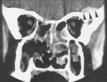
geneous low density solid lesion in the right lacrimal gland measuring 15x6mm (Fig. 2), polipoid solid lesion in the paranasal sinuses and nasal cavity, extending through na-sopharynx, predominantly on the left side with destruction in the sinus walls (Fig. 3).
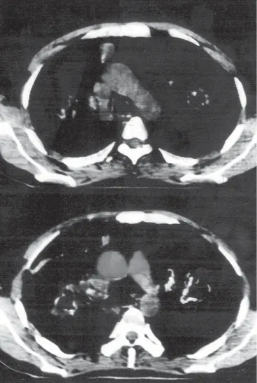
In the high resolution lung CT; there were thick walled irregular cavitary lesions and nodulary infiltrates, the big-gest measuring 55 x 43 mm in the both lung paranchyme (Fig. 4), and multiple mediastinal, bilateral hilar and perivascular lymphadenopathies (Fig. 5).
Abdominal ultrasonography showed splenic inhomog-enity and large anechoic fields adjusting splenic infarcts. With patency of the splenic artery and vein, CT showed splenic inhomogenity with widespread hypodense lesions.
Nasal mass biopsy was performed and revealed WG. Treatment with prednisolone and cyclophosphamide was initiated and the patient was discharged from the hospital.
Discussion
The typical triad of WG consist of; necrotizing granu-lomatous inflammation of the upper and lower respiratory tracts, systemic vasculitis of small arteries and veins; focal glomerulonephritis.The upper respiratory tract is the area most often involved at initial presentation and generally precedes pulmonary or renal involvement (1). WG effects respiratory system 95%, paranasal sinuses 90%, kidneys 85%, nasal cavity and nasophaynx 65%, less frequently orbita, middle ear, joints, muscles, nervous system and gas-trointestinal system (2,3). Primary involvement occurs in upper and lower respiratory tracts and kidneys. Neurologi-cal involvement seen primarily as cranial neuropathies occur in about 34% of cases. Other commonly affected organs in-clude skin and salivary glands. A definitive diagnosis is ob-tained by establishing the presence of a necrotizing granu-lomatous vasculitis usually by lung, upper airway or kidney biopsy. C-ANCA has been shown to have a high sensitivity for WG (5). In our case c-ANCA was found high.
Pulmonary system involvement occurs 45% of patients at presentation and in 90% during the course of the dis-ease. Cough, hemoptysis and pleuritis are the most com-mon pulcom-monary signs. Classic chest radiographic findings include bilateral paranchymal nodes (+/- cavitation) or airway disease simulating pneumonia. Less common find-ings include hilar or mediastinal adenopathy and pleural effusion, pleural thickening or effusion, laryngeal or tra-cheobronchial stenosis (6,7). Cavitation usually occures in nodules larger than 2cm (6). In our case the smallest cavi-tary lesion was in 1.5 cm in diameter. Hilar adenopathy and mediastinal masses are distinctly rare in patients with Figure 1. Dural based homogeneously contrast enhanced solid nodular
lesion (black arrowheads) in the left frontal lobe and 4mm cranial extension through the right posterior ethmoidal cell wall destruction.
Figure 2. Hypodens mass in the right lacrimal gland (black arrowheads).
Figure 3. Polipoid mass in the paranasal sinuses and nasal cavity with
Ankara Üniversitesi Tıp Fakültesi Mecmuası 2006; 59(2)
56 Wegener granulomatozis; multisystem involvement
WG. George et al (8). described 2%, mediastinal and hilar involvement in 302 patients. In our case there are multiple mediastinal and hilar lymphadenopathies.
Ocular involvement occurs in up to 25-50% of patients with WG. Opthalmological manifestations including both ocular (involving the globe) and orbital disease have been reported in 40-50% of WG patients. The principal types of opthalmologic involvement are granulomatous disease (causing an orbital inflammatory mass often with proptosis and/or optic nerve compression) and small vessel vasculitis (causing conjuctivitis, scleritis, uveitis, optic neuritis, optic nerve vasculitis, retinitis, central retinal artery ischemia or cranial nerve paresis) (9,10). Patients may have proptosis caused by a retroorbital pseudotumor or an extension sino-nasal disease into the orbit.(1). Orbital granulomas can be characterized as contigious (i.e secondary to extension of granulomatous disease from the nasal passages of paranasal sinuses) or focal arising primarily within the orbit. Usually extraconal and intraconal involvement occur together and c-ANCA titer was found high in orbital granolomatous mass lesion(11). In our case there were conjunctivities in both eyes and there was a lacrimal gland mass in the right eye and c-ANCA titres were high.
Neurologic involvement in WG is primarily in the peripheral nervous system and less frequently like cranial neuropathy (11-14). Incidence during the course of the disease is up to 20%. Peripheral neuropathy is the most common single neurologic feature with 16% incidence and mononeuritis multiplex. The incidence of cranial neu-ropathy is 6%. Cranial nerves II, VI and VII are most com-monly affected (1). Granulumatous inflammation of the meninges occures rarely (2-8%) (4,12,14). Drachman (12) reviewing 104 cases of WG discounted three mechanisms for neurologic involvement. Direct invasion from the nasal
granuloma, granulomatous lesions remote from the nasal granuloma and vasculitis of the nervous system. These may produce peripheral neuropathy or cerebral infarcts and hemorrages, dural thickening may be diffuse, nodular or plaque like (15). Meningeal involvement , seen as enhance-ment, is less common but is reported (14,16). In our case there was direct invasion from granuloma in the posterior ethmoidal cell, primary nodular frontal granulomatous le-sion related to dura and intensive content enhancement in tentorium cerebelli secondary to dural granulomatous thickening.
Splenic infarcts secondary to vasculitis may occur in WG (15-17). In our case splenic infarcts were established with performing US and CT.
As a result; a WG case was described with paranasal sinus, orbital, cranial, meningeal, lung and splenic in-volvement. In these settings with presence of ANCA, WG should be considered in differential diagnosis.
Figure 4. Irregular cavitary and solid lesions in both lung parenchyme.
Journal of Ankara University Faculty of Medicine 2006; 59(2)
57
N. Yıldırım, S. Bostanoğlu, E. Akar et al. References
1. Clark WJ, Broumand V, Ruskin JD et al: Erythematous, granular, soft tissue lesion of the gingiva. J Oral Maxillofac Surg 1998; 56:962-7.
2. Weinberger LM, Cohen ML, Remler BF et al. Intracranial Wegener’s Granulomatosis. Neurology 1993; 43:1831-1834. 3. Provenzale JM, Mukherji S, Allen NB et al. Orbital involvement
by Wegener’s Granulomatosis. AJR 1996; 166:929-934.
4. Hoffman GS, Kerr GS, Leavitt RY et al. Wegener Granulomatosis: an analysis of 158 patient. Ann Intern Med 1992; 116:488-498. 5. Provenzale JM, Allen NB. Wegener Granulomatosis. AJNR 1996;
17:785- 792.
6. Wadsworth DT, Siegel MJ, Day DL. Wegener’s Granulomatosis in childeren: chest radioghraphic manifestations. AJR 1994;163:901-904. 7. Weir IH, Muller NL, Chiles C, et al. Wegener’s Granulomatosis:
findings from computed tomography of the chest in 10 patients. Can Assoc Radiol J 1992; 43:31-34.
8. Geoge TM, Cash JM, Farver C et al. Mediastinal mass and hilar adenopathy: rare thoracic manifestations of Wegener’s Granulomatosis. Artritis Rheum 1997; 40:1992-1997. 9. Bullen CL, Liesegang TJ, McDonald TJ et al. Ocular
complication of Wegener’s Granulomatosis. Ophthalmology 1997; 90:279-290.
10. Haynes B, Fishman ML, Fauci AS et al. Ocular complications of Wegener’s Granulomatosis: fifteen years experience and review of the literature. Am J Med 1997; 63:131-141.
11. Drahman DD. Neurologıcal complications of Wegener’s Granulomatosis. Arch Neurol 1993; 8:145-155.
12. Fauci AS, Haynes BF, Katz P et al. Wegener’s Granulomatosis: prospective clinical and therapeutic experience with 85 patients for 21 years. Ann Intern Med 1983; 98:76-85.
13. Andersen JM, Jamiesen DG, Jefferson JM. Non-healing granuloma and the nervous system. Q J Med 1975; 44:309-323. 14. Nishino H, Rubino FA, De Remee RA et al. Neurologıcal
involvement in Wegener’s Granulomatosis: an analysis of 324 consecutive patients at the Mayo Clinic. Ann Neurol 1993; 33:4-9. 15. Kalaitzoglou I, Drevenlengas A, Palladas P et al. MRI Appearance
of pulmonary Wegener’s Granulomatosis with concomitant splenic infarction. Eur Radiol 1998; 8:367-370.
16. Spranger M, Schwab S, Meinck HM. Meningeal involvement in Wegener's granulomatosis confirmed and monitored by positive circulating antineutrophil cytoplasm in cerebrospinal fluid. Neurology 1997; 48:263-5
17. Mc Hugh K, Manson D, Eberhard BA et al. Splenic necrosis in Wegener’s Granulomatozis. Pediatr Radiol 1991; 21:588-589.