Macro anatomical investigations of the cranial cervical ganglion
in roe deer (Capreolus capreolus)
Murat KABAK1, Burcu ONUK1
1 Department of Anatomy, Faculty of Veterinary Medicine, University of Ondokuz Mayıs, Samsun.
Summary: In this study, left and right cranial cervical ganglia (ganglion cervicale craniale) of 9 (6 male, 3 female) roe deer, weighing 20-30 kg were inspected macro anatomically. The cranial cervical ganglion (CCG) was found at ventral of the atlas, caudal of the pharynx, medial retropharyngeal lymph node and levator veli palatini muscle, cranial to the divergence place to final branches of common carotid artery. The internal carotid nerve and jugular nerve ramified from the cranial end of CCG. The jugular nerve gave branches that merged with the vagus and glossopharyngeal nerves. The internal carotid nerve varied among cadavers regarding the number of branches (2, 3 or 4). The CCG gave thin nerve branches that reached to various anatomical structures including wall of the pharynx, the first cervical, accessory, hypoglossal and vagus nerves. The external carotid nerves, with one or two branches, also originated from different areas of CCG. The nerves ramified as two branches from CCG and formed a plexus at the ventral of the caudal part of CCG. This plexus gave thin branches to the nearby anatomical structures at that region. In conclusion, nerves ramifying from CCG of the roe deer varied in number among cadavers. The number and course of these nerves, especially external carotid nerves, were observed different than other species.
Key words: Anatomy, cranial cervical ganglion, roe deer.
Karacada (Capreolus capreolus) ganglion cervicale craniale’nin makroanatomik olarak incelenmesi
Özet: Bu çalışmada, 20-30 kg canlı ağırlığında 9 adet (6 erkek ve 3 dişi) karacanın (Capreolus capreolus) sağlı sollu ganglion cervicale craniale’si makro-anatomik olarak incelendi. Ganglion cervicale craniale’nin, atlas’ın ventral’inde, pharynx, ln. retropharyngeus medialis ve m. levator veli palatini’nin caudal’inde, a. carotis communis’in son kollarının cranial’inde yer aldığı görüldü. Ganglion cervicale craniale’nin cranial ucundan; n. caroticus internus ve n. jugularis’in ayrıldığı, n. jugularis’den de n. vagus ve n. glossopharyngeus’a sinir kollarının çıktığı belirlendi. Kadavralar arasında farklı sayıda olan n. caroticus internus’un 2, 3 yada 4 koldan oluştuğu saptandı. Ganglion cervicale craniale pharynx, n. vagus, n. accessorius, n. hypoglossus ve 1. servikal sinir (n. cervicalis primus) gibi farklı anatomik yapılara ince sinir kolları vermekteydi. Nn. carotici externi’nin ganglion’un farklı bölgelerinden bir yada iki kol olarak başlangıç aldığı görüldü. İki kol olarak başlangıç alan nn. carotici externi’nin, ganglion’un caudal yarımının ventral’inde bir plexus şekillendirdiği, bu plexus’tan çevredeki anatomik yapılara ince kollar ayrıldığı belirlendi. Sonuç olarak, karacanın ganglion cervicale craniale’sinden ayrılan sinirlerin sayısı materyaller arasında değişkendi. Bu sinirlerin, özellikle de nn. carotici externi’nin, sayısı ve seyri diğer türlerden farklı bulundu.
Anahtar sözcükler: Anatomi, ganglion cervicale craniale, karaca.
Introduction
Post-ganglionic nerve fibers origins from the cranial cervical ganglion (CCG) and carries sympathetic innervation to head. The flat-oval, shuttle, or spindle formed CCG is located at the cranial extremity of the sympathetic trunk, caudo-medial of the jugular process, ventral of the tympanic bulla and atlantooccipital joint, and medial of the final branches of the common carotid artery (5, 6, 10, 11, 21). The internal and external carotid and the jugular nerves are the major nerve trunks of CCG. Additionally, CCG also provides nerve branches to the last four cranial nerves, the first cervical nerve, and occasionally to the second, third and fourth cervical nerves, thyroid gland, larynx, pharynx, the common carotid artery and the carotid body as well (2, 6, 10).
Nerves ramifying from CCG are usually accompanied by the vessels and establish the perivascular plexus. Some other independent nerve fibers intermingle with the cranial nerves and transmit sympathetic impulses (9, 17, 21).
Numerous studies in various experiments have dealt with the morphology (3, 4) and development (5, 20) of CCG, the nerves originating from this ganglion, its relationship with the adjacent nerves or arteries (2, 7, 10, 19, 22), and the nerve endings (1) in the ganglion. Although shape, localization and nerve branches of this ganglion have been examined in different species (2, 3, 10, 11, 21), there is no such data available for CCG in roe deer. The purpose of this study, therefore, is to describe the dimensions and localization of this ganglion and the nerve branches originating from it, in roe deer.
Materials and Methods
In this study, 9 adult (6 male and 3 female) roe deer (Capreolus capreolus) heads, weighing between 20-30 kg were examined. Materials were provided from the roe deers referred to our faculty surgery clinics with firearm injuries, that could not survived. The heads were divided into two halves from the median plane by an electric saw and fixated in 10% formalin. Each half of the head was later considered as one cadaver totalling 18 cadavers. The CCG’s of the cadavers were dissected by Olympus SZ61 TRC stereomicroscope and their dimensions were measured by the callipers compass (Mitutoyo,Tokyo, Japan). Upon determination of the nerve origins, the ganglion was hypothetically divided into a cranial and a caudal half. The photographs were taken using an Olympus C-5060 digital camera. The nomenclature adheres to that given in Nomina Anatomica Veterinaria (8).
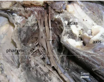
Figure 1: Medial view of cranial cervical ganglion in roe deer. a) cranial cervical ganglion, b) medial retropharyngeal lymph node, c) levator veli palatini muscle, d) common carotid artery, e) ascending pharyngeal artery, f) internal carotid artery, g) internal carotid nerve, h) external carotid nerves, i) jugular nerve, j) the branch extented from CCG to vagus nerve, k) the branch extented from CCG to cranial laryngeal nerve of vagus nerve, l) vagus nevre, m) glossopharyngeal nerve, n) pharyngeal ramus of vagus nerve, o) cranial laryngeal nerve of vagus nerve, p) accessorial nerve, r) first cervical nerve, s) lingual artery, t) sympathetic trunk, u) hypoglossal nerve, v) pharyngeal ramus of glossopharyngeal nerve, w) external carotid artery. Şekil 1: Karacada ganglion cervicale craniale’nin medial’den görünümü.
a) ganglion cervicale craniale, b) ln. retropharygeus medialis, c) m. levator veli palatini, d) a. carotis communis, e) a. pharyngea ascendes, f) a. carotis interna, g) n. caroticus internus, h) nn. carotici externi, i) n. jugularis, j) ganglion’dan n. vagus’a uza-nan kol, k) ganglion’dan n. vagus’un n. laryngeus cranialis’ine uzanan kol, l) n. vagus, m) n. glossopharyngeus, n) n. vagus’un ramus pharyngeus’u, o) n. vagus’un n. laryngeus cranialis’i, p) n. accessorius, r) 1. servikal sinir, s) a. lingulis, t) tr. sympathicus, u) n. hypoglossus, v) n. glossopharyngeus’un ramus pharyngeus’u, w) a. carotis externa.
Results
The CCG (Figures 1/a, 2/a, 3/a) was located at ventral of the atlas, dorsal of pharynx, medial retropharyngeal lymph node (Figure 1/b) and levator veli palatini muscle (Figure 1/c), cranial of the divergence place to final branches of the common carotid artery (Figures 1/d, 2/b), and was in between the ascending pharyngeal (Figures 1/e, 2/c) and the internal carotid (Figures 1/f, 2/d, 3/b) arteries. The ganglion was oval in shape. The mean dimensions of its length, width, and thickness were 13.84 ± 0.50 mm, 3.67 ± 0.15 mm and 3.07 ± 0.14 mm, respectively.
The origin, number, and termination of the nerve branches originating from CCG are presented in Table 1. Branches of CCG comprised the internal carotid nerve (Figures 1/g, 2/e, 3/c), external carotid nerves (Figures 1/h, 2/f), jugular nerve (Figures 1/i, 3/d), and connecting branches to the pharynx (Figure 2/g), vagus (Figures 1/j, 2/h), glossopharyngeal, cranial laryngeal (Figures 1/k, 2/i), the first cervical, accessory nerves, and vessels in the carotid body region.
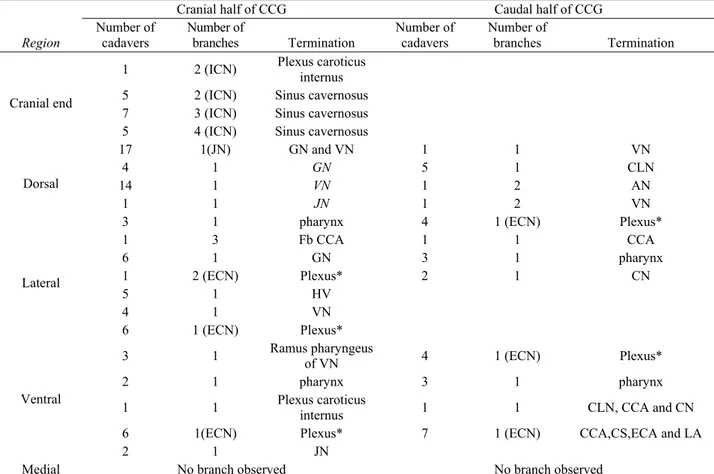
The internal carotid nerve (Figures 1/g, 2/e, 3/c) gave branches, different in number among cadavers: 2 in 6 cadavers, 3 in 7 cadavers (Figure 1), and 4 in 5 cadavers (Figure 3). All of these branches, except in one cadaver, entered the cranial cavity through the petrooccipital fissure running along with the internal carotid artery without forming internal carotid plexus. From the remaining one cadaver, internal carotid nerve which diverged from the cranial end of ganglion as two branches demonstrated a different course. The thick and dorsal one of these branches formed internal carotid plexus around internal carotid artery together with a thin branch seperated from the ventral of cranial part of CCG. The other branch in this cadaver which forms internal carotid nerve and courses ventrally, entered cavum cranii together with internal carotid artery, without participating the mentioned plexus. The jugular nerve (Figures 1/i, 3/d) originated as a single nerve at the dorsal of cranial end of the ganglion in all cadavers except one. The jugular nerve split into 2 branches near the jugular foramen or almost at the cranial end of CCG, giving off fibres that merged with the vagus nerve (Figures 1/l, 2/j, 3/e) and the glossopharyngeal nerve (Figures 1/m, 2/k, 3/f). Only in one cadaver, this nerve formed two branches originating from ventral and dorsal of the cranial part of CCG. Different from the jugular nerve, the one thin branch ramifying dorsally from the cranial part of the ganglion reached the vagus nerve (in fourteen cadavers) and glossopharyngeal nerve (in four cadavers).
From the lateral of cranial part of CCG, one branch extented to pharynx (in three cadavers), one branch to hypoglossal nerve (in five cadavers), and one branch extented to vagus nerve (in four cadavers). In another cadaver, three branches originating laterally from the
cranial part of the ganglion reached to a region between the final branches of the common carotid artery. Besides, in eleven cadavers, a nerve branch (ramus sinus caroticus) (Figures 2/l, 3/g) arising from the glossopharyngeal nerve reached to the same location. In seven cadavers, this branch (ramus sinus caroticus) of glossopharyngeal nerve joined to the plexus (Figure 2*) formed by branches of the external carotid nerve.
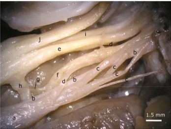
Figure 2: The nerve branches originating from caudal part of cranial cervical ganglion in roe deer.
a) cranial cervical ganglion, b) common carotid artery, c) ascending pharyngeal artery, d) internal carotid artery, e) internal carotid nerve, f) external carotid nerves, g) the branch extented from CCG to pharynx, h) the branch extented from CCG to vagus nerve, i) the branch extented from CCG to cranial laryngeal nerve of vagus nerve, j) vagus nerve, k) glossopharyngeal nerve, 1) branch reached from glossopharyngeal nerve to carotid sinus (ramus sinus carotici), m) pharyngeal ramus of vagus nerve, n) cranial laryngeal nerve of vagal nerve, o) first cervical nerve, p) lingual artery, r) branch extented from plexus to lingual and external carotid arteries, s) branch extented from plexus to external carotid artery and last branches of common carotid artery, t) branch extented from plexus to lingual artery, u) branch extented from plexus to common carotid artery, v) sympathetic trunk, w) hypoglossal nerve, x) branch reached from ascending pharyngeal artery to medial retropharyngeal lymph node, y) occipital artery, z) external carotid artery, *) the plexus formed by branches of the external carotid nerves. Şekil 2: Karacada ganglion cervicale craniale’nin caudal kısmından orijin alan sinir kolları.
a) ganglion cervicale craniale, b) a. carotis communis, c) a. pharyngea ascendes, d) a. carotis interna, e) n. caroticus internus, f) nn. carotici externi, g) ganglion’dan pharynx’e uzanan kol, h) ganglion’dan n. vagus’a uzanan kol, i) ganglion’dan n. vagus’un n. laryngeus cranialis’ine uzanan kol, j) n. vagus, k) n. glossopharyngeus, l) n. glossopharyngeus’tan sinus caroticus’a uzanan kol (ramus sinus carotici), m) n. vagus’un ramus pharyngeus’u, n) n. vagus’un n. laryngeus cranialis’i, o) 1. servikal sinir, p) a. lingualis, r) plexus’tan a. lingualis ve a. carotis externa’ya uzanan kol, s) plexus’tan a. carotis externa ve a. carotis communis’in son kollarına uzanan kol, t) plexus’tan a. lingualis’e uzanan kol, u) plexus’tan a. carotis communis’e uzanan kol, v) tr. sympathicus, w) n. hypoglossus, x) a. pharyngea ascendes’ten ln. retropharyngeus medialis’e uzanan kol, y) a. occipitalis z) a. carotis externa, *) nn. carotici externi’nin oluşturduğu plexus.
Figure 3: The nerve branches originating from cranial end of cranial cervical ganglion in roe deer
a) cranial cervical ganglion, b) internal carotid artery, c) internal carotid nerve, d) jugular nerve, e) vagus nerve, f) glossopharyngeal nerve, g) branch reached from glossopharyngeal nerve to carotid sinus, h) pharyngeal ramus of vagus nerve, i) accessorial nerve, j) hypoglossal nerve, k) medial retropharyngeal lymph node.
Şekil 3: Karacada ganglion cervicale craniale’nin cranial ucundan başlangıç alan sinir kolları.
a) ganglion cervicale craniale, b) a. carotis interna, c) n. caroticus internus, d) n. jugularis, e) n. vagus, f) n. glossopharyngeus, g) n. glossopharyngeus’tan sinus caroticus’a uzanan kol, h) n. vagus’un ramus pharyngeus’u, i) n. accessorius, j) n. hypoglossus, k) ln. retropharyngeus medialis.
From the ventral of cranial part of CCG, one branch originated to pharyngeal ramus of vagus nerve (Figures 1/n, 2/m, 3/h) in three cadavers, and one branch originated to pharynx in two cadavers. In addition, one branch arising from the ventral of the cranial part of CCG, entered into internal carotid plexus. Also, in six cadavers, one branch (external carotid nerve) started from the same region of CCG and formed a plexus with another external carotid nerve originating from lateral of the cranial part of CCG.
A nerve branch observed in six cadavers originating from the dorsal of the caudal part of CCG, extended to the vagus nerve in one cadaver and to the cranial laryngeal nerve (Figures 1/o, 2/n) of the vagus nerve in five cadavers. From the dorsal of the caudal part of CCG, two branches joined with the accessory nerve (Figures 1/p, 3/i) in one cadaver, and with the vagus nerve in another cadaver.
From the lateral of caudal part of CCG, one branch extented to common carotid artery (in one cadaver), to the first cervical nerve (Figures 1/r, 2/o) (in three cadavers) and to pharynx (in three cadavers).
A nerve branch observed in three cadavers originating from ventral of the caudal part of CCG, extended to the pharynx. In another cadaver, one branch originating from ventral of caudal part of the ganglion
ramified three nerve branches. These branches reached to cranial laryngeal nerve of vagus nerve, to common carotid artery and to the first cervical nerve.
External carotid nerve (Figures 1/h, 2/f) ramified from ganglion as one or two branches. The origin from the ganglion of this nerve was fairly different. In seven cadavers, a single (Figure1/h) branch ramified from the ventral of the caudal part of CCG. This branch (external carotid nerve) gave thin branches to the carotid sinus, lingual artery (Figures 1/s, 2/p), common carotid artery and the last branches of that. Ganglion exit regions of the two external carotid nerves were rather variable. In six cadavers, one branch seperated from the lateral of the cranial part of the ganglion, where the other branch seperated from the ventral border. In one cadaver, external carotid nerve consist of two nerve branches originated from only the lateral of cranial part of CCG. In four cadavers, (Figure 2) external carotid nerves originated from the lateral and also ventral of the caudal part of the ganglion. These branches forms a plexus (Figure 2*) at the ventral of ganglion, as in the other cadavers with two branches. From this plexus, thin
branches (Figure 2) ramified to pharynx, cranial laryngeal nerve of vagus nerve, external carotid artery (Figure 2/r-s), lingual artery (Figure 2/t), common carotid artery (Figure 2/u) and the last branches of that (figure 2/s). Nerve branches going to arteries formed a perivascular plexus around the mentioned arteries.
Discussion and Conclusion
The post-ganglionic nerve fibers which arises from the neurons in CCG, transfer sympathetic innervation to the smooth muscles, glands and mucosa of the head. Lesions at the ganglions results with reduction of melatonin release in pineal gland (13, 15) and Horner’s syndrome (12, 14, 16, 18). The localization of this ganglion and the nerve origins emerging from it, has a crucial importance. In this study, it was observed that the form and localization of the ganglion was similar to other species (5, 6, 10, 19, 21).
In camel, CCG is 15-20 mm long, 4-6 mm wide, and 3 mm thick (2). In sheep, CCG is approximately 15 mm in length, 5 mm in width, and 3 mm in thickness while it is 8 mm long, 3 mm wide, and 3 mm thick in
Table 1: Nerve branches orinigating from the cranial cervical ganglion (CCG) of roe deer: origin, number and termination Tablo 1: Karacada ganglion cervicale craniale’den ayrılan sinir kolları: başlangıç, sayı ve sonlanması
Cranial half of CCG Caudal half of CCG
Region Number of cadavers Number of branches Termination Number of cadavers Number of branches Termination
1 2 (ICN) Plexus caroticus internus 5 2 (ICN) Sinus cavernosus 7 3 (ICN) Sinus cavernosus Cranial end
5 4 (ICN) Sinus cavernosus
17 1(JN) GN and VN 1 1 VN
4 1 GN 5 1 CLN
14 1 VN 1 2 AN
Dorsal
1 1 JN 1 2 VN
3 1 pharynx 4 1 (ECN) Plexus*
1 3 Fb CCA 1 1 CCA 6 1 GN 3 1 pharynx 1 2 (ECN) Plexus* 2 1 CN 5 1 HV 4 1 VN Lateral 6 1 (ECN) Plexus*
3 1 Ramus pharyngeus of VN 4 1 (ECN) Plexus*
2 1 pharynx 3 1 pharynx
1 1 Plexus caroticus internus 1 1 CLN, CCA and CN
6 1(ECN) Plexus* 7 1 (ECN) CCA,CS,ECA and LA
Ventral
2 1 JN
Medial No branch observed No branch observed
ICN) Internal carotid nerve JN) Jugular nerve GN) Glossopharyngeal nerve VN) Vagus nerve HV) Hypoglossal nerve CN) Cervical nerve AN) Accessory nerve ECA) External carotid artery ECN) External carotid nerve Fb CCA) at the level seperation of the final branches of CCA CCA) Common carotid artery CLN) Cranial laryngeal nerve LA) Lingual artery PLEXUS*) Thin branches reach from this plexus to pharynx,.CLN, Fb CCA,CCA,ECA and LA
goat (6). The yak’s CCG is 19.72 mm in length, 7.65 mm in width and 4.55 mm in thickness (21). Width and thickness dimensions of CCG demonstrated in this study are similar to that of Getty (6) reported for goat. However, ganglion length was between that of sheep and goat, but more near to sheep.
Three major branches originating from CCG were the internal and external carotid and jugular nerves. The origin and course of the jugular and internal carotid nerves corresponded with those of other domestic animals (2, 10, 11, 19, 21). However, there were numerical differences in literature associated with the branches of these nerves. The internal carotid nerve ramified as 2 branches from the cranial end of CCG in literature (3, 6, 11). These branches form the internal carotid plexus, circumscribes the internal carotid artery and run along with this artery towards the cranial cavity (6, 17, 21). Our results revealed that the internal carotid nerve in the roe deer possessed 2, 3 or 4 branches, similar to pig (10). More importantly, these branches running along with the internal carotid artery do not form a plexus around the artery. These results are generally concordant with the results of Kabak et al., (10) in pig, Kabak (11) in guinea pig and Cui-Seng et al. (2) in camel, however in one cadaver, formation of a plexus around internal carotid artery by the dorsal branch of internal carotid nerve is similar to literature (6, 17, 21).
In literature, it is indicated that external carotid nerve diverges at the caudal part of ganglion to one (2, 3, 6, 11, 19), two or three branches (10). In this study, external carotid nerve had a single branch in seven cadavers and two branches in eleven cadavers. Origin of external carotid nerves which solitarly exits from the ganglion and ramifies as two branches in four cadavers, was observed to originate from ventral and lateral of the caudal part of CCG similar to literature (2, 3, 10, 11, 19). The nerve existing as two branches in seven cadavers, originates from the lateral and ventral of the cranial part of CCG in the six and only the lateral in the remaining one cadaver. This finding has not been notified in any literature yet. In addition, the other important finding in this study is the formation of a plexus from the unition of these nerves starting as two seperate branches, at the ventral of the caudal part of the ganglion and ramifying of thin branches to the nearby anatomical structures from this plexus. Origin and course of the jugular nerve was generally similar to literature (2, 3, 10, 11, 19). Difference in the origin of the nerve was described in only one cadaver.
The observations on the communicating branches from the CCG to the last four cranial nerves, the pharynx, the vessels near to the ganglion and to the cervical nerves are in accordance with the literature reports (2, 7, 10, 17, 19). This study exposed a branch extending between
CCG and cranial laryngeal nerve, as was the case observed in the rat (7), the pig (10) and the guinea pig (11). As stated in literature (3, 21, 22), in five cadavers, there was a connection between the hypoglossal nerve and the ganglion.
The branches connecting CCG with the cervical nerves are either a single branch (2, 6, 17, 19) or two-three branches (10), or four branches (7) in domestic animals. In this study, it was a single branch in three cadavers.
In conclusion, no sexual difference was observed in this study which determined the location of CCG, major nerves ramified from the ganglion and thin nerve branches reaching to anatomical structures of the region, in roe deer. While presenting some new findings, the results are generally in accordance with current literature.
References
1. Abdel-Magied EM (1995): Fine structure of nerve
endings and junctions in the superior cervical ganglion of the camel (Camelus dromedarius). Anat Histol Embryol,
24,117-121.
2. Cui-Sheng, Wang JL, Xie ZM (1998): The gross
anatomy of the cranial cervical ganglion and its branches in the Bacterian camel (Camelus bactrianus). Vet Res
Commun, 22, 1-5.
3. Çakir A (2001). The morphology of cranial cervical
ganglion in the New Zealand Rabbit. Vet J Ankara Univ,
48, 83-87.
4. Ebbesson SOE (1968): Quantitative studies of superior
cervical sympathetic ganglia in a variety of primates including man. I. The ratio of preganglionic fibers to ganglionic neurons. J Morphol, 124, 117-132.
5. Fioretto ET, de Abreu RN, Castro MFDS, Guidi WL, Ribeiro AACM (2007): Macro- and microstructure of the
superior cervical ganglion in dogs, cats and horses during maturation. Cells Tissues Organs, 186, 129-140.
6. Getty R (1975): Sisson and Grossman’s The Anatomy of
The Domestic Animals. In: King AS, Respiratory system,
5th ed., W.B. Saunders Company, Philadelphia.
7. Hedger JH, Webber RH (1976): Anatomical study of the
cervical sympathetic trunk and ganglia in the albino rat (Mus norvegicus albinos). Acta Anat, 96, 206-217.
8. Frewein J, Habel RE (2005): Nomina Anatomica
Veterinaria, 5th. ed. Prepared by the International Committee
on Veterinary Gross Anatomical Nomenculature (I.C.V.G.A.N) and authorized by the General Assembly of the World Association of Veterinary Anatomists (W.A.V.A.). Hannover.
9. Jenkins TW (1972): Autonomic nervous system. 138. In: Functional Mammalian Neuroanatomy. Philadelphia Lea&Febiger.
10. Kabak M, Orhan İÖ, Hazıroğlu RM (2005): Macro
anatomical investigations of the cranial cervical ganglion in domestic pig (Sus scrofa domesticus). Anat Histol
Embryol, 34, 199-202.
11. Kabak M (2007): The Gross anatomy of the cranial
cervical ganglion in the guinea pig (Cavia porcellus). Vet
12. Kara CO, B Topuz (2002): Horner’s syndrome after
excision of cervical sympathetic chain schwannoma.
Otolaryngol Head Neck Surg, 127, 127-128.
13. Karasek M, Zielinska A, Marek K, Swietoslawski J (2002): Effect of superior cervical ganglionectomy on the
ultrastructure of pinealocytes in the Djungarian hamster (Phodopus sungorus): quantitative study. Neuro
Endocrinol Lett, 23, 443-446.
14. Lembo TM, Wright KC, Cromeens DM, Price RE (2001): Iatrogenic Horner's syndrome in an experimental
pig. Contemp Top Lab Anim Sci, 40, 33-35.
15. Maurel DL, Ben Saad MM, Roch G, Siaud P (2002):
Testicular activity is restored by melatonin replacement after suprachiasmatic nucleus lesion or superior cervical ganglionectomy in mink. J Pineal Res, 32, 15-20.
16. Melian C, Morales M, Espinosa de los Monteros A, Peterson ME (1996): Horner's syndrome associated with
a functional thyroid carcinoma in a dog. J Small Anim
Pract, 37, 591-593.
17. Miller ME, Chistensen GC, Evans HE (1965):
Autonomic nervous system. 634-635. In: Anatomy of the
Dog. W.B. Saunders Company, Philadelphia.
18. Morgan RV, Zanotti SW (1989): Horner's syndrome in
dogs and cats: 49 cases (1980-1986). J Am Vet Med
Assoc, 194, 1096-1099.
19. Özgel Ö, Kurtul I, Dursun N (2004): On the gross
anatomy of the cranial cervical ganglion of the donkey (Equus asinus) in Turkey. Vet Res Commun, 28, 261-266.
20. Rubin E (1985): Development of the rat superior cervical
ganglion: initial stages of synapse formation. J Neurosci,
5, 697-704.
21. Shao BP, Ding YP,Xie ZH, Yu HX, Brand-Saberi B, Wang JL (2007): The cranial cervical ganglion and its
branches in yak (Bos grunniens). Vet J, 173, 174-177
22. Tseng CY, Lue JH, Lee SH, Wen CY, Shieh JY (2001):
Evidence of neuroanatomical connection between the superior cervical ganglion and hypoglossal nerve in the hamster as revealed by tract-tracing and degeneration metods. J Anat, 198, 407-421.
Geliş tarihi: 02.03.2009 / Kabul tarihi: 08.05.2009
Address for correspondence
Asc. Prof. Dr. Murat Kabak
Department of Anatomy, Faculty of Veterinary Medicine, University of Ondokuz Mayıs,
55139 Kurupelit, Samsun – TURKEY Telephone: 90 (0) 362 312 19 19- 2806