Burcu Enneli
1, S. Özbaş Turan
2,
B. Uslu
3, J. Akbuğa
2S1 Yeditepe University, Faculty of Pharmacy, Kayışdağı, Istanbul, Turkey.
2 Marmara University, Faculty of Pharmacy, Department of Pharmaceutical Biotechnology, 34668, Haydarpaşa, Istanbul, Turkey.
3 Van Gynaecological and Children Illnessess Hospital, Van, Turkey.
Abstract
In this study, the complex forms of AsODNs (phosphodiester and phosphorothioate forms) were prepared by using a cationic polymer called chitosan. The effect of chitosan mol wts and chitosan/AsODN ratio on the complex properties was studied. Afterwards, the physicochemical properties such as zeta-potential and particle size of complexes were measured and the in vitro antisense activities of the appropriate formulations were investigated. Full complexation was observed with phosphorothioate AsODN at the ratio of 10:1 (+/-), however phosphodiester AsODN could not be formed full complex with chitosan. The antisense activity of 100:1 (chitosan/AsODN) complexes which have approximately 200 nm particle size, was measured based on the inhibition of β-galactosidase by the antisense containing formulations into the b-Gal transfected HeLa cell lines. Eventually, both of
these complex forms protected AsODNs from potential enzymatic degradations. In 100:1 (+/-) complex formulations, high antisense activity (88-90%) was observed. As a result, chitosan can be considered as a suitable carrier especially for phosphorothioate modified AsODNs. Molecular weight of chitosan and chitosan/AsODN mass ratio had no importance inin vitro inhibition. This study can form the basis for the forthcoming studies related with carrier systems of AsODNs that will be done with chitosan polymer.
Key words: Antisense oligonucleotide, chitosan,
complex, carrier, gene expression.
Introduction
Antisense oligonucleotides (AsODNs) are short, single-stranded, synthetic molecules that have complementary sequences of targeted mRNA or pre-mRNA by which the selective hybridization yields the inhibition of gene expression. These molecules have the potential to provide greater therapeutic benefit over traditional drugs for the treatment of viral infections, cancers, multiple sclerosis, hepatitis, kidney transplantation and ulcerative colitis (Agrawal et al., 1997; Dias et al., 2002; Kurreck et al., 2003; Lee et al., 2003; Lysik et al., 2003). However, the therapeutic applications of oligonucleotide-based therapies are limited by the instability of these molecules towards intra- and extracellular nucleases in biological fluids, short half-life in vivo and insufficient cellular uptake (Brus et al., 2004; Roth et al., 2005; Peng et al., 2006). Chemical modifications have been introduced to increase the stability of AsODNs and their ability to penetrate the plasma membrane but these modifications have also resulted in a variety of non-antisense activities such as sequence independent protein interaction. Viral or non-viral AsODN carrier systems have being developed in order to increase cellular penetration of AsODNs. Viral
In vitro antisense activity of chitosan/AsODN complexes
SCorrespondence Author;
Marmara University, Faculty of Pharmacy, Department of Pharmaceutical Biotechnology 34668 Haydarpaşa, İstanbul, TURKEY
vectors have high transduction efficiency but their therapeutic usage is limited because of high toxicity and immunogenicity (Philips et al., 1997). Therefore, in recent years non-viral AsODN carrier systems have been developed using natural or synthetic cationic polymers and lipids (Guillem et al., 2002; Hussain et al., 2002; Lindner et al., 2006).
Chitosan [β(1-4) 2-amino 2-deoxy β-D glucan], is a deacetylated form of chitin, an abundant polysaccharide present in crustacean shells. It is hydrophilic and positively charged, so it can easily interact with negatively charged antisense oligonucleotides (Prasitsilp et al., 2000; Gao et al., 2005). It is a biodegradable, biocompatible and non-toxic biopolymer (Hirano et al., 1988; Rolland et al., 1998). Though chitosan has been evaluated as a carrier for plasmid DNA (pDNA) into various cell types and experimental animals (Aral et al., 2000; Akbuğa et al., 2003; Issa et al., 2006), there is little information on its use for the delivery of AsODN. Gao et al. (2004) prepared oligonucleotide complexes with galactosylated low molecular weight chitosan for the hepatocyte targeting delivery and obtained high in vitro transfection results. Ferreiro et al., (2003) prepared AsODN complexes with different types of polycations, chitosan glutamate and chloride salts in order to stabilize oligonucleotides in the gastrointestinal tract against nuclease degradation when applied orally. Springate et al., (2005) prepared controlled release delivery system for AsODNs based on complexed chitosan/AsODN dispersed in a biodegradable polymeric paste for intratumoral treatment of solid tumors. In vitro controlled release and in vivo efficacy has been produced over 4 weeks by the complexation of clustering AsODNs with chitosan. Föger et al. (2006) incorporated 30 mer AsODNs against malarial topoisomerase II gene in depolymerized chitosan nanoparticles for anti-malarial treatment and these chitosan nanoparticles exhibited strong inhibitory effect on the parasite growth. However there is no detailed study on the characterization and antisense effect of chitosan/AsODN complexes.
In this study, 15-mer phosphodiester (PO)-and phosphorothioate (PS)-AsODNs against β-galactosi-dase enzyme gene have been complexed with chitosan polymer and the effect of chitosan mol
weight and ratios on complex formation were investigated. Complex formation was displayed by agarose gel electrophoresis and transmission electron microscopy (data not shown). Zeta-potential and particle sizes of the complexes were measured, stability against serum nuclease degradation and in vitro antisense activity were determined.
Materials and methods
Materials
Phosphorothioate-modified (PS)- and phosphodiester (PO)- antisense oligonucleotides (AsODNs, 15 mers) were designed to be antisense to sequence of β-galactosidase enzyme gene (Fillion et al., 2001). All antisense oligonucleotides were synthesized by MWG Biotech (Germany) and were of HPLC grade. Low molecular weight chitosan (70 kDa) was obtained from Sigma (USA), low molecular weight chitosan (150 kDa) and medium molecular weight chitosan (400 kDa) were obtained from Fluka (Germany). Triton X-100 was obtained from E. Merck (Germany). O-nitrophenyl-D-galactopyranoside (ONPG) and β-galactosidase were obtained from Sigma (USA). The pSV-β-galactosidase control vector (pSV-β-Gal) containing SV40 early promoter, enhancer and Lac Z gene is supplied by Promega Corporation, USA.
β-Gal expressed HeLa cell line was obtained from American Type Culture Collection (ATCC CLL–2) and was grown in DMEM (Dulbecco’s Modified Eagle’s Medium) (Sigma, USA) containing 10 % fetal bovine serum (FBS), 0.1 % antibiotic solution [penicillin (10.000 units/mL), streptomycin (10 mg/mL) and amphotericin B (10 mg/mL)]. Cells were maintained at 37°C, 5.0 % CO2 in a 95 % air humidified atmosphere (Heto-Holten, Denmark).
Preparation of chitosan/AsODN complexes
AsODNs were dissolved in tris-EDTA buffer (TE, pH 8.00) and their quantities and qualities were determined spectrophotometrically at 260 and 280 nm (Shimadzu UV-Biospec 1601, Japan) and by electrophoresis in agarose gel. Chitosan with different molecular weight, was dissolved in 1.0 % acetic acid to form solutions of 0.25 % pipetting to form
complexes at various ratios (w/w) (+/-) 0.5:1, 1:1, 2:1, 4:1, 6:1, 10:1, 20:1, 50:1, 80:1, 100:1 and 200:1), vortexed rapidly for 3-5 seconds and left for 1 hour in room temperature for chitosan/AsODN complexes to completely form. Agarose gel elec-trophoresis was carried out to display the complexes.
Agarose gel electrophoresis of complexes
Chitosan/AsODN complexes were applied into a 2.0 % agarose gel containing tris-boric acid-EDTA buffer (TBE) and ethidium bromide (0.5 µg/mL) at constant current (80 mA) (Horizontal gel apparatus system, ATTO, Japan). Complexes were visualized under UV-light and their gel appearance was photographed by Kodak, Digital Science DC290 Camera and 1D Image Analysis Software.
Determination of particle size and surface charge
Surface charge of AsODNs, chitosan polymer and prepared complexes were measured by Zeta-sizer (Malvern Instruments, UK). The measurements were carried out at room temperature in phosphate buffered saline (PBS, pH 7.4). The results were calculated by Malvern PCS Version 4.41 (1992) software. Particle sizes of complexes were measured by particle sizer entegrated Malvern Instruments (UK) and the measurements were carried out at room temperature with a scattering angle of 90°C.
Stability of chitosan/AsODN complexes in DNAase I
DNAase I enzyme (1.0 U/µg AsODN) was added into the free AsODN solutions and prepared complexes, then the mixtures were incubated at 37°C. After incubation of 5., 15., 30., 60. minutes and 2., 24., 48. hours; digestion was terminated with 5 µL of 0.5 M EDTA per 10 µL of samples. Afterwards, agarose gel electrophoresis was carried out with the samples taken.
In vitro antisense effect of chitosan/AsODN complexes
HeLa cells were seeded at the density of 5.0x10⁴
cells/well in 24-well plates with 1 mL of complete medium (DMEM containing 10 % FBS) and incubated
for 24 hours prior to transfection. Transfections were performed on cells that were approximately 70 % confluence. Before transfection, the complete medium was removed and serum free DMEM was added on the cells. The naked AsODN and chitosan/AsODN complexes (containing 2 µg of AsODN) were diluted in DMEM and then were added on the cells. After incubated at 37°C for 6 hours, DMEM containing 10 % serum was added to the wells. After 48 hours of pDNA transfection, the medium containing free complexes was removed. The cells were rinsed twice with PBS, and then 200 µL of Triton X-100/lysis buffer was added on each well. Freezing and thawing process is applied twice for lysis of all cells, 25 µL of cell lysate was pipetted into wells of a 96-well plate, and then 135 µL of Buffer A/µ-mercaptoethanol solution was added into each well, waited for 15 minutes at 37°C. 50 µL of ONPG solution was added and incubated at 37°C for 19 hours. When faint yellow color appeared, 90 µL of Na2CO3solution (1M) was added into each well in order to stop the reaction. The absorbance of the samples were read at 420 nm in UV spectrophotometer (Shimadzu UV Biospec–1601, Japan) (µ-Gal assay was performed according to Promega corporation Technical Bulletin No: 097).
Statistical analysis
All experiments were performed at least three times. The statistical significance of the results was evaluated by Student’s t-test.
Results and discussions
Physical characterization of chitosan/AsODN complexes
In the present study, we evaluated the ability of chitosan, to carry antisense oligonucleotides [phosphorothioate (PS)- and phosphodiester (PO)-] and investigated the effect of formulation factors such as molecular weight of chitosan and chitosan/AsODN ratio on antisense efficacy of chitosan/AsODN complexes.
Firstly we tested the ability of chitosan to form complexes with AsODN as a function of the AsODN chitosan mass ratio. Agarose gel electrophoresis
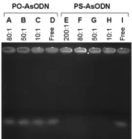
technique was applied to assess the complexation properties of AsODN with chitosan and to determine the presence of uncomplexed (free) antisense oligonucleotides. Chitosan complexes were generally prepared with respect to the ratio between free phosphate in the DNA in earlier studies. However the amount of free amino in the chitosan used in this study is not known previously therefore, instead of N/P ratios, relative weight ratios were used in the preparation of complexes. Different chitosan/AsODN mass ratios, as mentioned in method, have been tested however full complexations were not obtained until the ratio 10:1 therefore gel photograph of these samples are not given gel photograph of complexes in 10:1, 50:1, 80:1 and 200:1 mass ratios is given.
Partial complexation was observed at lower PS-AsODN and chitosan mass ratio (lower than 10:1), therefore gel photograph of these complexes is not shown. Full complexes could be formed at the ratio of 10:1 (+/-) by using PS-AsODNs (lanes E-H) however uncomplexed free AsODNs were observed at all ratios used when complexes were prepared with PO-AsODNs (Figure 1, lanes A-C). It can be said that the mass ratio of PS-AsODN to chitosan is crucial and between the PS-AsODN and chitosan formed full complexes at mass ratios higher than 10:1.
Partial complexes formed between chitosan and PO-AsODNs at all used ratios because in addition to brightness in agarose gel, there are uncomplexed AsODNs in gel. The amount of uncomplexed free oligonucleotide in gel reduced at mass ratio of 200:1 (+/) and complex formation reached optimum level at mass ratio of 200:1 (+/-) (Figure 1).
It is known that the molecular weight of the chitosan has a major effect on its biological and physico-chemical properties (Sato et al., 2001; Huang et al., 2004). Gene-transfer efficiency can be promoted by the modulation of the molecular weight of chitosan. However, in our study, complex formed ability of chitosan did not change with its molecular weight, but the structure of antisense oligonucleotides more important in complex formation with chitosan.
As seen in Figure 2. full complexes were formed between PS-AsODN and chitosan having different
Figure 1. Confirmation of the complex formation between
phosphorothioate (PS) and phosphodiester (PO) chitosan/ AsODN (medium molecular weight, 0.25%) by gel electropho-resis. A. PO chitosan/AsODN (80:1) complex B. (50:1) complex C. (10:1) complex D. Free PO-AsODN E. PS chitosan/AsODN (200:1) complex F. (80:1) complex G. (50:1) complex H. (10:1) complex I. Free PS AsODN.
Figure 2. Electrophoretic control of PS (A) and PO (B)
chi-tosan/AsODN complexes prepared different mol. wt. (70, 150 and 400 kDa) and concentration of chitosan. Lane 1; prepared with 0.25% (w/v) medium molecular weight (400 kDa) chitosan. Lane 2; prepared with 0.25% (w/v) low molecular weight (150 kDa) chitosan. Lane 3; prepared with 0.25% (w/v) low molecular weight (70 kDa) chitosan. Lane 4; Free AsODN. Ratio of the complexes; (Lane 4) Free AsODN (Lane 1-3) 125:1.
molecular weights but partial complexes were obtained with PO-AsODN as mentioned above. All chitosans with different mol. wts behaved similary in their ability to complex with PS-AsODN. PO-AsODN formed partial complexes with chitosan having different molecular weight. Therefore PO-AsODN was excluded from particle size, surface charge and stability studies.
Junghans et al. (2001) prepared protamine and spermidine (spermine) phosphodiester (PO) and phosphorothioate (PS) complex by adsorption, they reported that complexes consisting of the more lipophilic phosphorothioate oligonucleotides were more stable and they showed no aggregation although their surface charge was lower.
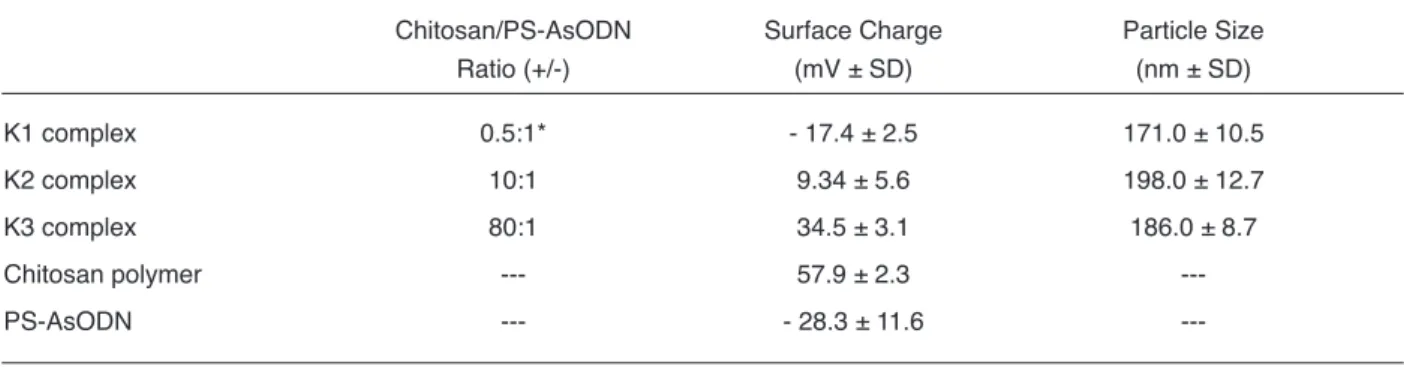
Particle sizes of chitosan/PS-AsODN complexes are given in Table 1. Particle sizes of chitosan complexes, change between 171.0 ± 10.5 nm and 198.0 ± 12.7 nm. There is statistically meaningful particle size difference between complexes however there is no statistically significant correlation between ratio of chitosan and the particle size of the complexes (p>0.05) (Table 1). This result shows similarity with the report of Föger et al. (2006). Gao et al. (2005) prepared complexes using galactosylated chitosan, but they obtained large complexes.
Free AsODNs have negative surface charge whereas chitosan polymer has high positive surface charge value (57.9 ± 2.3 mV) (Table 1). When looked at the surface charge values of complexes, only the
complexes with mass ratio of 0.5:1 (+/-) has negative surface charge indicating that the positive charge of chitosan polymer used in the complex is not enough to cover the negative charge of oligonucleotides of this ratio. However, the complexes prepared at mass ratios of 10:1 and 100: 1 (+/-) have positive surface charges (Table 1). There is significant difference between the surface charges of complexes (p<0.05). Additionally, surface charges of the complexes increase with the increasing amount of chitosan in the complexes and there is statistically significant relationship between the amount of chitosan and the surface charge of the complexes prepared (p<0.05).
Stability of chitosan/AsODN complexes in DNase I
The stability of chitosan/PS-AsODN complexes was studied by gel electrophoresis after incubation with DNAase I or serum for 48 hours. Free AsODN started to be degraded within 5 minutes and disappeared in 15 minutes (Figure. 3).
However, significant protection of AsODNs against the enzymatic degradation was demonstrated with chitosan complex at the mass ratio of 20:1 (+/-). After the incubation of chitosan/AsODN complexes with DNAase I enzyme, uncomplexed AsODN degraded rapidly but complex started to be decomposed after 24 hours. AsODN which is dissociated from complexes is protected against the degradation effects of nucleases (Figure 3).
Table 1. Surface charges and particle sizes of free oligonucleotides, chitosan polymer and complexes.
Chitosan/PS-AsODN Surface Charge Particle Size Ratio (+/-) (mV ± SD) (nm ± SD) K1 complex 0.5:1* - 17.4 ± 2.5 171.0 ± 10.5 K2 complex 10:1 9.34 ± 5.6 198.0 ± 12.7 K3 complex 80:1 34.5 ± 3.1 186.0 ± 8.7 Chitosan polymer 57.9 ± 2.3 ---PS-AsODN - 28.3 ± 11.6 ---*Partial complexation
Thus, the result presented in this study showed that chitosan exerts a certain protection against enzymatic degradation, one of the major obstacles for successful antisense therapy.
In vitro antisense effect of chitosan/AsODN complexes
To verify the ability of chitosan polymer to deliver and increase antisense efficiency of oligonucleotides, HeLa cells were transfected with chitosan/pSV-β-Gal complexes, and then chitosan/AsODN complexes were applied. β-galactosidase assay was performed to evaluate the transfection efficiency of chitosan polymer and antisense activity of antisense oligonucleotides.
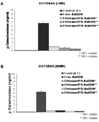
Figure 4 shows the antisense activity of complexes prepared low (Figure 4A) mol wt and medium (Figure 4B) mol wt of chitosan. Use of chitosan antisense oligonucleotides complexes, β-Gal expression decreased remarkable (92 %).
According to the results that is shown in Figure 4, there is no significant difference between the complexes prepared with neither LMW (70 kDa) nor MWM (400 kDa) chitosan when compared according to the antisense activities in HeLa cells (p>0.05). Similarly, there is no significant activity difference
between PS- and PO-AsODNs (approximately both have 90% antisense activity in 100:1 and 200:1 chitosan complexes). Moreover the ratio of chitosan/AsODN was not affected on the antisense activity of complexes.
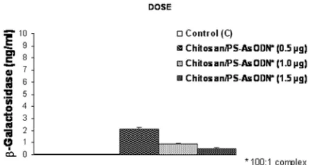
Secondly the effect of complex doses on β-Gal inhibition was investigated. As seen in Figure 5, complexes having different doses of AsODN such as 0.5, 1.0 and 1.5 µg were applied to HeLa cells and antisense effect was determined. Maximum antisense activity (90 %) was observed with 1.5 µg of AsODN containing the chitosan/PS-AsODN complex (100:1).
We studied the time-dependent antisense effect of complexes formed with PS-AsODN. As shown in Figure 6, PS-AsODN delivered with chitosan produced a rapid response and β-galactosidase levels declined rapidly with as much as 49 % of β-Gal lost in the first hour. The antisense effects were maximal between 8
Figure 4. β-galactosidase inhibition activity of the chitosan/AsODN (PS and PO AsODN) complexes prepared with (A) low molecular weight (LMW) and (B) medium molecular weight (MMW) chitosan.
Figure 3. Degradation profile of free AsODN against DNAase I
and 24 hours. Maximum antisense activity (88 %) was observed after 24 hours of chitosan/AsODN complex transfection into cells.
In this study, the effect of different factors on chitosan delivery properties of antisense oligonucleotide was investigated, because there are fewer studies focusing on the delivery of AsODNs using chitosan. Chirila et al., (2002) noted that most important polymers being studied are too large and have high surface charge densities for AsODN deliver, because of the large difference between the sizes of the two macromolecules, complexation of these polymers with AsODN is a difficult problem. The complexes of cationic polymers with AsODN are so stabile that these complexes are taken up by the cells without release the AsODN, which therefore are not able to show antisense activity (Chirila et al., 2002;
Gao et al., 2005). However in our studies complexes were prepared using chitosan as polymer and remarkable antisense effect was obtained with the chitosan/AsODN complexes.
We studied various combinations of chitosan molecular weights and AsODN molecules (PO- and PS) for their ability to elicit an antisense response. Comparable complex formation was observed between chitosan PS-AsODNs and PO-AsODNs.
PS-AsODNs indicated strong binding and full complexation with chitosan while partial complexation was shown between PO-AsODNs and chitosan. Similar result was reported by Sundaram et al. (2007) for PEI/AsODN complexes. They suggested that both AsODNs have similar charge densities due to phosphate groups; however PS- are known to be more hydrophobic than PO-and their backbone differ only by a single atom; oxygen is replaced by sulphur in the PS-backbone and studies report that the sulphur atom increases its strengthening the interaction with lower charge density groups in protein (Sundaram et al., 2007; Tan et al., 2007).
However both AsODNs showed similar in vitro antisense effect in our study. Molecular weight of chitosan and chitosan/AsODN mass ratio had no affect on in vitro inhibition. Our studies demonstrated that chitosan can be used as a potential carrier without making modification or depolimerization for antisense oligonucleotide.
There are some studies indicating that unmodified AsODNs can form stable complexes with chitosan polymer and there is inhibition effect of these complexes. However, the inhibition effect of these studies is very low when compared with the inhibition results of our study.
This work reports that chitosan can form stable nano-complexes with AsODNs by complexation. Complexes formed are spherical of 200 nm in diameter with homogeneous nanoparticle structure. Chitosan/AsODN complexes show significantly higher antisense efficiency in HeLa cells. Transfection efficiency depends on the chitosan/AsODN (+/-) ratio and dose of AsODN strongly. Association of AsODN
Figure 5. Dose-dependent inhibition of the β-galactosidase gene in HeLa cells after application of chitosan/AsODN complexes containing in different doses of AsODN.
Figure 6. Time-dependent inhibition of β-Gal in HeLa cells after application of chitosan/AsODN complexes (β-Gal amount after the post-transfection 1hr, 4 hrs, 8 hrs and 24 hrs).
with cationic polymers such as chitosan represents an alternative to chemically modifying AsODNs in order to enhance stability and antisense effect. Our studies suggest that chitosan polymer is an effective vector for antisense oligonucleotides and can be used in antisense therapy to improve the protein degradation. The in vivo delivery investigation will be carried out in further studies.
Acknowledgements
The authors are grateful for financial support of Marmara University Scientific Research Projects Association (BAPKO, Project no: SAĞ-YLS 270306-0034).
References
Agrawal S and Iyer RP. Perspectives in antisense therapeutics. Pharmacol Ther. 76:151-160, 1997. Akbuğa J, Aral C, Özbaş-Turan S, Kabasakal L and
Keyer-Uysal M. Transfection efficiency of chitosan microspheres: Effect of DNA topology. STP Pharm Sci. 13(2):99-103, 2003.
Aral C, Özbaş-Turan S, Kabasakal L, Keyer-Uysal M and Akbuğa J. Studies of effective factors of plasmid DNA-loaded chitosan microspheres I. plasmid size, chitosan concentration and plasmid addition techniques. STP Pharm Sci. 10(1):83-88, 2000. Brus C, Kleemann E, Aigner A, Czubayko F and Kissel
T. Stabilization of oligonucleotide-polyethylenimine complexes by freeze-drying: physicochemical and biological characterization. J Control Release. 95:119-131, 2004.
Chirila TV and Constable IJ. The use of synthetic polymers for delivery of therapeutic antisense oligodeoxy-nucleotides. Biomaterials 23:321-342, 2002.
Dias N and Stein CA. Potential roles of antisense oligonucleotides in cancer therapy. The example Bcl-2 antisense oligonucleotides. Eur J Pharm Biopharm. 54:263-269, 2002.
Ferreiro MG, Crooke RM, Tillman L, Hardee G and Bodmeier R. Stability of polycationic complexes of an antisense oligonucleotide in rat small intestine homogenates. Eur J Pharm Biopharm. 55:19-26, 2003.
Fillion P, Desjardins A, Sayasith K and Lagacé J. Encapsulation of DNA in negatively charged liposomes and inhibition of bacterial gene expression with fluid liposome-encapsulated antisense oligonuc-leotides. Biochim Biophys Acta. 1515:44-54, 2001. Föger F, Noonpakdee W, Loretz B, Joojuntr S,
Salvenmoser W, Thaler M and Bernkop-Schnürch A. Inhibition of malarial topoisomerase II in plasmodium falciparum by antisense nanoparticles. Int J Pharm. 319:139-146, 2006.
Gao S, Chen J, Dong L, Ding Z, Yang Y and Zhang J. Targeting delivery of oligonucleotide and plasmid DNA to hepatocyte via galactosylated chitosan vector. Eur J Pharm Biopharm. 60:327-334, 2005. Guillem VC, Tormo M, Moret I, Benet I, Garcia-Conde J,
Crespo A and Aliño SF. Targeted oligonucleotide delivery in human lymphoma cell lines using a polyethylenimine based immunopolyplex. J Control Release 83:133-146, 2002.
Hirano S, Seino H, Akiyama Y and Nonaka I. Biocompatibility of chitosan by oral and intravenous administration. Polym Eng Sci. 6:897-901, 1988. Huang M, Khor E and Lim LY. Uptake and cytotoxicity of
chitosan molecules and nanoparticles, Effects of molecular weight and degree of deacetylation. Pharm Res. 21(2):344-353, 2004.
Hussain M, Belae G, Hughes M and Akhtar S. Co-delivery of an antisense oligonucleotide and 5-fluorouracil using sustained release poly (lactide-co-glycolide) microsphere formulations fo potential combination therapy in cancer. Int J Pharm. 234:129-138, 2002.
Issa M M, Köping-Häggård M, Tømmeraas K, Vårum KM, Christensen BE, Strand S P and Artursson P. Targeted gene delivery with trisaccharide-substituted chitosan oligomers in vitro and after lung administration in vivo. J Control Rel. 115:103-112, 2006.
Junghans M, Kreuter J and Zimmer A. Phosphodiester and phosphorothioate oligonucleotide condensation and preparation of antisense nanoparticles. Biochimica et Biophsica Acta (BBA)-Protein Structure and Molecular Enzymology. 1544(1-2):177-188, 2001.
Kurreck J. Antisense technologies. Improvement through novel chemical modifications. Eur J Biochem. 270:1628-1644, 2003.