Annals of Medical Research
DOI: 10.5455/annalsmedres.2019.10.596 2020;27(1):233-6
Original Article
Effects of bilateral knee arthroplasty on sagittal
spinopelvic balance in patients with primer degenerative
osteoarthritis
Sefa Giray Batibay1, Turgut Akgul2, Huseyin Koca3, Savas Camur3, Ozcan Kaya4, Serkan Bayram2, Necdet Saglam3 1Ankara Occupational and Environmental Diseases Hospital, Department of Orthopedics and Traumatology, Ankara, Turkey
2Istanbul University, Faculty of Medicine, Department of Orthopedics and Traumatology, Istanbul, Turkey 3Umraniye Education and Research Hospital, Department of Orthopedics and Traumatology, Istanbul, Turkey 4Istinye University, Faculty of Medicine, Department of Orthopaedic and Traumatology, Istanbul, Turkey Copyright © 2020 by authors and Annals of Medical Research Publishing Inc.
Abstract
Aim: We aimed to evaluate the change in sagittal spinopelvic parameters in knee osteoarthritis patients without prior degenerative spinal disease following bilateral knee arthroplasty. Knee osteoarthritis often causes knee flexion contracture and the flexion contracture may affect posture and lumbar lordosis.
Material and Methods: We performed a retrospective study by evaluating the records of patients who have undergone bilateral knee arthroplasty in our clinic between 2012 and 2016 in patients due to degenerative bilateral knee osteoarthritis and minimum 12 months follow up. Patients with prior hip surgery, prior spine surgery, rheumatic disease and postoperative flexion contracture were excluded. Lumber lordosis, sacral slope, pelvic tilt and pelvic incidence were measured on lateral lumbosacral view in supine position pre-postoperatively and compared with paired T test. Extension deficit was also measured preoperatively.
Results: This study includes 83 patients (77 females and 6 males) who underwent bilateral knee arthroplasty and matched the inclusion and exclusion criteria. The average age at the time of first total knee arthroplasty was 64.4 years. Mean follow up was 37.3(12-66) months. The mean lumber lordosis was 43.9° preoperatively and 49.6° postoperatively. The increase in lumbar lordosis angle was statistically significant (p: 0,001). These changes were only related to the degree of extension deficit but not to age and postoperative follow-up time. Sacral slope, pelvic incidence and pelvic tilt did not change significantly.
Conclusion: Bilateral knee arthroplasty increased lumbar lordosis patients with primer degenerative osteoarthritis especially who had high extension deficit. Sacral slope and pelvic tilt were decreased postoperatively but not significantly changed.
Keywords: Lumbar lordosis; bilateral knee arthroplasty; knee-spine; primer osteoarthritis; extension deficit
Received: 01.10.2019 Accepted: 31.12.2020 Available online: 18.02.2020
Corresponding Author: Serkan Bayram, Istanbul University, Faculty of Medicine, Department of Orthopedics and Traumatology,
Istanbul, Turkey E-mail: [email protected]
INTRODUCTION
Knee osteoarthritis often causes knee flexion contracture and in response, other parts of the body compensate for the contracture. Sagittal imbalance leads to adaptive changes in the pelvis, hip, and knee joints through a compensatory mechanism. Knee flexion contracture may affect posture and lumbar lordosis. Murata et al. reported that loss of extension occurring due to arthritic changes was correlated with decrease in lumbar lordosis. (LL)(1). Spinopelvic parameters are not only affected by changes in the spinal sagittal parameters that can affect the pelvic parameters but also lower extremity problems. Lee et al.
reported that change of the flexion contracture after total knee arthroplasty affect the spinopelvic parameters (2). In our study, we aimed to evaluate sagittal spinopelvic parameters in a population with knee osteoarthritis without degenerative spinal disease and the change in these parameters following bilateral knee arthroplasty.
MATERIAL and METHODS
We performed a retrospective study by evaluating the records of 651 patients who were operated in our clinic between 2012 and 2016 for knee osteoarthritis. The patients were included in the study after approval was
Ann Med Res 2020;27(1):233-6
received from the Ethics Committee (2017/1482). Informed consent was obtained from all individual participants included in the study.
Exclusion criteria of the study; 1-Previous surgery to the hip 2-Previous surgery to the spine
3-Hip contracture along with hip arthrosis,
4-Deviation of lumbar spine of ≥10° in the coronal plane and presence of symptomatic spinal degenerative disease, 5-Postoperative flexion contracture of the knee joint, 6-Patients with rheumatologic diseases.
Inclusion criteria of the study; 1-Primer degenerative osteoarthritis, 2-Bilateral knee arthroplasty,
3-Minumum one year follow-up postoperatively
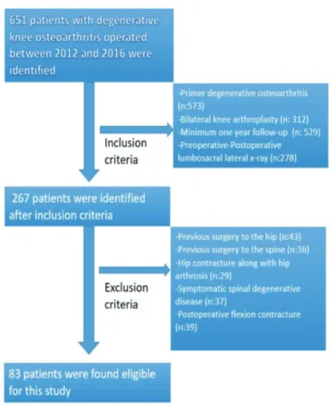
4- Preoperative-postoperative lumbosacral lateral x-ray (Figure 1).
Figure 1. The exclusion and inclusion criteria of the patients.
Eighty-three patients who met the above criteria were included this study. Lumbosacral views were obtained for all patients preoperatively to evaluate for spinal deformities for the purpose of spinal anesthesia. None of the patients underwent bilateral knee arthroplasty in
both knees simultaneously. Patients with TKA performed in only one knee were excluded to negate the effects of the other knee on spinopelvic parameters. Patients were operated first on the knee with the greater extension deficit. All patients underwent cruciate-retaining Genesis II knee arthroplasty (Smith and Nephew, Memphis, USA) with a medial parapatellar approach. The degree of contracture in the knee joints, presence of lumbosacral complaints and presence of flexion contracture of the hip joint in the study patients were evaluated with physical examination by the same observer using a goniometer. None of the patients had chronic back pain and hip flexion contracture (Figure 2).
Figure 2. A: Preoperative B: Postoperative, Lumbosacral view
Radiological evaluation: The measurements were performed on standard lateral lumbosacral radiographs taken in supine position obtained preoperatively and at the last follow-up. All radiographs showed the region between T12 (thoracic spine) and the hip joint. Digital analyses of all the radiographic measurements were done using a digital software program (Extreme-pacs). Parameters of the spine, including the lumbar lordosis (LL), were measured using the Cobb’s angle method. The angle between the superior endplates of L1 and inferior endplates of L5 was used as the lumbar lordotic angle. Pelvic parameters included in this study were the sacral slope (SS), the pelvic tilt (PT) and the pelvic incidence (PI). The SS is equal to the angle between the sacral plate and the horizontal axis. The Pelvic tilt equals to the angle between the line connecting the midpoint of the sacral plate to the hip axis) and the vertical plane. Pelvic incidence is the angle between the vertical axis to the sacral plate and the line connecting the midpoint of the sacral plate to the hip axis. In other words, the sum of sacral slope and pelvic tilt is equal to the pelvic incidence. 234
All radiological measurements were performed by the same observer (Figure 3).
Figure 3. A: Preoperative B: Postoperative, Bilateral knee views.
Statistical analysis
The paired Student’s t-test was used for comparing quantitative results pre- and post-operatively with normal distribution. Correlations were evaluated using the Pearson correlation test (2-tailed). P values less than 0.05 were considered significant. SPSS v.20.0 (SPSS Inc., Chicago, IL, USA) was used for statistical analysis.
RESULTS
This study includes 83 patients (77 females and six males) who underwent bilateral knee arthroplasty and matched the inclusion and exclusion criteria. The average age at
the time of first total knee arthroplasty was 64.4 (52-77) years. Mean follow up was 37.3 (12-63) months. There was no simultaneous bilateral TKA procedure. The mean time to the opposite knee arthroplasties was 6.3 (4-18) months. Right knee was operated in 47 patients first, and left in 36 patients.
The mean extension deficit was 11.9 (5-30), while 26 patients had no extension deficit.
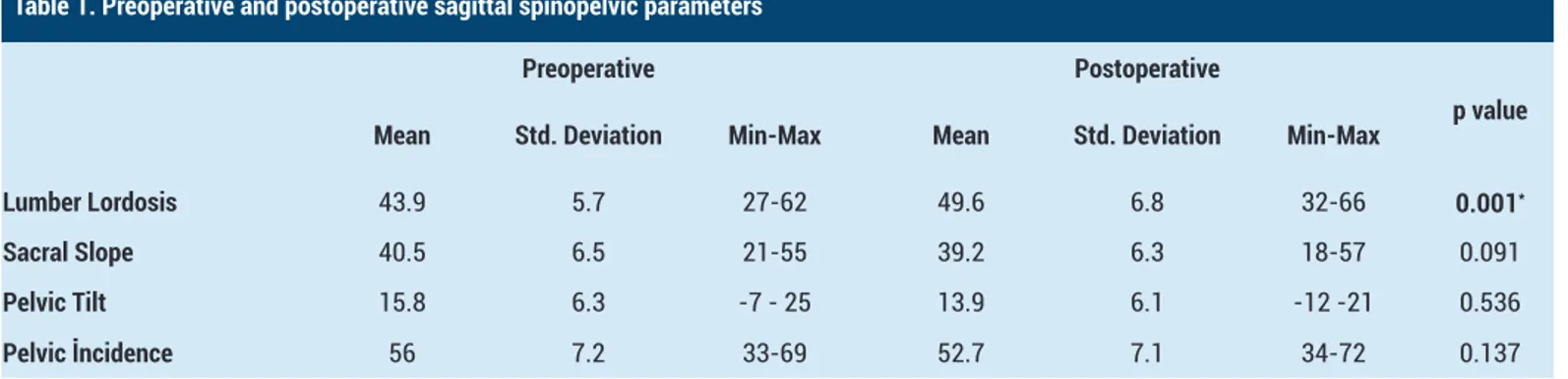
The mean lumber lordosis was 43.9° preoperatively and 49.6° postoperatively. The increase in lumbar lordosis angle was statistically significant (p: 0.001). The mean sacral slope angle was 40.5°, preoperatively and 39.2° postoperatively. The mean sacral slope decreased post-operatively, but the decrease was not statistically significant. (p: 0,091). The mean pelvic tilt angle was 15.8°, preoperatively and 13.9° postoperatively. The mean pelvic tilt angle decreased at postoperative, but the decrease was not statistically significant (p: 0,536). The mean pelvic incidence angle was 56°, preoperatively and 52.7° postoperatively. The mean pelvic incidence decreased postoperatively, but the decrease was not statistically significant (p: 0.137) (Table 1).
In correlation analysis, there was significant correlation between extension deficit and lumber lordosis (p: 0,001 and r: 0.67). The other parameters were not significantly correlated with the extension deficit. In patients with no extension deficit, lumber lordosis was 44.7 degrees preoperatively and 45.3 postoperatively (p: 0.06).
Table 1. Preoperative and postoperative sagittal spinopelvic parameters
Preoperati̇ve Postoperati̇ve
p value
Mean Std. Deviation Min-Max Mean Std. Deviation Min-Max
Lumber Lordosis 43.9 5.7 27-62 49.6 6.8 32-66 0.001*
Sacral Slope 40.5 6.5 21-55 39.2 6.3 18-57 0.091
Pelvic Tilt 15.8 6.3 -7 - 25 13.9 6.1 -12 -21 0.536
Pelvic İncidence 56 7.2 33-69 52.7 7.1 34-72 0.137
In correlation analysis, there was no significant correlation between spinopelvic parameters and either age or follow-up time.
DISCUSSION
Spinopelvic parameters are related to degenerative changes in the joints of the lower extremity. In this study we found that, lumber lordosis significantly increased after bilateral knee arthroplasty in patients with primary degenerative osteoarthritis. The changes in the spine in the sagittal plane are compensated by the pelvis in the early period, but the pelvis fails to compensate further sagittal plane imbalance and flexion position of the knees may occur. Obeid et al. found a strong correlation between
flexion deformity in the knees and lordosis in the lumber spine (5). Ferrero et al. showed a relationship between spinal deformities and lower limb compensation and pelvic translation (6).
In a study, decrease in lumbar lordosis was reported in an asymptomatic population aged more than 60 years (8). Another similar study reported a decrease in lumbar lordosis above the age of 40 years (9). In a study performed by Jalai et al. involving 778 patients, the relationship between adult spinal deformity and compensatory mechanisms in the lower extremities were evaluated and significant correlations between sagittal spinopelvic parameters and compensatory lower-limb mechanisms were highlighted depending on patient age (7). In our study Ann Med Res 2020;27(1):233-6
236 we also evaluated the correlation of sagittal parameters. Post-operative changes did not correlate with age.
In a study of 554 patients by Jalai et al. (277 obese and 277 non-obese patients), the relationship between compensation mechanisms and body mass parameters were studied. It was reported that in obese patients, compensation mechanism was through lower extremities, whereas in non-obese patients, it was through pelvic mechanisms. This study also found that negative impact of obesity on the ability to compensate for sagittal malalignment through pelvic retroversion (7).
There is only one study in the literature on the effects of knee arthroplasty on sagittal spinopelvic parameters. In their study, Lee et al. brought the knees of the patients to the flexion position and examined the changes in the lumbar spine (2). Because the loss of extension occurs over time in patients with gonarthrosis, there may be differences in the responses of pelvic parameters. They divided their patients into two groups as patients with an extension deficit below or above 10° following total knee replacement and evaluated the changes in the pelvic parameters through radiographs obtained after at least 12 months postoperatively. In their comparative study on patients with gonarthrosis, Lee et al. found no significant relationship between knee extension limitation and changes in the lumbar lordotic angle. In this study found that sacral slope significantly increased after total knee arthroplasty, especially in patients with high extension deficit. There is no similar study in the literature with respect to the sample size and the follow-up duration that examines changes in spinopelvic parameters after total bilateral knee replacement.
There are some weaknesses in our study. First of all, this is a retrospective study. Because of that, we could not measure the functional outcomes. Our study did not include an assessment of the body mass index of the patients, and therefore, we were unable to assess the impact of these on the results. We evaluated only sagittal spinopelvic parameters, coronal parameters were not evaluated. Lastly, we evaluated the parameters on lateral lumbosacral view in supine position. The parameters may be different from those in standing position.
CONCLUSION
In this study, we found that lumber lordosis significantly increased postoperatively in patients with degenerative osteoarthritis. These changes were not correlated with age and follow-up time but only with the degree of extension deficit. The other sagittal spinopelvic parameters did not change significantly after bilateral knee arthroplasty.
Competing interests: The authors declare that they have no competing interest.
Financial Disclosure: There are no financial supports.
Ethical approval: This study was approved by the institutional review board/Ethics Committee of Umraniye Training and Research Hospital.
Sefa Giray Batibay ORCID: 0000-0002-6226-6651 Turgut Akgul ORCID: 0000-0002-0704-3797 Huseyin Koca ORCID: 0000-0001-7487-7217 Savas Camur ORCID: 0000-0001-5899-6910 Ozcan Kaya ORCID: 0000-0002-4547-3042 Serkan Bayram ORCID: 0000-0001-7651-1200 Necdet Saglam ORCID: 0000-0001-5747-9903
REFERENCES
1. Murata Y, Takahashi K, Yamagata M, et al. The knee-spine syndrome. Association between lumbar lordosis and extension of the knee. J Bone Joint Surg Br 2003;85:95-9.
2. Lee SM, Yoon MG, Moon MS, et al. Effect of correction of the contractured flexed osteoarthritic knee on the sagittal alignment by total replacement. Asian Spine J 2013;7:204-11.
3. Roussouly P, Gollogly S, Berthonnaud E, et al. Classification of the normal variation in the sagittal alignment of the human lumbar spine and pelvis in the standing position. Spine 2005;30:346-53.
4. Labelle H, Roussouly P, Berthonnaud E, et al. The importance of spino-pelvic balance in L5–S1 developmental spondylolisthesis: a review of pertinent radiologic measurements. Spine 2005;30:27-34. 5. Obeid I, Hauger O, Aunoble S, et al. Global analysis
of sagittal spinal alignment in major deformities: correlation between lack of lumbar lordosis and flexion of the knee. European Spine J 2011;20:681. 6. Ferrero E, Liabaud B, Challier V. et al. Role of pelvic
translation and lower-extremity compensation to maintain gravity line position in spinal deformity. Journal of Neurosurgery: Spine 2016;24:436-46. 7. Jalai CM, Cruz DL, Diebo BG , et al. Full-body analysis
of age-adjusted alignment in adult spinal deformity patients and lower-limb compensation. Spine 2017;42:653-61.
8. Korovessis PG, Stamatakis M, Baikousis A. Reciprocal angulation of vertebral bodies in the sagittal plane in an asymptomatic Greek population. Spine 1998;23:700-4.
9. Gelb DE, Lenke LG, Bridwell KH, et al. Ananalysis of sagittal spinal alignment in 100 asymptomatic middle and older aged volunteers. Spine 1995;20:1351-8. Ann Med Res 2020;27(1):233-6