RESEARCH ARTICLE
Effects of dexmedetomidine and dexketoprofen on the
conduction block of rat sciatic nerve
*Correspondence to: Sengal Bagci Taylan, PhD, [email protected]. orcid:
0000-0002-8158-4487 (Sengal Bagci Taylan) doi: 10.4103/1673-5374.268926 Received: December 14, 2018 Peer review started: December 28, 2018 Accepted: September 4, 2019 Published online: November 8, 2019 Sengal Bagci Taylan1, *, Hulagu Bariskaner2
1 Department of Medical Services and Techniques, Vocational School of Health Services, Hakkari University, Hakkari, Turkey 2 Department of Pharmacology, School of Medicine, Selcuk University, Konya, Turkey
Funding: This study was supported by Scientific Committee Foundation (No. 13102007) of Selçuk University, Konya, Turkey (to HB).
Abstract
Dexmedetomidine is a selective α2-adrenoceptor agonist that is used because of its sedative, anxiolytic, and analgesic effects. Dexketoprofen, which is used as an analgesic, is a nonselective nonsteroidal anti-inflam-matory drug (NSAID). The use of dexmedetomidine and dexketoprofen as adjuvants to local anesthetics for the peripheral nerve is gradually increasing. In this study, we aimed to investigate the effects of different doses of dexmedetomidine and dexketoprofen on conduction block of rat sciatic nerve. The isolated sciatic nerve from adult rats was transferred to a nerve chamber. The compound action potentials (CAPs) were recorded from stimulated nerve with electrophysiological methods. Dexmedetomidine (n = 8) and dexketo-profen (n = 8) were administered in the chamber with cumulative concentrations of 10–9 to 10–5 M, and the
CAPs were recorded for 5 and 10 minutes. The CAP parameters were calculated. Both dexmedetomidine and dexketoprofen significantly depressed all CAP parameters in a dose-dependent manner compared with the control group, i.e., the group in which rats did not receive treatment. CAP parameters showed there was no significant difference in nerve conduction inhibition between dexmedetomidine and dexketopro-fen. Higher doses of dexmedetomidine suppressed the conduction in the fast-conducting fibers; however, dexketoprofen was found to suppress the conduction in the slow-conducting fibers in a time-dependent manner and suppress the conduction in the medium- and slow-conducting fibers in a dose-dependent manner. These findings suggest that dexmedetomidine and dexketoprofen exhibit better anesthetic effects on peripheral nerve through different ways of action. The experimental procedures were approved by the Necmettin Erbakan University on January 30, 2013 (approval No. 2013-024).
Key Words: compound action potentials; dexketoprofen; dexmedetomidine; maximum depolarization; nerve chamber; nerve fibers; sciatic nerve
Chinese Library Classification No. R453; R364; R741
Introduction
The α2-adrenoceptors that are found in the central nervous system, peripheral nerve and autonomic ganglia are the tar-get receptors of many drugs (Kosugi et al., 2010). Agonists of α2-adrenoceptor inhibit the conduction of nerve action potentials and can therefore contribute to an increase in the effects of local anesthetic agents (Kosugi et al., 2010). Dexmedetomidine, an α2 agonist for clinical anesthesia, is known to have various effects, such as anesthesia, analgesia, sedation, and vasoconstriction (Peng and Zhang, 2015). Ad-ditionally, dexmedetomidine has a potent anti-inflammatory effect (Li et al., 2017).
The use of dexketoprofen, a nonselective nonsteroidal an-ti-inflammatory drug (NSAID), is gradually increasing (Moore and Barden 2008; Miranda et al., 2012; Kara et al., 2014). Dex-ketoprofen is a preferred analgesic due to its faster onset of ac-tion and because it has fewer side effects than other NSAIDs (Moore et al., 2015; Hanna and Moon, 2018).
Rat sciatic nerve contains both sensory and motor nerve fibers, including nerve fibers with fast, medium and slow conduction velocities. The first appears as the activity of the fastest conducting fibers when a current pulse of the threshold stimulus intensity is applied to nerve. It is accepted that all the fibers are stimulated when a supramaximal stimulus is ap-plied to the nerve (Katsuki et al., 2006). The compound action
potentials (CAPs) recorded from supramaximal stimulus-in-duced rat sciatic nerve carry information about the activity of all the fibers forming the nerve. A change in the shape of the CAP may occur due to changes in the conduction velocity of the fibers (Gracias et al., 2011; Sousa et al., 2015).
The purpose of this study was to investigate the effects of different doses of dexmedetomidine and dexketoprofen on the conduction block of rat sciatic nerve.
Materials and Methods
Animals
The experimental procedures were approved by the Nec-mettin Erbakan University on January 30, 2013 (approval No. 2013-024) and conducted in accordance with the Guide for the Care and Use of Laboratory Animals (Konya/Tur-key). Male Wistar albino rats (n = 24), aged 4–6 months and weighing 350 ± 50 g, were used in our study. Rats were provided with food and water ad libitum and were housed in a thermoneutral environment (22 ± 2°C) and relative hu-midity under a 12-hour light/dark cycle. Rats were randomly divided into three groups: a control group (n = 8), a dexme-detomidine group (n = 8) and a dexketoprofen group (n = 8).
Isolated rat sciatic nerve and experimental procedures
(Pental Sodium 0.5 g injectable flacon, İ. E ULUGAY, Istan-bul, Turkey) at 30 mg/kg. The sciatic nerve from the right hind limb of each rat was carefully isolated with a glass hook, and 5–6 cm of the nerve was removed. Nerve tissues were immediately transferred into the nerve chamber, which is a three-compartment system (nerve chamber, stimulating and recording Ag-AgCl electrodes) (Grass Model SIU5, Sequim, Washington, USA). The nerve chamber was superfused with a fresh Krebs solution (119 mM NaCl, 20 mM NaHCO3, 4.8
mM KCI, 1.8 mM CaCl2, 1.2 mM KHPO4, 1.2 mM MgSO4,
and 10 mM glucose, gassed with a mixture 95% O2 and 5%
CO2). The ambient temperature was maintained at 37°C with
a pH of 7.4.
The CAP recordings were obtained from the sciatic nerve using a suction method, as previously described (Wijesing-he et al., 1991). Stimulations were applied to t(Wijesing-he proximal ends of the nerve trunk. Square wave pulses were delivered as supramaximal stimulations with a frequency of 1 Hz and duration of 200 µs by a stimulator (Grass Model S88, Sequim, Washington, USA) through a stimulation isolation unit (Grass Model SIU5, Sequim, Washington, USA). CAP recordings were obtained from the distal end of the nerve trunk (tibial branch) using a suction electrode. During the experiments, the testing equipment was kept in a Faraday’s cage to avoid external noise. Preamplified (Grass Model CP511, Sequim, WA, USA) CAP signals were digitized at a 50-kHz sampling rate by an Analog/Digital converter (Ad-vantech Model PCL1710, Cincinnati, OH, USA), acquired with the BiosigW data acquisition software (Bios, New Orle-ans, LA, USA), and saved for future analysis on a hard drive.
Dexmedetomidine and dexketoprofen were added to the nerve chamber at cumulative doses of 10–9 M, 10–8 M, 10–7 M,
10–6 M, and 10–5 M with a volume of 0.1 mL. To investigate
the acute effects of dexmedetomidine and dexketoprofen, the CAPs were recorded for 5 and 10 minutes after each dose was administered. Dexmedetomidine (Precedex 200 µg/2 mL, Meditera, Izmir, Turkey), dexketoprofen (Arveles 50 mg/2 mL ampule, UFSA İlaç, Istanbul, Turkey), pentobarbital (Pental Sodium 0.5 g injectable flacon, İ. E ULUGAY, Istanbul, Tur-key). Lower concentrations were obtained from the stock solu-tions that were made with distilled water. All chemicals were supplied by Sigma-Aldrich (Steinheim, Germany).
Analysis procedure
Several parameters were calculated to evaluate the effects of 10–9 M to 10–5 M doses of dexmedetomidine and
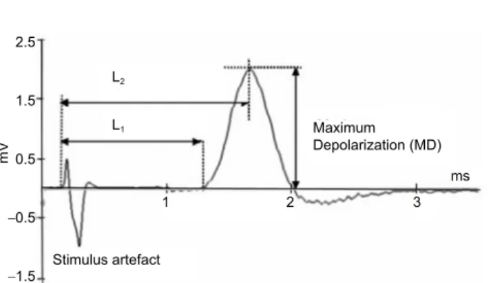
dexketo-profen at 5 and 10 minutes after dosing. The analysis provid-ed information regarding the electrophysiological changes in the sciatic nerve as a result of drug administration. Evalua-tion showed that on an example of a CAP recording (Figure
1), MD (maximum depolarization) and AUC (area under the
curve) are proportional to the number of stimulated nerve fibers in that nerve. Therefore, these values were calculated and are expressed as a percentage value relative to the
con-The latency periods were calculated, with L1 measuring the
time between the start of CAP and stimulation time and L2
measuring the time between the start of CAP and MD. These periods were used in the following equations (Eq {1} and Eq {2}) to calculate the conduction velocity of the fast-conduct-ing nerve fibers (VCAP) and the conduction velocity of the slow- and medium-conducting nerve fibers (VMD), respec-tively (Dalkilic and Pehlivan, 2002). The distance was taken as the optimum value (Δx) of 50 mm between the stimula-tion electrode and the recording electrode.
VCAP = Δx/ΔtL1 {1}
VMD = Δx/ΔtL2 {2} Statistical analysis
SPSS 18.0 software package was used for the statistical analysis of the recorded data. Two-way analysis of variance was used to calculate the difference in parameters between experimental groups. Paired samples t-test with Bonferroni correction was used for the repeated measures to evaluate the difference between groups. A level of P < 0.05 was con-sidered statistically significant. All experimental parameters are expressed as the mean ± SEM (standard error of mean).
Results
CAP records
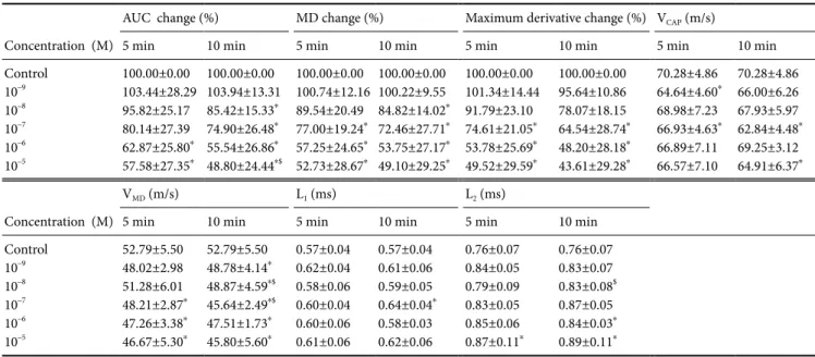
Both dexmedetomidine and dexketoprofen significantly depressed all CAP parameters in a dose-dependent (Tables
1 and 2) and reversible manner compared to those of the
control group. CAP records for both drugs returned to base-line after 15 minutes. An example of CAP records from each drug group recorded from the rat sciatic nerve and using previously calculated parameters that best reflected the dif-ference between the groups are presented in Figure 2, with the same temporal axis as the stimulus artifacts.
MD values
MD values were compared between dexmedetomidine and dexketoprofen at 5 and 10 minutes are shown in Figure 3. The CAP peak values recorded for the dexmedetomidine
2.5 1.5 0.5 –0.5 –1.5 mV Stimulus artefact Maximum Depolarization (MD) 1 2 3 ms L1 L2
Figure 1 Demonstration of the measurement parameters on a sample recording of the compound action potentials.
Table 1 Effects of different concentrations of dexmedetomidine on CAP parameters at 5 and 10 minutes
Concentration (M)
AUC change (%) MD change (%) Maximum derivative change (%) VCAP (m/s)
5 min 10 min 5 min 10 min 5 min 10 min 5 min 10 min
Control 100.00±0.00 100.00±0.00 100.00±0.00 100.00±0.00 100.00±0.00 100.00±0.00 70.28±4.86 70.28±4.86 10–9 103.44±28.29 103.94±13.31 100.74±12.16 100.22±9.55 101.34±14.44 95.64±10.86 64.64±4.60* 66.00±6.26 10–8 95.82±25.17 85.42±15.33* 89.54±20.49 84.82±14.02* 91.79±23.10 78.07±18.15 68.98±7.23 67.93±5.97 10–7 80.14±27.39 74.90±26.48* 77.00±19.24* 72.46±27.71* 74.61±21.05* 64.54±28.74* 66.93±4.63* 62.84±4.48* 10–6 62.87±25.80* 55.54±26.86* 57.25±24.65* 53.75±27.17* 53.78±25.69* 48.20±28.18* 66.89±7.11 69.25±3.12 10–5 57.58±27.35* 48.80±24.44*$ 52.73±28.67* 49.10±29.25* 49.52±29.59* 43.61±29.28* 66.57±7.10 64.91±6.37* Concentration (M) VMD (m/s) L1 (ms) L2 (ms)
5 min 10 min 5 min 10 min 5 min 10 min
Control 52.79±5.50 52.79±5.50 0.57±0.04 0.57±0.04 0.76±0.07 0.76±0.07 10–9 48.02±2.98 48.78±4.14* 0.62±0.04 0.61±0.06 0.84±0.05 0.83±0.07 10–8 51.28±6.01 48.87±4.59*$ 0.58±0.06 0.59±0.05 0.79±0.09 0.83±0.08$ 10–7 48.21±2.87* 45.64±2.49*$ 0.60±0.04 0.64±0.04* 0.83±0.05 0.87±0.05 10–6 47.26±3.38* 47.51±1.73* 0.60±0.06 0.58±0.03 0.85±0.06 0.84±0.03* 10–5 46.67±5.30* 45.80±5.60* 0.61±0.06 0.62±0.06 0.87±0.11* 0.89±0.11*
Data are expressed as the mean ± SEM. Sample size: 6, experiment repeats: 3. Two-way analysis of variance was used to compare parameters between experimental groups. *P < 0.05 represents the significance compared to the control, and $P < 0.05 represents the significance compared to that at 5 minutes in the same drug concentration group. AUC: Area under the curve; CAP: compound action potential; L1: latency period 1,
the time between the start of CAP and stimulation time; L2: latency period 2, the time between the start of CAP and maximum depolarization; MD: maximum depolarization; VCAP: conduction velocity of the fast-conducting nerve fibers; VMD: conduction velocity of the slow- and
medium-conducting nerve fibers.
Table 2 Effects of different concentrations of dexketoprofen on CAP parameters at 5 and 10 minutes
Concentration (M)
AUC change (%) MD change (%) Maximum derivative change (%) VCAP (m/s)
5 min 10 min 5 min 10 min 5 min 10 min 5 min 10 min
Control 100.00±0.00 100.00±0.00 100.00±0.00 100.00±0.00 100.00±0.00 100.00±0.00 73.75±2.53 73.75±2.53 10–9 102.34±16.51 134.04±8.55* 102.40±13.87 101.24±12.37 100.33±16.03 95.78±24.77 67.66±3.84* 67.66±6.75* 10–8 109.74±5.32* 119.78±16.32*$ 95.38±17.33 87.91±18.97 89.72±26.03 83.54±28.35 69.42±4.89* 72.03±4.39* 10–7 90.97±15.36 97.76±31.90 80.09±17.20* 69.68±23.28*$ 74.67±22.22* 65.55±27.77* 68.36±2.36* 70.59±3.49*$ 10–6 72.24±26.37* 78.58±42.95 61.06±23.81* 53.33±29.62*$ 55.68±26.93* 47.35±31.63* 68.92±4.14* 67.59±3.20* 10–5 61.94±33.87* 74.02±38.63$ 49.04±27.77* 48.70±25.67* 43.03±28.22* 43.35±26.90* 68.09±4.23* 69.30±3.71* Concentration (M) VMD (m/s) L1 (ms) L2 (ms)
5 min 10 min 5 min 10 min 5 min 10 min
Control 53.11±1.53 53.11±1.53 0.54±0.02 0.54±0.02 0.78±0.06 0.78±0.06 10–9 49.25±2.75 47.98±5.39 0.59±0.03 0.59±0.06 0.81±0.04 0.84±0.09 10–8 49.15±5.9 49.52±5.48 0.58±0.04* 0.55±0.03 0.82±0.111 0.82±0,10 10–7 48.58±2.33* 49.26±2.81* 0.58±0.02* 0.56±0.03*$ 0.82±0.04 0.81±0.05 10–6 48.13±1.97* 46,19±2.43* 0.58±0.03* 0.59±0.03* 0.83±0.03 0.87±0.04* 10–5 46.75±2.21* 46.75±2.16* 0.58±0.03* 0.058±0.03* 0.86±0.04* 0.86±0.04*
Data are expressed as the mean ± SEM. Sample size: 6, experiment repeats: 3. Two-way analysis of variance was used to compare parameters between experimental groups. *P < 0.05 represents the significance compared to the control, and $P < 0.05 represents the significance compared to that at 5 minutes in the same drug concentration group. AUC: Area under the curve; CAP: compound action potential; L1: latency period 1,
the time between the start of CAP and stimulation time; L2: latency period 2, the time between the start of CAP and maximum depolarization;
MD: maximum depolarization; VCAP: conduction velocity of the fast-conducting nerve fibers; VMD: Conduction velocity of the slow- and
minutes, respectively. There was no significant difference in MD values that were calculated from the CAP records be-tween the two drug groups (P > 0.05).
AUC values
At the highest dose of dexmedetomidine and dexketoprofen (10–5 M), the AUC values that were calculated from the CAP
records were decreased at 5 and 10 minutes when compared to those of the control group (Figure 4). Compared to those of the controls, the AUC values of the dexmedetomidine group (10–5 M) at 10 minutes were more greatly reduced
than those of the dexketoprofen group, but the difference was not statistically significant (P > 0.05). For the 10–6 and
10–5 M doses, a statistically significant decrease in the AUC
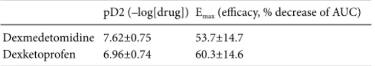
values was found at 5 and 10 minutes for the dexmedeto-midine group, but only at 5 minutes for the dexketoprofen group, compared with those of the control group (P < 0.05). The inhibitory concentration 50 (IC50) values were calcu-lated with semilogarithmic sigmoidal dose-response curves using the decrease in AUC value (%) and potency, pD2 (–log IC50), and were found to be 7.62 ± 0.75 for dexmedetomi-dine and 6.96 ± 0.74 for dexketoprofen (Table 3). There were no statistically significant differences in potency (pD2) and efficacy (Emax) values between dexmedetomidine and
dexke-toprofen (P > 0.05).
jor surgery (Combettes et al., 2010; He et al., 2015).
In our study, the effects of the α2 agonist dexmedetomi-dine and the NSAID dexketoprofen, both of which are used for pain control and have an anti-inflammatory effect, on peripheral nerve conduction were examined. Results from this study showed that dexmedetomidine and dexketopro-fen caused a dose-dependent suppression of all CAP param-eters. The suppressed CAPs returned to baseline 15 minutes after washing. In our study, the effects of dexmedetomidine and dexketoprofen on the sciatic nerve were investigated only in vitro. If further studies are performed in vivo, the ef-fects of dexmedetomidine and dexketoprofen on all systems can be examined or their effects on other peripheral or cen-tral nerves can be examined to provide more information about use of dexmedetomidine and dexketoprofen in pain management.
The peak values (MDs) of CAP curves decrease with an increase in dexmedetomidine dose, and as a result, AUC was decreased. The changes of MDs of CAP curves were significant at high doses (10–6 and 10–5) were significant. Oda
et al. (2007) reported that dexmedetomidine inhibited volt-age-dependent Na+ channels. Additionally, in a study using
a patch-clamp method on cardiac cells, dexmedetomidine blocked the persistent sodium current induced by veratri-dine (Stoetzer et al., 2016). Another study reported that in frog sciatic nerve, high doses of dexmedetomidine (5 × 10⁻⁴
M) reduced the value of MD, but this effect might not be related to adrenoceptors (Kosugi et al., 2010). In our study, reduction of MD may be the result of inhibition of sodium channels that are active in the rising phases of CAPs.
The suppression of fast-conducting fibers causes an increase in L1 values, while the suppression of slow-con-ducting fibers causes an increase in L2 values (Dalkilic and Pehlivan, 2002). In the dexmedetomidine group, both laten-cy periods increased, and consequent conduction velocity decreased compared to those of the controls. At the doses of 10–6 and 10–5 M, there were significant differences in L2
parameters between two drug groups and the control group (P < 0.05). In this study, dexmedetomidine showed dose-de-pendent activity on slow-conducting fibers. At high doses (10–7, 10–6 and 10–5 M) of dexmedetomidine, a significant
reduction in the maximum derivative was observed when compared to the controls. The maximum derivative is relat-ed to the maximum rate of change in the rising phase of the CAP over time and gives information about Na+ channels
(Katsuki et al., 2006). Additionally, fast-conducting fibers make a greater contribution during the CAP rising phase. For this reason, the changes in the maximum derivatives can be interpreted as changes in the fast-conducting fibers (Dalkilic et al., 2009). Therefore, dexmedetomidine at high doses suppresses the fast-conducting fibers in the rat sciatic nerve. These results show similarities to previous studies. Butterworth and Strichartz (1993) reported that clonidine,
Table 3 pD2 and Emax values for dexmedetomidine and dexketoprofen at 10 minutes
pD2 (–log[drug]) Emax (efficacy, % decrease of AUC)
Dexmedetomidine 7.62±0.75 53.7±14.7
Dexketoprofen 6.96±0.74 60.3±14.6
Data are expressed as the mean ± SEM. AUC: Area under the curve; Emax: efficacy, maximum response of drug; pD2: potency of the drug.
Maximum derivative, conduction velocity values
Treatment with three highest doses of both drugs (10–7,
10–6 and 10–5 M) caused a statistically significant (P < 0.05)
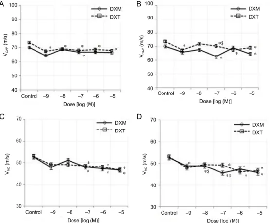
decrease in the maximum derivative of CAP at 5 and 10 minutes compared to those of the control group (Figure 5). In addition, the reduction in conduction velocity value was significantly different (P > 0.05) between the two drugs. In contrast, the conduction velocity of the fast-conducting fi-bers, the medium- and slow-conducting fibers was decreased for each drug at all doses and times (Figure 6) compared to those of the controls.
Discussion
A recent study reported that dexmedetomidine is used as an analgesic adjuvant in peripheral nerve block with an
inflam-0 0 0.4 ms 0.2 mV 0.4 ms 0.2 mV Control 10–9 M 10–8 M 10–7 M 10–6 M 10–5 M Control 10–9 M 10–8 M 10–7 M 10–6 M 10–5 M A B
Figure 2 The dose-dependent effects of dexmedetomidine and dexketoprofen on the compound action potentials of the rat sciatic nerve recorded at 10 minutes.
(A) Dexmedetomidine; (B) dexketoprofen.
A B 120 100 80 60 40 20 0 120 100 80 60 40 20 0 Control –9 –8 –7 –6 –5 Control –9 –8 –7 –6 –5
Dose [log (M)] Dose [log (M)]
MD change (%) MD change (%) DXM DXT DXM DXT * * * * * * * * **$ **$
Figure 3 Percent changes in the dose-dependent MD of dexmedetomidine and dexketoprofen at 5 and 10 minutes.
(A) At 5 minutes, (B) at 10 minutes. Sample size: 6, experiment repeats: 3, two-way analysis of variance was used to compare parameters between experimental groups. *P < 0.05 represents the significance compared to the control, and $P < 0.05 represents the significance compared to that of 5 minutes). DXM: Dexmedetomidine; DXT: dexketoprofen; MD: maximum depolarization.
A 160 B 140 120 100 80 60 40 20 0 Control –9 –8 –7 –6 –5 Control –9 –8 –7 –6 –5
Dose [log (M)] Dose [log (M)]
AUC change ( %) AUC change ( %)
DXM DXT DXMDXT 160 140 120 100 80 60 40 20 0 * * * ** * * * *$ $ *$ * $
Figure 4 Percent changes in the dose-dependent AUC of dexmedetomidine and dexketoprofen at 5 and 10 minutes.
(A) At 5 minutes; (B) at 10 minutes. Sample size: 6, experiment repeats: 3. Two-way analysis of variance was used to compare the parameters be-tween experimental groups. *P < 0.05 represents the significance compared to the control, and $P < 0.05 represents the significance compared to that at 5 minutes. AUC: Area under the curve; DXM: dexmedetomidine; DXT: dexketoprofen.
Dexmedetomidine is used in combination with a local an-esthetic to increase the amount of time that peripheral nerve conduction is blocked (Brummett et al., 2011). A meta-anal-ysis of neuraxial adjuvant anesthesia and analgesia indicates that dexmedetomidine is a good adjuvant for local anesthe-sia, causing the extension of the duration of postoperative pain relief and lowering pain intensity (Wu et al., 2014).
An-local anesthetic due to the An-local vasoconstriction caused by dexmedetomidine (Yabuki et al., 2014). The results of our in
vitro study suggest that dexmedetomidine directly inhibits
neuronal conduction.
According to the AUC analysis in our study, dexmedeto-midine and dexketoprofen have similar effects on the CAP parameters recorded from rat sciatic nerve, but
dexketopro-A B
Control –9 –8 –7 –6 –5 Dose [log (M)]
Maximum derivative change (%)
DXM DXT DXM DXT 120 100 80 60 40 20 0 120 100 80 60 40 20 0
Maximum derivative change (%)
Control –9 –8 –7 –6 –5 Dose [log (M)] * * * * * * * * * * * *
Figure 5 Percent changes in the dose-dependent maximum derivative of dexmedetomidine and dexketoprofen at 5 and 10 minutes.
(A) At 5 minutes; (B) at 10 minutes. Sample size: 6, experiment repeats: 3. Two-way analysis of variance was used to compare parameters between experimental groups. *P < 0.05 represents the significance compared to the control, and $P < 0.05 represents the significance compared to that at 5 minutes. DXM: Dexmedetomidine; DXT: dexketoprofen .
100 90 80 70 60 50 40 Control –9 –8 –7 –6 –5 Dose [log (M)] VCAP (m/s) Control –9 –8 –7 –6 –5 Dose [log (M)] Control –9 –8 –7 –6 –5 Dose [log (M)] Control –9 –8 –7 –6 –5 Dose [log (M)] 100 90 80 70 60 50 40 70 60 50 40 30 70 60 50 40 30 VCAP (m/s) VMD (m/s) VMD (m/s) DXM DXT DXM DXT DXMDXT DXM DXT * * * * * * * * * * * * * * * * * * * ** * *$ * *$ *$ A B C D
Figure 6 Percent changes in the dose-dependent VCAP and
VMD of dexmedetomidine
and dexketoprofen at 5 and 10 minutes.
(A) VCAP at 5 minutes; (B)
VCAP at 10 minutes; (C) VMD
at 5 minutes; (D) VMD at 10
minutes. Sample size: 6, ex-periment repeats: 3. Two-way analysis of variance was used to investigate the difference in parameters between ex-perimental groups. *P < 0.05 represents the significance compared to the control, and $P < 0.05 represents the sig-nificance compared to that at 5 minutes. DXM: Dexmedeto-midine; DXT: dexketoprofen; VCAP: conduction velocity of
the fast-conducting nerve fi-bers; VMD: conduction velocity
of the slow- and medium-con-ducting nerve fibers.
ever, a significant inhibition (55.90 ± 30.57%) was observed at the lowest dose (10–9 M) compared to that at the highest
dose (10–5 M), resulting in the dose-dependent suppression
of the conduction velocity for all fiber types distributed in the nerve bundle. Mazario et al. (1999), in a study involving rats, found that dexketoprofen inhibited single motor unit (SMU) records, which showed electrical and mechanical stimulation even at a very low dose (25 nmol/kg).
Most dexketoprofen studies to date have investigated the analgesic potency of dexketoprofen, its use in combination with other drugs, the usefulness of preemptive or postoper-ative administration, its use in the management of chronic pain, and its effectiveness for reducing opioid drug con-sumption (Yucel et al., 2013; Kelsaka et al., 2014; Kaye et al., 2018), as well as the effects on the SMU records generated by electrical or mechanical stimulation (Mazario et al., 2001; Gaitan and Herrero, 2002). However, most of the studies were conducted in vivo, and our study was the first to report the effect of dexketoprofen on rat sciatic nerve in vitro.
In this study on sciatic nerve, we observed no significant difference between dexmedetomidine and dexketoprofen in terms of inhibiting nerve conduction. However, we report that dexmedetomidine at higher doses suppresses the con-duction of fast-conducting fibers, whereas dexketoprofen has a time-dependent effect on slow-conducting fibers and a dose-dependent effect on medium- and fast-conducting fibers.
Author contributions: Concept and design of study, definition of
in-tellectual content, literature retrieval, data analysis, statistical analysis, manuscript preparation and editing: SBT, HB; experimental studies and data acquisition: SBT; manuscript review: SBT. Both authors approved the final version of this paper.
Conflicts of interest: The authors have no conflict of interest to declare. Financial support: This study was supported by the Scientific Committee
Foundation (No. 13102007) of Selçuk University, Konya, Turkey (to HB).
Institutional review board statement: The experimental procedures
were approved by the Necmettin Erbakan University on January 30, 2013 (approval No. 2013-024).
Copyright license agreement: The Copyright License Agreement has
been signed by all authors before publication.
Data sharing statement: Datasets analyzed during the current study are
available from the corresponding author on reasonable request.
Plagiarism check: Checked twice by iThenticate. Peer review: Externally peer reviewed.
Open access statement: This is an open access journal, and articles
are distributed under the terms of the Creative Commons Attribu-tion-Non-Commercial-ShareAlike 4.0 License, which allows others to remix, tweak, and build upon the work non-commercially, as long as appropriate credit is given and the new creations are licensed under the identical terms.
Open peer reviewers: Alonzo D Cook, Brigham Young Unversity, USA;
Lorenzo Di Cesare Mannelli, University of Florence, Italy.
References
Abdallah FW, Brull R (2013) Facilitatory effects of perineural dexmedetomidine on neuraxial and peripheral nerve block: a systematic review and meta-analysis. Br J Anaesth 110:915-925.
Bagry H, de la Cuadra Fontaine JC, Asenjo JF, Bracco D, Carli F (2008) Effect of a continuous peripheral nerve block on the inflammatory response in knee ar-throplasty. Reg Anesth Pain Med 33:17-23.
Brummett CM, Hong EK, Janda AM, Amodeo FS, Lydic R (2011) Perineural dex-medetomidine added to ropivacaine for sciatic nerve block in rats prolongs the duration of analgesia by blocking the hyperpolarization-activated cation current. Anesthesiology 115:836-843.
Butterworth JF, Strichartz GR (1993) The alpha 2-adrenergic agonists clonidine and guanfacine produce tonic and phasic block of conduction in rat sciatic nerve fibers. Anesth Analg 76:295-301.
Combettes E, Benhamou D, Mazoit JX, Beloeil H (2010) Comparison of a bupiva-caine peripheral nerve block and systemic ketoprofen on peripheral inflamma-tion and hyperalgesia in rats. Eur J Anaesthesiol 27:642-647.
Dalkiliç N, Pehlivan F (2002) Comparison of fiber diameter distributions deduced by modeling compound action potentials recorded by extracellular and suction techniques. Int J Neurosci 112:913-930.
Dalkilic N, Tuncer S, Bariskaner H, Kiziltan E (2009) Effect of tramadol on the rat sciatic nerve conduction: a numerical analysis and conduction velocity distribu-tion study. Yakugaku Zasshi 129:485-493.
Gaitan G, Herrero JF (2002) Subeffective doses of dexketoprofen trometamol enhance the potency and duration of fentanyl antinociception. Br J Pharmacol 135:393-398.
Gracias NG, Cummins TR, Kelley MR, Basile DP, Iqbal T, Vasko MR (2011) Vaso-dilatation in the rat dorsal hindpaw induced by activation of sensory neurons is reduced by paclitaxel. Neurotoxicology 32:140-149.
Grosu I, Lavand’homme P (2015) Continuous regional anesthesia and inflamma-tion: a new target. Minerva Anestesiol 81:1001-1009.
Hanna M, Moon JY (2018) A review of dexketoprofen trometamol in acute pain. Curr Med Res Opin 24:1-14.
He Y, Li Z, Zuo YX (2015) Nerve blockage attenuates postoperative inflammation in hippocampus of young rat model with surgical trauma. Mediators Inflamm 2015:460125.
Kara I, Apiliogullari S, Bagcı Taylan S, Bariskaner H, Celik JB (2014) The effects of dexketoprofen on duration of analgesia to a thermal stimulus when compared with a systemic control in a rat sciatic nerve block with levobupivacaine. Fun-dam Clin Pharmacol 28:205-210.
Katsuki R, Fujita T, Koga A, Liu T, Nakatsuka T, Nakashima M, Kumamoto E (2006) Tramadol, but not its major metabolite (mono-O-demethyl tramadol) depresses compound action potentials in frog sciatic nerves. Br J Pharmacol 149:319-327. Kaye AD, Cornett EM, Hart B, Patil S, Pham A, Spalitta M, Mancuso KF (2018)
Novel pharmacological nonopioid therapies in chronic pain. Curr Pain Head-ache Rep 22:31.
Kelsaka E, Güldoğuş F, Cetinoğlu E (2014) Effect of intravenous dexketoprofen use on postoperative analgesic consumption in patients with lumbar disc surgery. Agri 26:82-86.
Kosugi T, Mizuta K, Fujita T, Nakashima M, Kumamoto E (2010) High concentra-tions of dexmedetomidine inhibit compound action potentials in frog sciatic nerves without alpha(2) adrenoceptor activation. Br J Pharmacol 160:1662-1676. Li J, Wang H, Dong B, Ma J, Wu X (2017) Adding dexmedetomidine to ropivacaine for femoral nerve block inhibits local inflammatory response. Minevra Anestesi-ol 83:590-597.
Mazario J, Gaitan G, Herrero JF (2001) Cyclooxygenase-1 vs. cyclooxygenase-2 inhibitors in the induction of antinociception in rodent withdrawal reflexes. Neuropharmacology 40:937-946.
Mazario J, Roza C, Herrero JF (1999) The NSAID dexketoprofen trometamol is as potent as mu-opioids in the depression of wind-up and spinal cord nociceptive reflexes in normal rats. Brain Res 816:512-517.
Miranda HF, Romero MA, Puig MM (2012) Antinociceptive and anti-exudative synergism between dexketoprofen and tramadol in a model of inflammatory pain in mice. Fundam Clin Pharmacol 26:373-382.
Moore RA, Barden J (2008) Systematic review of dexketoprofen in acute and chronic pain. BMC Clin Pharmacol 8:11.
Moore RA, Gay-Escoda C, Figueiredo R, Toth-Bagi Z, Dietrich T, Milleri S, Tor-res-Lagares D, Hill CM, García-García A, Coulthard P, Wojtowicz A, Matenko D, Peñarrocha-Diago M, Cuadripani S, Pizà-Vallespir B, Guerrero-Bayón C, Bertolotti M, Contini MP, Scartoni S, Nizzardo A, et al. (2015) Dexketoprofen/ tramadol: randomised double-blind trial and confirmation of empirical theory of combination analgesics in acute pain. J Headache Pain 16:541.
Oda A, Iida H, Tanahashi S, Osawa Y, Yamaguchi S, Dohi S (2007) Effects of al-pha2-adrenoceptor agonists on tetrodotoxin-resistant Na+ channels in rat dorsal root ganglion neurons. Eur J Anaesthesiol 24:934-941.
Peng W, Zhang T (2015) Dexmedetomidine decreases the emergence agitation in infant patients undergoing cleft palate repair surgery after general anesthesia. BMC Anesthesiol 15:145.
Sousa DG, Sousa SD, Silva RE, Silva-Alves KS, Ferreira-da-Silva FW, Kerntopf MR, Menezes IR, Leal-cardoso JH, Barbosa R (2015) Essential oil of Lippia alba and its main constituent citral block the excitability of rat sciatic nerves. Braz J Med Biol Res 48:697-702.
Stoetzer C, Reuter S, Doll T, Foadi N, Wegner F, Leffler A (2016) Inhibition of the cardiac Na+ channel α-subunit Nav1. 5 by propofol and dexmedetomidine. Naunyn Schmiedebergs Arch Pharmacol 389:315-325.
Wijesinghe RS1, Gielen FL, Wikswo JP Jr (1991) A model for compound action potentials and currents in a nerve bundle III: A comparison of the conduction velocity distributions calculated from compound action currents and potentials. Ann Biomed Eng 19:97-121.
Wu HH, Wang HT, Jin JJ, Cui GB, Zhou KC, Chen Y, Chen GZ, Dong YL, Wang W (2014) Does dexmedetomidine as a neuraxial adjuvant facilitate better anesthe-sia and analgeanesthe-sia? A systematic review and meta-analysis. PLoS One 9:e93114. Yabuki A, Higuchi H, Yoshitomi T, Tomoyasu Y, Ishii-Maruhama M, Maeda S,
Mi-yawaki T (2014) Locally injected dexmedetomidine induces vasoconstriction via peripheral alpha-2A adrenoceptor subtype in guinea pigs. Reg Anesth Pain Med 39:133-136.
Yucel E, Kol IO, Duger C, Kaygusuz K, Gursoy S, Mimaroglu C (2013) Ilioingui-nal-iliohypogastric nerve block with intravenous dexketoprofen improves post-operative analgesia in abdominal hysterectomies. Braz J Anesthesiol 63:334-339.
P-reviewers: Cook AD, Di Cesare Mannelli L; C-Editor: Zhao M; S-Editor: Li CH; L-Editor: Song LP; T-Editor: Jia Y