Asian Pacific Journal of Cancer Prevention, Vol 16, 2015
351
DOI:http://dx.doi.org/10.7314/APJCP.2015.16.1.351 Neutrophil/Lymphocyte Ratio Findings and Larynx Carcinoma in TurkeyAsian Pac J Cancer Prev, 16 (1), 351-354
Introduction
There is increasing evidence about the relationship between inflammation and cancer. Chronic inflammation was found be associated with colorectal carcinoma (Mariani et al., 2014). Chronic inflammation occurring due to laryngopharyngeal reflux was supposed to be related with laryngeal carcinoma development (Galli et al., 2006). Systemic inflammation reflected by neutrophile/ lymphocyte (N/L) ratio is studied in different cancer groups and found to be related with prognosis and survival (Halazun et al., 2009; Proctor et al., 2012).
The most important factors for prognosis in larynx carcinoma are; ‘T’ stage of the tumor ‘N’ presence and the presence of distant metastasis ‘M’ which are included in the criteria of American Joint Committee on Cancer (AJCC) (Edge et al., 2010). There are also other factors as the presence of cartilage invasion on magnetic resonance imaging (MRI) and p53 mutation in histopathological examination (Castelijns et al. 1996; Vielba et al., 2002).
A routine complete blood count (CBC) is a readily available test that is routinely performed, thus N/L ratio may be easily calculated before the treatment of larynx carcinoma. Therefore in this study we aimed to examine the potential prognostic role of N/L ratio in larynx carcinoma by comparing the preoperative CBC data of the patients with different tumor size and stage as well as with the healthy control group.
1Gazi University Faculty of Medicine Department of Otorhinolaryngology, Ankara, 2Medipol University Faculty of Medicine Department of Otorhinolaryngology, Istanbul, Turkey *For correspondence: [email protected]
Abstract
Background: To identify the potential prognostic role of the neutrophil/lymphocyte (N/L) ratio in larynx carcinoma. Materials and Methods: Oncologic archive charts of patients with a larynx carcinoma diagnosis between the years 2010 and 2013 were retrospectively reviewed. The inclusion criterion was to be available with hemogram test prior to diagnostic procedure. Patients undergoing septorinoplasty comprised the control group. Results: There were 65 cases in the study and 42 cases in control group meeting inclusion criteria. In general a non-significant increase in N/L ratio was observed with increasing tumor size and stage (p>0.05) in larynx carcinoma. The N/L ratio was found to be significantly higher in larynx carcinoma compared to control group (p=0.004). Conclusions: In conclusion, the N/L ratio was shown to be significantly increased in larynx carcinomas compared to control group. Further studies are needed to assess any prognostic role.
Keywords: Neutrophil/lymphocyte ratio - larynx carcinoma - Turkey
RESEARCH ARTICLE
Neutrophil-lymphocyte Ratio Findings and Larynx Carcinoma:
a Preliminary Study in Turkey
Mehmet Duzlu
1*, Recep Karamert
1, Hakan Tutar
1, Furkan Karaloglu
1, Melih
Sahin
1, Rasit Cevizci
2Materials and Methods
This study was conducted in the Otorhinolaryngology Department of Gazi University Faculty of Medicine in Ankara, Turkey. The approval was taken from the local ethics committee. The oncologic follow-up charts of the patients between the years 2010-2013 were retrospectively reviewed. The patients with the diagnosis of larynx carcinoma of any stage who were available with hemogram test prior to diagnostic suspension-laryngoscopy procedure were enrolled in the study. CBC parameters including; neutrophile, lymphocyte, monocyte, white blood cell (WBC) and platelet (Plt) count, hemoglobin level (Hb) were recorded and the N/L ratio were calculated.
In addition the demographics of the patients, tumor size (T) the presence of local (N) or distant metastasis (M), histopathological type and occurrence of loco regional or distant metastasis during follow up were reviewed in detail. Tumors were staged according to the TNM classification of 7th edition of AJCC criteria. The patients lacking definitive histopathologic diagnosis in the computer network archive were excluded from the study. Forty-five randomly selected patients from the surgical archive chart who had undergone septorinoplasty at our institute in the same time period comprised the control group whom were also available with preoperative hemogram test.
Mehmet Duzlu et al
Asian Pacific Journal of Cancer Prevention, Vol 16, 2015
352
Statistical analysis
SPSS version 15.0 package program (Illinois, Chicago) was used for statistical analysis. Continuous variables were tested for normality using Kolmogorov-Smirnov or Shapiro-Wilk’s test depending on number of subjects, histograms and P-P plots. Kruskal-Wallis and Mann-Whitney U test were used for group comparisons. In all the tests p<0.05 was considered to be significant statistically.
Results
There were 65 patients with the diagnosis of larynx carcinoma who have satisfied inclusion criteria. Of these 65 individuals 64 (98.4%) were male and only one patient (1.6%) was female. However gender ratio in septorinoplasty group was 1:1 (21 male and female a total of 42 patients). The age of the patients in the study group were ranging between 39 and 80 years old (mean: 60.22±8.74). The mean age in control group was 29.05±10.34 years (ranging between 17-64). The predominant histopathologic type for larynx carcinoma was squamous cell carcinoma (SCC, 60 cases 92.3%),
only 5 patients (7.7%) was differently diagnosed with basaloid variant of SCC.
Primarily the hemogram parameters were compared between larynx carcinoma and control group as shown in Table 1. There was an increase in WBC count and N/L ratio in the study group compared to control group. The difference in N/L ratio between larynx carcinoma and control group was significant statistically (p=0.004). The N/L ratio was compared between males and females in the control group and found to be 2.01±0.85 and 2.07±0.97, respectively. There was no difference between genders (p=0.815).
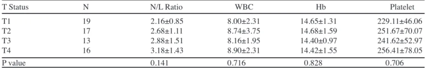
Secondly N/L ratio was compared between different T status of the tumor as well as the different stages of larynx carcinoma as shown in Table 2 and Table 3a-b. However there wasn’t a statistical significance (p=0,141). There wasn’t any significant change in the other hemogram parameters such as WBC count, Hb levels or Plt count with the increasing T status neither (Table 2).
N/L ratio was shown to be increased with increasing stage as it was mean 2.12 and 3.08 in stage I and IV respectively only exception was the decrement from Stage II to III as shown in Table 3a. The same difference was also Table 1. Comparison of Hemogram Parameters between Larynx Carcinoma and Control Group
Group N N/L Ratio WBC Hb Platelet Larynx carcinoma 65 2.70±1.25 8.45±2.73 14.55±1.37 244.23±62.44 Control 42 2.04±0.90 7.42±1.72 14.18±1.62 248.91±46.40 P value 0.004 0.097 0.301 0.334
Table 2. Comparison of Hemogram Parameters between Different T Status in Larynx Carcinoma
T Status N N/L Ratio WBC Hb Platelet T1 19 2.16±0.85 8.00±2.31 14.65±1.31 229.11±46.06 T2 17 2.68±1.11 8.74±3.75 14.68±1.59 251.67±70.07 T3 13 2.88±1.51 8.16±1.95 14.40±0.97 241.62±52.97 T4 16 3.18±1.43 8.90±2.31 14.42±1.55 256.41±78.05 P value 0.141 0.716 0.828 0.706
Table 3a. Comparison of Hemogram Parameters between Different Stages of Larynx Carcinoma
Stage N N/L Ratio WBC Hb Platelet I 19 2.12±0.79 7.96±2.27 14.75±1.32 226.67±46.95 II 12 2.97±1.07 8.02±2.27 14.58±1.40 263.85±77.85 III 15 2.73±1.53 8.00±1.51 14.70±1.16 238.19±50.57 IV 19 3.08±1.37 9.56±3.95 14.21±1.57 254.17±72.45 P value 0.081 0.517 0.648 0.424
Table 3b. Comparison of Hemogram Parameters between Early and Advanced Stages of Larynx Carcinoma
Stage N N/L Ratio WBC Hb Platelet Early (I,II) 31 2.45±0.99 7.98±2.08 14.68±1.33 241.06±62.32 Advanced (III, IV) 34 2.92±1.43 8.87±3.18 14.43±1.40 247.12±63.35 P value 0.259 0.217 0.361 0.655
Table 4. Comparison of Hemogram Parameters in the Presence or Absence of Lymph Node Metastasis
N status N N/L Ratio WBC Hb Platelet N- 48 2.61±1.16 8.10±2.14 14.61±1.37 245.71±61.15 N+ 17 2.93±1.51 9.43±3.85 14.38±1.39 240.05±67.73 P value 0.511 0.119 0.339 0.526
Asian Pacific Journal of Cancer Prevention, Vol 16, 2015
353
DOI:http://dx.doi.org/10.7314/APJCP.2015.16.1.351 Neutrophil/Lymphocyte Ratio Findings and Larynx Carcinoma in Turkey observed between early and advanced stages (Table 3b).However the difference was not significant statistically. No significant difference revealed in the other hemogram parameters with the increasing stage (Table 3a-b).
N/L ratio was also shown to be increased in the presence of regional lymph node metastasis (Table 4). It was 2.61±1.16 and 2.93±1.51 in the absence and presence of metastasis respectively (p=0.511 ).
In addition loco regional recurrence was observed in 14 cases. The mean N/L ratios in recurrent and non-recurrent cases were 2.83±1.45 and 2.66±1.20, respectively (p=0.646).
Discussion
In this study N/L ratio was studied for the first time for larynx carcinoma prognosis. In general N/L ratio was found to be increased with tumor size, stage, lymph node involvement which are already included TNM classification. Therefore it could be used to estimate tumor prognosis in the beginning of the treatment. However the differences were not significant statistically which may be due to limited number of our study group (65 cases). Likewise no significant difference was observed in the N/L ratio of the patient’s developing loco regional recurrence (p>0.05). However we were hypothesizing N/L ratio to be higher in the recurrent cases. Kum et al. first studied N/L ratio in larynx carcinoma. They found a significantly elevated N/L ratio in laryngeal SCC compared to benign and pre-malign laryngeal lesions and postulated that N/L ratio would be a useful inflammatory marker to differentiate benign and malign laryngeal lesions (Kum et al., 2014). We also found a significantly higher N/L ratio (p<0.05) in larynx carcinoma compared to control group whom were randomly selected from the patients undergoing septorhinoplasty. Therefore it may be useful in diagnosis and follow up especially in case of recurrence suspicion or differentiation of malign and pre-malign lesions.
As seen in the literature review N/L ratio were commonly studied for cardiovascular diseases previously. In the study of Sen et al. elevated N/L ratio revealed to be a significant indicator for three-year mortality and major adverse cardiac events in ST-segment elevation myocardial infarction (Sen et al., 2013). Likewise elevated N/L ratio was found to be associated with higher long term-mortality in the patients undergoing percutaneous coronary intervention (Duffy et al., 2006).
As inflammation is known to have a crucial role in cancer development (Balkwill et al., 2001), there is also a substantial increase in the publications searching the relationship between N/L ratio and cancer prognosis. Balkwill F, Mantovani A (2001). Inflammation and cancer: back to Virchow? Lancet, 357, 539-45. Lee et al. studied preoperative inflammatory parameters in the patients undergoing gastric surgery. They showed that elevated N/L ratio was associated with poorer prognosis (Lee et al., 2013). Ohno et al. showed better disease-free survival rates with N/L ratio lesser than 2.7 in clear cell renal cell carcinoma (Ohno et al., 2012). Kemal et al. (2014) found that N/L ratio and platelet-to-lymphocyte
ratio was significantly higher in case of lung carcinoma. Pretreatment N/L ratio was found to be an independent predictor of survival outcome for patients with metastatic nasopharyngeal carcinoma (Jin et al., 2013). Teramukai et al. (2009) postulated that paraneoplastic production of myeloid growth factors by cancer cells was one obvious cause of neutrophilia in case of cancer. However, the exact mechanism underlying the association between increased N/L ratio and cancer development and/or poor prognosis is not fully understood, yet.
However it must be kept in mind that N/L ratio may be affected by many other conditions as cardiovascular diseases as we mentioned above, renal or hepatic dysfunction abnormal thyroid function tests, previous history of focal or systemic inflammation (Balta et al., 2013).Therefore in the present study preoperative CBC records were taken into account in order to prevent possible local inflammation due to larynx biopsy procedure.
There are some limitations of the current study. Five-year disease free and overall survival ratios were not included in this preliminary report. A multivariate analysis including all risk factors should be done to identify if N/L ratio would be an independent risk factor for larynx carcinoma prognosis.
In conclusion N/L ratio was shown to be increased in larynx carcinoma compared to healthy control group. Also a non significant elevated N/L ratio was observed in advanced stages and tumor size. However further studies with larger study groups including treatment response and surveillance should be carried out to indicate the possible role of N/L ratio in larynx carcinoma prognosis.
Acknowledgements
We thank a lot to Prof.Dr.Nur Aksakal from Gazi University Faculty of Medicine Department of Public Health for her assistance in statistical analysis.
References
Balkwill F, Mantovani A (2001). Inflammation and cancer: back to Virchow? Lancet, 357, 539-45.
Balta S, Demirkol S, Sarlak H, Kurt O (2013). Comment on ‘Elevated preoperative neutrophil/lymphocyte ratio is associated with poor prognosis in soft-tissue sarcoma patients’: neutrophil to lymphocyte ratio may be predictor of mortality in patients with soft-tissue sarcoma. Br J Cancer,
108, 2625-6.
Castelijns JA, van den Brekel MW, Tobi H, et al (1996). Laryngeal carcinoma after radiation therapy: Correlation of abnormal MR imaging signal patterns in laryngeal cartilage with the risk of recurrence. Radiology, 198, 151-5. Duffy BK, Gurm HS, Rajagopal V, et al (2006). Usefulness of an
elevated neutrophil to lymphocyte ratio in predicting long-term mortality after percutaneous coronary intervention. Am J Cardiol, 97, 993-6.
Galli J, Cammarota G, Volante M, et al (2006). Laryngeal carcinoma and laryngo-pharyngeal reflux disease. Acta Otorhinolaryngol Ital, 26, 260-3.
Edge S, Byrd DR, Compton CC, et al (2010). In 7th.ed. AJCC cancer staging manual. Springer pp 57-68.
Halazun KJ, Hardy MA, Rana AA, et al (2009). Negative impact of neutrophil-lymphocyte ratio on outcome after
Mehmet Duzlu et al
Asian Pacific Journal of Cancer Prevention, Vol 16, 2015
354
liver transplantation for hepatocellular carcinoma. Ann Surg, 250, 141-51
Jin Y, Ye X, He C, Zhang B, Zhang Y (2013). Pretreatment neutrophil-to-lymphocyte ratio as predictor of survival for patients with metastatic nasopharyngeal carcinoma. Head Neck, doi: 10.1002/hed.23565 [Epub ahead of print]. Kemal Y1, Yucel I, Ekiz K, et al (2014). Elevated serum
neutrophil to lymphocyte and platelet to lymphocyte ratios could be useful in lung cancer diagnosis. Asian Pac J Cancer Prev, 15, 2651-4.
Kum RO, Ozcan M, Baklaci D, et al (2014). Elevated neutrophil-to-lymphocyte ratio in squamous cell carcinoma of larynx compared to benign and precancerous laryngeal lesions. Asian Pac J Cancer Prev. 15, 7351-5.
Lee DY, Hong SW, Chang YG, Lee WY, Lee B (2013). Clinical significance of preoperative inflammatory parameters in gastric cancer patients. J Gastric Cancer, 13, 111-6. Mariani F, Sena P, Roncucci L (2014). Inflammatory pathways
in the early steps of colorectal cancer development. World J Gastroenterol, 20, 9716-31.
Ohno Y, Nakashima J, Ohori M, et al (2012). Follow up of neutrophil-to-lymphocyte ratio and recurrence of clear cell renal cell carcinoma. J Urol, 187, 411-7.
Proctor MJ, McMillan DC, Morrison DS, et al (2012). A derived neutrophil to lymphocyte ratio predicts survival in patients with cancer. Br J Cancer, 107, 695-9
Sen N, Afsar B, Ozcan F, et al (2013). The neutrophil to lymphocyte ratio was associated with impaired myocardial perfusion and long term adverse outcome in patients with ST-elevated myocardial infarction undergoing primary coronary intervention. Atherosclerosis, 228, 203-10.
Teramukai S1, Kitano T, Kishida Y, et al (2009). Pretreatment neutrophil count as an independent prognostic factor in advanced non-small-cell lung cancer: an analysis of Japan Multinational Trial Organization LC00-03. Eur J Cancer,
45, 1590-8.
Vielba R, Bilbao J, Ispizua A, et al (2003). p53 and cyclin D1 as prognostic factors in squamous cell carcinoma of the larynx. Laryngoscope, 113, 167-72.