www.biodicon.com Biological Diversity and Conservation
ISSN 1308-8084 Online; ISSN 1308-5301 Print
4/3 (2011) 14-18
Research article/Araştırma makalesi
Anatomical characteristics of Bellevalia mathewii Özhatay & Koçak (Liliaceae)
Süleyman DOĞU
*1, Muhittin DİNÇ
2, Ayvaz
ÜNAL
11
Department of Science, Ahmet Keleşoğlu Faculty of Education, Selçuk University, 42090 Konya, Turkey
2Department of Biology, Ahmet Keleşoğlu Faculty of Education, Selçuk University, 42090 Konya, Turkey
Abstract
Bellevalia mathewii Özhatay & Koçak is a stenoendemic species growing in South Anatolia. In the present
study, anatomical features of this species were determined. The studies were carried out on tranverse sections of scapes
and leaves, and surface sections of the leaves. According to the results, the leaves are equifacial and amphistomatic with
anomocytic stomata. There is 2-3-layered palisade parenchyma under each epidermis, and richly developed 7-9-layered
spongy parenchyma between the palisades. Some spongy mesophyll cells include raphide crystals. Vascular bundles are
located in equal intervals in spongy parenchyma. The lower epidermal cells lack raphide crystals, but some of the upper
ones have plenty of them. In the scape, the cortex is multilayered and the vascular bundles are located in two rows.
Key words: Liliaceae, Bellevalia mathewii, Anatomy, Turkey
---
* ---
Bellevalia mathewii Özhatay & Koçak (Liliaceae)’nin anatomik özellikleri
Özet
Bellevalia mathewii Özhatay & Koçak
Güney Anadoluda yayılış gösteren dar yayılışlı bir endemiktir. Bu
çalışmada, bu türün anatomik özellikleri belirlenmiştir. Çalışma, skayp ve yaprak enine kesitleri ile yaprak yüzeysel
kesitleri üzerinde yapılmıştır. Sonuçlara göre, yapraklar ekvifasial ve amfistomatik olup anamositik stomalıdırlar. Alt ve
üst epidermisin altında 2-3-tabakalı palizad parankiması ve aralarında iyi gelişmiş 7-9-tabakalı sünger tabakası
bulunmaktadır. Bazı sünger parankiması hücreleri rafit kristalleri içermektedir. Sünger parankiması içinde eşit
aralıklarla lokalize olmuş iletim demetleri yer almaktadır. Alt epidermal hücreler rafit kristallerinden yoksun iken, bazı
üst epidermal hücreler bol miktarda rafit içermektedir. Skaypta, korteks çok tabakalı olup, iletim demetleri iki sırada
dizilmişlerdir.
Anahtar kelimeler: Liliaceae, Bellevalia mathewii, Anatomi, Türkiye
1. Introduction
Bellevalia Lapeyr., a genus of spring-flowering bulbous plants in the family Liliaceae, mostly occurs in the
Mediterranean and the central-west Asiatic region (Govaerts, 1996). The genus was represented by 18 species in Turkey
(Wendelbo, 1984). But, B. latifolia Feinbrun was reduced to a synonym of B. olivieri (Baker) Wendelbo later
(Wendelbo, 1985).
Three more species was added in Turkish Flora (Özhatay, 2000). B. pycnantha (K.Koch) Losinsk. was reduced
to a synonym of B. paradoxa Boiss. and a new Bellevalia species was described from Turkey later (Johnson, 2003;
Persson, 2006). The total number of Bellevalia species has now reached to 21 in Turkey.
Bellevalia is closely related the genera Hyacintella Schur, Hyacinthus L. and Muscari Mill. The genus is
distinguished from these genera by the relative characters such as capsule and perianth shape. In addition, there are
some taxonomical problems in seperating Bellevalia species (Wendelbo, 1984). Therefore, some anatomical pecularities
*
Corresponding author / Haberleşmeden sorumlu yazar: Tel.: +903323238220-5556; Fax.: +903323238223; E-mail: [email protected]
such as the distribution and shape of calcium oxalate crystals in plant tissues may be useful as additional diagnostic
characters in interspecific classification (Kahraman et al., 2010).
Bellevalia mathewii Özhatay & Koçak a perennial geophyte, is geographically distributed in South Anatolia
and closely related to B. dubia (Guss.) Roemer & Schultes, B. clusiana Griseb. and B. tauri Feinbrun, and only
distinguished from them by its dense raceme and light blue corolla. The anatomical pecularities of B. mathewii have not
been provided before. The present study aims to provide anatomical properties of B. mathewii for the first time.
2. Materials and methods
Bellevalia mathewii specimens were collected from Antalya (C4 ANTALYA: Alanya, Çayarası mevkii, Tarla
kenarı, 1050 m, 25.04.2009, S. Doğu 1993 & M.Dinç). The samples were put in 70 % alcohol for anatomical studies.
Anatomical studies were carried out on 10 samples. In these samples, leaves and scape cross-sections were studied with
the lower and upper surface sections of the leaves. On average, twenty preparations were made of each type of sections.
The cross-sections were stained with basic fuchsine. All sections were covered by glycerin gelatin and made into
permanent slides as described by Vardar (1987). Preparats were observed through an Olympus BX-50 microscope and
photographed.
3. Results
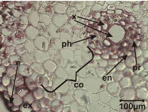
3.1. Root anatomy
The root is covered by the epidermis on the outermost surface. It is made up of a single layer of flattened,
rectangular, ovoid and squarish cells. Under the epidermis 2-3-layered exodermis is located. The cortex covers a large
area with 8-10 layers of mainly polygonal and orbicular parenhymatous cells. The single layered and regularly arranged
endodermis is present between the cortex and central cylinder. It is circular-shaped encircled composed of thick walled
cells, and encircled the central cylinder. The pericycle, which consists of single layered and thin walled cells, is located
under the endodermis. Its shape is similar to the endodermis. There are vascular elements under the area encircled by
the pericycle. The xylem elements constitute aster-shaped tissue. The phloem elements occur between the arms of the
xylem (Figure 1).
Süleyman DOĞU et al., Anatomical characteristics of Bellevalia mathewii Özhatay & Koçak (Liliaceae)
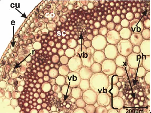
layered cortex consists of orbicular or hexagonal parenchymatic cells. Some parenchymatic cells include raphide
crystals. Under the cortex, the sclerenchymatic tissue constitute a circular band along the transection of the scape. The
vascular bundles are distributed on two rows. The vascular bundles on outer row are smaller than the inner ones and
partly or completely sunk into sclerenchymatic tissue. The parenchymatic cells fill up the area under the
sclerenchymatic band. The vascular bundles on inner row are larger and located in the parenchymatic cells (Figure 2).
Figure 2. The transverse section of the scape of Bellevalia mathewii. e: epidermis, co: cortex, sc: scleranchyma, ph:
phloem, x: xylem, vb: vascular bundle, r: raphide crystals
3.3 Leaf anatomy
The upper and lower epidermis are uniseriate and covered by a cuticle. The stomata are visible in some
transections of the leaf. The upper cuticle is thinner than the lower cuticle. The leaf is amphistomatic and equifacial.
Beneath both the surfaces of the mesophyll is present 2-3 layered palisade parenchyma. The spongy parenchyma is
6-8-layered. It consists of nearly orbicular large cells, and is located between the palisades. Vascular bundles are arranged in
a single row in spongy parenchyma. The vascular bundle in the midrib region is not conspicuously larger than the
others. Therefore, the midrib do not constitute a projecting part (Fig.3).
The leaves have anomocytic type stomata. The stomata lie at the same level as the epidermal cells. Namely,
they are mesomorphic type. On avarage, six stomata occur on both surfaces in the unit area under the x20 objective.
Abundant raphide type crystals are present in upper epidermal cells, but no crystals present in lower epidermal cells.
The number of the stomata is nearly the same as the upper and lower surface. The epidermal cells on both surfaces are
very long and narrow. They are 10 x as long as wide on avarage (Figures 4-5).
Figure 3. The transverse section of the leaf of Bellevalia mathewii. cu: cuticle, ue: upper epidermis, st: stoma, ph:
phloem, x: xylem, pp: palisade parenchyma, sp: spongy parenchyma, le: lower epidermis, r: raphide crystals
Süleyman DOĞU et al., Anatomical characteristics of Bellevalia mathewii Özhatay & Koçak (Liliaceae)