Summary
The aim of this study is to determine the importance of B-mode ultrasonography in the diagnosis of hepatic lipidosis in dairy cows and compare this mode of diagnosis with both the histologic examination of liver biopsy samples and investigation of some biochemical parameters associated with hepatic lipidosis. 15 Holstein cows with moderate hepatic lipidosis and 15 cows with severe hepatic lipidosis and 6 healtyh cows were used as a metarilas. Blood samples were obtained from all cows and analyzed. Liver samples were obtained by biopsy in cattle. The ultrasonographic examination of liver was performed on animal. Serum glutamate dehydrogenase (GDH) and aspartate amino transferase (AST) concentrations were increased in cows with moderate hepatic lipidosis. Serum sorbitol dehydrogenase (SDH), GDH, and AST concentrations were increased in cows with severe hepatic lipidosis. Ultrasonographic examination revealed an increase in diffuse echogenicity of the liver in cows with moderate and severe hepatic lipidosis cows, but the increase was little in moderate hepatic lipidosis. Both serum GDH and SDH levels were found to be increased in severe hepatic lipidosis. However, only the serum GDH level was elevated in moderate hepatic lipidosis. Therefore, both ultrasonographic examination and measurement of specific liver enzymes seem to be beneficial in the diagnosis of hepatic lipidosis.
Keywords: Hepatic lipidosis, Liver enzymes, Liver ultrasonography, Dairy cow
Sütçü Sığırların Hepatik Lipidozisinin Teşhisinde B-Mode
Ultrasonografik Muayene ve Serum Sorbital Dehidrogenaz ve
Glutamete Dehidrogenaz Düzeylerinin Önemi
Özet
Bu çalışmanın amacı; sütçü sığırların yağlı karaciğer sendromunun teşhisinde B-mode ultrasonografinin önemini belirlemek, hastalığın tanısında ultrasonografik muayene ile karaciğer biyopsi örneklerinin histolojik bulguları ve bazı biyokimyasal parametre sonuçlarını karşılaştırmaktır. Bu çalışmada 15 orta hepatik lipidozisli, 15 şiddetli hepatik lipidozisli ve 6 sağlıklı Holştein ırkı sütçü sığır kullanıldı. Bütün sığırlardan kan alındı ve analiz edildi. Sığırların karaciğerinin ultrasonografik muayenesi yapıldı. Hepatik lipidozisli sığırlardan karaciğer biyopsisi alındı. Orta dereceli hepatik lipidozisli sığırlarda serum glutamate dehidrogenaz (GDH) ve aspartat aminotransferaz (AST) seviyeleri artarken, şiddetli hepatik lipidozisli sığırlarda serum sorbital dehidrogenaz (SDH), GDH ve AST seviyeleri arttı. Ultrasonografik maueyenede; orta ve şiddetli hepatik lipidozisli sığırlarda karaciğerde diffuz ekojenite artışı gözlemlenmekle birlikte, orta dereceli hepatik lipidozis olgularında ekojenite artışı daha azdı. Şiddetli hepatik lipidozisde SDH ve GDH enzim düzeylerinde, orta dereceli hepatik lipidozisde ise sadece GDH enzim düzeyinde artış belirlendi. Hem ultrasonografik muayene hem de karaciğerin spesifik enzim ölçümü hepatik lipidozisin teşhisinde faydalı olduğu düşüncesine varıldı.
Anahtar sözcükler: Hepatik lipidozis, Karaciğer enzimleri, Karaciğer ultrasonu, Sütçü sığır
The Importance of Concentrations of Sorbitol Dehydrogenase and
Glutamate Dehydrogenase and B-Mode Ultrasonographic
Examination in The Diagnosis of Hepatic Lipidosis
in Dairy Cows
[1]Mahmut OK * İsmail ŞEN * Hasan GÜZELBEKTEŞ * Murat BOYDAK **
Cenk ER * Uğur AYDOĞDU * Ramazan YILDIZ ***
[1] * ** ***
This study was supported by the University of Selcuk, Scientific Research Project Office (SUBAP No: 10401062) Selcuk University, Faculty of Veterinary Medicine, Department of Internal Medicine, TR-42079 Konya - TURKEY Selcuk University, Faculty of Veterinary Medicine, Department of Histology, Konya, TR-42079 Konya - TURKEY Mehmet Akif Ersoy University, Faculty of Veterinary Medicine, Department of Internal Medicine, TR-15047 Burdur - TURKEY
Makale Kodu (Article Code): KVFD-2012-8146
İletişim (Correspondence)
+90 332 2233584INTRODUCTION
Liver plays a pivotal role in provision of energy for periparturient dairy cows from various precursors 1. A
combination of a deep negative energy balance and fatty liver is a common feature in high producing dairy cows after parturition2. Fatty liver syndrome or hepatic lipidosis
is characterized by infiltration of triacylglycerol (TAG) in the liver3. Fatty liver is a metabolic disorder that is caused
by excessive mobilization of body fat, which occurs in high yielding dairy cows in which the demands for energy exceed the supply 4. Over conditioning is a major risk factor
for developing fatty liver 5. Fatty liver is associated with
decreased health status, well-being, milk yield, reproductive performance, and reduced immune response. Severe and moderate fatty liver develops in approximately 15% and 35% of dairy cows, respectively 6. Fatty liver occurs primarly
in the 1st month of lactation in dairy cows 7,8.
Blood profiles and changes in body condition score have been used to monitor metabolic imbalance around parturition and, in early lactation, to investigate problem 9,10
as well as to predict the risk of diseases such as displaced abomasum 11. Parameters used to monitor imbalance in
energy metabolism have included glucose, non-esterified fatty acids (NEFA), betahydroxybutyrate (BHBA), and change in body condition score 12,13. Fatt liver develops
when hepatic availability of lipogenic and glucogenic products is imbalanced. Thereby the oxidation capacity of fatty acids is exceeded and since hepatic secretion of lipids is inherited, low excess hepatic lipids are stored as TAG in the liver tissue 6,14. In domestic animals, serum enzyme
test are grouped into those that indicate hepatocelluler leakage due to hepatocyte damage 15-17. Hepatocelluler
enzymes, particularly AST, GDH and SDH may be useful in monitoring the hepatic lipidosis that commonly occurs at parturition 5,18. The hepatocelluler leakage enzymes-AST
and cholestatic enzymes-GGT activities have been used to evaluate the liver 19. Blood cholesterol concentration is
related to feed intake and low cholesterol concentrations have been associated fatty liver post-partum 20.
Fatty liver can be diagnosed as reliable only by determining TAG content biochemical or histological analysis of a liver puncture biopsy sample. Biopsies are impracticable for on-farm diagnosis because they cause temporary discomfort to cow, pose risk of infection, and can be lethal if a major blood vessel is punctured21.
Therefore, a non-invasive technique would be very useful. Ultrasonograph has been used routinely for about long time as a diagnostic procudure in hepatic disease of cows 22.
Ultrasound imaging followed by digital analysis of sono-grams has potential to non-invasively detect fatty liver and estimate liver TAG content 23. Haudum et al.24 reported
that ultrasonography has proven useful for the evaluation of hepatic triacylglycerol content in dairy cows. However, detection of fatty liver is more difficult because it results in
smaller cages of hepatic echostructure 22.
The aim of this study is to evaluate the importance of B-mode ultrasonography in the diagnosis of hepatic lipidosis in dairy cows and compare this mode of diagnosis with both the histologic examination of liver biopsy samples and investigation of some biochemical para- meters (Especially SDH and GDH) associated with hepatic lipidosis.
MATERIAL and METHODS
Animals and Clinical Examination
The institutional ethical committee approved this prospective study. In this study, 15 Holstein cows with moderate hepatic lipidosis and 15 Holstein cows with severe hepatic lipidosis in the first 4 weeks of lactation were used. Cows were aged 3-7 years, and the mean daily milk yield was 25 kg. In the reference group, 6 clinically healthy postparturient Holstein cows from a local dairy farm were used. These cows were aged 3-6 years, were in the first 2-4 weeks of lactation, and were reported by the owner to have a daily milk yield ranging from 23 to 27 kg on milking twice daily. Of 15 cows with moderate and severe hepatic lipidosis, 10 showed left abomasal displacement, and of 10 cows with moderate and severe hepatic lipidosis, 5 had ketosis. Routine physical examination, including simultaneous auscultation and percussion of the abdomen, ballottement of the abdomen for a splashing sound to indicate the presence of an air-fluid interface in a large viscus, palpation per rectum, urinary examination, and ultrasonographic examination, were performed on each cow.
Ultrasonographic Examination of Abomasum and Liver
The ultrasonographic examination of the abomasum is performed on the left side of the standing animal. Ultrasonographic examination of the abomasum was performed at the 10th and 13th intercostal spaces on the
left side, and the area was examined ventrally to dorsally using a real-time 3.5-5.0-MHz convex transducer 25,26. The
liver was examined by ultrasonography from the caudal to the cranial region, beginning from the caudal region to the last rib on the right side and ending at the fifth intercostal space, and from the dorsal to ventral region in every inter- costal space using a real-time 3.5-5.0-MHz convex trans-ducer 22. Ultrasonographic examination of the liver was
performed in all cows. Detection of Ketosis
Detection of ketosis; urine was collected with a catheter or free flow urine was collected from all cows and a drop was applied to reagent strips (Multistix® 10 SG; Bayer). Results were scored as positive or negative depending on whether or not there was a change in color from white to purple.
Treatment
A right flank laparotomy was performed under regional analgesia on cows with a presumptive clinical diagnosis of LDA, and the diagnosis was confirmed during surgery using established criteria 27. Cows with LDA were hospitalized
for 1 day after surgery and then discharged. The clinical outcome was investigated at least 2 weeks after surgery by communicating with the owner via telephone. Cows with ketosis were treated. The treatment protocol for cows with ketosis was as follows: intravenous administration of 1.000 mL of 30% serum dextrose (Dekstrose®, Eczacibasi, Baxter) for 3 days, administration of 200 IU insulin (NPH, Humulin®, Lilly Ilac), intramuscular administration of 10 mg of dexamethasone (Devamed®, Topkim Ilac) for 3 days, and oral administration of 150 mL of propylene glycol (Bovical®, Bioteknik) for 4 days. Cows with moderate and severe hepatic lipidosis received ancillary treatment for hepatic lipidosis.
Blood Sample Collection
Blood samples were collected from the jugular vein immediately before surgery in cows with displaced abomasum and before treatment in cows with ketosis and from healthy cattle. An aliquot of blood was placed into glass tubes for serum biochemical analysis. The tubes were centrifuged after clotting, and the serum was harvested and stored at -20°C until analysis.
Biochemical Analyses
Serum SDH concentration was determined by sandwich ELISA (USCN Life Science Inc. Cat No: E91495 Bo, Wuhan, China). The manufacturer of this assay reported the limit of detection in bovine serum as 20 µL and the reference range as 1.56-100 U/L. Serum GDH concentration was determined by sandwich ELISA (USCN Life Science Inc. Cat No: E90293Bo,Wuhan, China). The manufacturer of this assay reported the limit of detection in bovine serum as 100 µL and the reference range as 7.8-500 U/L. Serum AST, ALT, GGT, and ALP activities, as well as serum glucose, cholesterol, triglyceride, blood urea nitrogen (BUN), creatinine, total protein, albumin, Mg+, P+, and Ca++ concentrations
were measured with an automatic analyzer (BT 3000 plus, Biotecnical Inc, SPA, Via lizenca, 18 00155, Rome, Italy). The serum insulin level was measured by an immunoassay system (Invitrogen immunoassays kit # PL2820085, Advia Center XP, Siemens, REVM). Blood Na+, K+, and Cl- concentrations
were measured by using ion-selective electrodes.
Liver Sample Collection and Histological Examination Liver biopsy samples were obtained preoperatively in cows with LDA and cows with ketosis percutaneously through the right 11th to 12th intercostal space. Liver biopsy
samples were not obtained from healthy cattle. Liver biopsy samples were placed in Baker’s formal-Ca solution and fixed in paraffin for at least 16 h. From each fixed liver sample,
12-mm sections were cut and stained with oil Red O and Sudan Black B. The sections were examined under light microscopy as described 28, and the percentage volume of
visible fat in hepatic parenchymal cells was estimated by a stereological point counting method. The extent of fat infiltration in the liver was categorized as mild (<10%, <10 µm2/100 µm2), moderate (10%-20%, 10-20 µm2/100 µm2),
and severe (>20%, >20 µm2/100 µm2) on the basis of the
percentage volume of visible fat 29.
Statistical Analyses
Statistical analyses were performed using a package program (SPSS 15.0), as reported by Akgül30. One-way
analysis of variance (ANOVA) and Tukey’s test were used for comparing the data (P<0.05).
RESULTS
Twenty cows with LDA did not have evidence of any other clinical disease. Cows with LDA had fair to moderate appetite and decreased rumen contraction frequency, defecation, and milk production. Ten cows with ketosis had depressed appetite, decreased rumen contraction frequency and milk production, dry feces, ketonuria, and ketonemia.
Serum GDH, and AST concentrations were increased in cows with moderate hepatic lipidosis and severe hepatic lipidosis as compared with in the controls (Table 1); however, serum SDH concentration was increased in cows with severe hepatic lipidosis. In addition, serum SDH, GDH, AST, and cholesterol levels were higher in cows with severe hepatic lipidosis than in cows with moderate hepatic lipidosis. In contrast, serum cholesterol, Ca++, Cl-, Na+, and
K+ levels were decreased in cows with moderate hepatic
lipidosis and in cows with severe hepatic lipidosis as compared with in the controls (Table 1). Serum total protein level was increased in cows with severe hepatic lipidosis as compared with in cows with moderate hepatic lipidosis and in the controls.
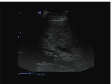
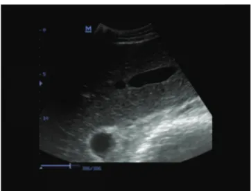
Increase in diffuse echogenicity of the liver was observed in cows with severe hepatic lipidosis (Fig. 1) and cows with moderate hepatic lipidosis, but the increase in the latter was little (Fig. 2). The liver appeared white on ultra- sonograms, and it was difficult to differentiate the liver from the surrounding tissue. Echogenicity of the liver was normal in healthy cows (Fig. 3). There was no significant difference between cows with ketosis and cows with LDA in terms of the liver fat percentage. There were 15 cows with moderate (10% - 20%) hepatic lipidosis and 15 with severe (20% - 48%) hepatic lipidosis.
DISCUSSION
The main finding of the present study was that SDH, GDH, and AST enzyme activities were increased in
post-parturient dairy cows with hepatic lipidosis. The second finding was that an increase in diffuse echogenicity of the liver was observed on ultrasonographic examination in cows with severe hepatic lipidosis and cows with moderate hepatic lipidosis, but the increase was little in the latter.
Hepatocelluler enzymes, particularly AST and GDH
may be useful in monitoring the hepatic lipidosis that commonly occurs at parturition 5,31. SDH and GDH are
liver-specific enzymes 16,17,32,33. Increased serum SDH and
GDH activity is suggestive of either hepatocyte death or sublethal hepatocyte injury. Sorbital dehydrogenase and GDH have been regarded by many as enzymes of choice for
Table 1. Serum biochemical parameters healthy lactating cows (control) and lactating cows with moderate and severe hepatic lipidosis. Tablo 1. Sağlıklı, orta ve şiddetli hepatik lipidozisli sütçü sığırların serum biyokimyasal parametreleri
Parameters Groups P
Control N=6 Moderate N=15 Severe N=15
SDH (UI/L) 18.93±4.32 a 42.63±5.28 a 75.78±9.75 b 0.000 GDH (UI/L) 101.33±8.78 a 132.08±9.93 ab 149.50±12.18 b 0.040 AST (UI/L) 71.33±6.30 a 139.33±13.28 b 155.07±15.19 b 0.007 ALT (U/L) 26.17±2.65 25.31±2.91 28.30±3.17 0.762 GGT (UI/L) 31.50±8.43 46.85±6.29 38.30±5.26 0.297 ALP (UI/L) 47.17±5.15 41.38±3.82 37.30±5.72 0.302 Insulin (µU/L) 0.35±0.07 0.42±0.29 0.50±0.34 0.752 Cholesterol (mg/dL) 176.10±89.03a 125.23±68.74ab 78.00±11.7b 0.36 Tryglyseride (mg/dL) 16.17±2.86 19.92±4.65 22.50±9.30 0.183 Glucose (mg/dL) 101.67±6.64 102.31±13.79 79.90±18.49 0.515 Urea (mg/dL) 29.50±0.99 39.00±4.17 32.70±4.96 0.329 Creatinin (mg/dL) 1.82±0.18 1.30±0.12 1.37±0.12 0.051 Total protein (g/dL) 7.08±0.14 a 8.65±0.31 a 10.81±0.84 b 0.001 Albumin (g/dL) 3.38±0.26 3.86±0.22 4.18±0.15 0.089 Magnesium(mg/dL) 2.00±0.05 1.67±0.17 2.09±0.11 0.101 Calcium (mg/dL) 10.05±0.31 a 8.49±0.25 b 9.12±0.27 ab 0.004 Phosphorus (mg/dL) 6.40±0.66 5.22±0.47 4.68±0.32 0.096 Chlor (mmol/L) 101.50±1.61a 94.23±1.76 b 96.10±1.66 ab 0.045 Sodium (mmol/L) 144.33±0.84 a 137.00±0.72 b 140.75±1.15 c 0.000 Potassium (mmol/L) 3.90±0.18 a 3.06±0.22 b 3.41±0.15 ab 0.043
SDH: Sorbitol dehydrogenase, GDH: Glutamate dehydrogenase, AST: Aspartate amino transferase, ALT: Alanin amino transferase, GGT: gamma-glutamyl transferase, ALP: Alkaline phosphatase, a-c means within row with different supercript differ (P<0.05)
Fig 1. Ultrasonogram of liver of a cow with severe hepatic lipidosis Şekil 1. Şiddetli hepatik lipidosisli bir inekte karaciğer ultrasonogramı
Fig 2. Ultrasonogram of liver of a cow with moderate with hepatic
lipidosis
use as indicators of the degree of hepatic necrosis in sheep, goats, and cattle 16,17,33-35. Braun et al.36 reported that SDH,
GDH, and GGT concentrations are increased in cows with liver tumors. Kalaitzakis et al.33 mentioned that AST, SDH,
GDH and ornitine carbamobyl transferase, activity was increased in cases of fatty liver syndrome because of the destruction of liver cells. The activities of hepatocellulerr leakage enzymes such as AST and cholestatic enzymes such as GGT have been used to evaluate the liver 19. Bogin
et al.37 found significantly increased AST levels in cows
with severe fatty liver. Sevinc et al.28showed that cows
with severe fatty liver had high AST and GGT activities. Hepatic lipidosis may contribute to the pathogenesis of abomasal displacement. Komatsu et al.38 indicated that
high levels of AST in cows with LDA were found in fatty liver degeneration. Kalaitzakis et al.39 reported that OCT
and GDH enzymes might be useful in diagnosis fatty liver in downer cows. In this study, serum GDH and AST concentrations were increased in cows with moderate and severe hepatic lipidosis compared with in the controls. However, only the serum SDH concentration was increased in cows with severe hepatic lipidosis. Increased SDH and GDH concentrations in cows with hepatic lipidosis may be related to hepatocyte death or sublethal hepatocyte injury. SDH, GDH, and AST levels were high in cows with moderate and severe hepatic lipidosis compared with in the control group. Although higher levels of SDH and GDH are generally reported in acute liver damage (within 4 to 24 hours of hepatic injury) 19,32. We found that higher levels of
these enzymes were found in cows with hepatic lipidosis. We understand that these enzymes are increased not only in acute liver damage but also in chronic liver damage 15,17,33.
Blood cholesterol concentration is related to feed intake 40 and low cholesterol concentration have been
associated fatty liver post-partum 20. Several authors 1,28,33
reported that triglyceride, cholesterol and HDL- cholesterol concentrations are decreased in the cows with fatty liver.
Sevinc et al.41 found that serum triglyceride and cholesterol
concentrations were decreased in the cows with moderate and severe fatty liver. In the present study, serum cholesterol level was decreased in cows with moderate hepatic lipidosis and in cows with severe hepatic lipidosis as compared with in the controls, but triglyceride level in cows with moderate and severe hepatic lipidosis was not different from healthy cows. The low cholesterol level can be thought to be caused by a fat infiltration in the liver and a low output of lipoprotein.
Madison and Trout 42 found that calcium when
present at a level of <1.2 mmol/L had a reducing effect on abomasal motility. They also brought attention to the fact that hypocalcemia cannot be a major causative factor for decreased abomasal motility with respect to the development of abomasal displacement. Some authors reported that hypocalcemia is a risk factor for abomasal displacement 43-45. Metabolic alkalosis is mentioned as a risk
factor for abomasal displacement 44. The fluid accumulating
in the distended abomasum indicates continuous secretion of hydrochloric acid. This sequestration of the chloride ion in the abomasum along with some abomasal reflux into the rumen results in metabolic alkalosis 46. During the
dilatation phase, which commonly lasts for several days, there is continuous secretion of hydrochloric acid, sodium chloride, and potassium into the abomasum; thus, the abomasum becomes gradually distended and does not evacuate its contents into the duodenum. This leads to dehydration and metabolic alkalosis with hypochloremia and hypokalemia 47. In this study, Ca++, Cl-, Na+, and K+ levels
were decreased in cows with moderate hepatic lipidosis and in cows with severe hepatic lipidosis as compared with in the controls. Decrease in the Ca++, Cl-, Na+, and
K+ concentrations may be related to metabolic alkalosis
resulting from abomasal displacement.
Nutritional stress in dairy cattle can be evaluated by determining the serum NEFA, BHBA, acetoacetate, cholesterol, and glucose and liver fat percentages, with the latter being regarded as the most accurate indicator of nutritional stress 15,16,48,49. Liver fat percentage is increased
in cows with abomasal displacement 50-52 and ketosis48. The
liver fat percentage in healthy cattle is typically 5%, and is increased to 8% in healthy cows shortly after calving and to 33% in cows with postparturient ketosis 48. In the present
study, cows with abomasal displacement and ketosis had moderate (10% - 20%) and severe (20% - 48%) hepatic lipidosis according to histopathologic evaluation. Increase in serum AST activities is consistently related to fatty liver 41,53.
Ultrasonography has been used routinely for a long time as a diagnostic procedure in hepatic disease of cows 22.
Ultrasound imaging followed by digital analysis of sono-grams has the potential to non-invasively detect fatty liver and estimate liver triacylglycerol content 23,24. Braun22
reported the ultrasonographic features of fatty livers as an Fig 3. Ultrasonogram of liver of healthy a cow
increase in the size of the liver, round margins, hyperechoic hepatic parenchyma near the abdominal wall, and decrease in the strength of the echo with increase in the distance from the abdominal wall, and poor visualization of hepatic blood vessels. In this study, increase in the diffuse echo-genicity of the liver was observed in cows with severe hepatic lipidosis (Fig. 1) and cows with moderate hepatic lipidosis (Fig. 2), but the increase in the latter was little. There was a good relationship between the ultrasonographic image findings and 20% and over fatty liver. Large vessels of the liver in cows with moderate or severe hepatic lipidosis were observed, but the small vessels of the liver were poorly imaged or not seen in cows with severe hepatic lipidosis (Fig. 1) and only faintly seen or poorly imaged in cows with moderate hepatic lipidosis (Fig. 2). The poor images of the small vessels may be attributable to the deposition of fat on the surface of vessels and the swelling of hepatocytes due to fat deposition in the cells. Otherwise, the cause of increase in diffuse echogenicity of the liver may be the diffuse fat deposition in the cells. Mohamed et al.54 reported that ultrasonography may provide good
images of focal fatty infiltration of the liver. There was also a significant relationship between serum liver-specific enzymes (SDH, GDH, and AST), histopathologic findings, and ultrasonographic imaging findings of the liver.
The results of our study showed that B-mode ultra-sonography is a valuable tool in the diagnosis of hepatic lipidosis. Both serum GDH and SDH levels were found to be increased in severe hepatic lipidosis. However, only the serum GDH level was elevated in moderate hepatic lipidosis. Therefore, both ultrasonographic examination and measurement of specific liver enzymes (SDH and GDH) seem to be beneficial in the diagnosis of hepatic lipidosis.
REFERENCES
1. Drackley JK, Overton TR, Douglas GN: Adaptations of glucose and
long-chain fatty acid metabolim in liver of dairy cows during the peri-parturient period. J Dairy Sci, 84, 100-112, 2001.
2. Herdt TH: Fatty liver in dairy cows. Vet Clin North Am: Food Anim Pract,
4, 269-287, 1988.
3. Gerloff BJ, Herdt TH, Emery RS: Relation of hepatic lipidosis to health
and performence in dairy cattle. J Am Vet Med Assoc, 88, 845-850, 1986.
4. Reid IM, Roberts CJ: Subclinical fatty liver in dairy cows. Iris Vet J, 37,
281-284, 1983.
5. Bobe G, Young JW, Beitz DC: Invited review: Pathology, etiology,
prevention and treatment of fatty liver in dairy cows. J Dairy Sci, 87, 3105-3124, 2004.
6. Rehage J, Starke A, Holtershinken M, Kaske M: Hepatic lipidosis.
Diagnostic tool and individual and herd risk factor. Congress XXIV World
Buiatrics, 15-19 October, Nice, France. pp 69-74, 2006.
7. Grummer RR: Etiology of lipid-related metabolic disorders in
periparturient dairy cows. J Dairy Sci, 76, 3882-3896, 1993.
8. Saber APS: Hepatic triacylglycerols and serum non-esterified fatty acids,
vit. E and selenium levels in cross breed cow in Tabiriz city of Azarbaijan province of Iran: An abattoir study. J Anim Vet Advance, 108, 1063-1068, 2011.
9. Oetzel GR: Monitoring and testing dairy herds for metabolic disease. Vet Clin North Am: Food Anim Pract, 20, 651-674, 2004.
10. Macrae AI, Whitaker DA, Burrough E, Dowell A, Kelly JM: Use of
metabolic profilies for the assesment of dietary adequacy in UK dairy herds. Vet Rec, 159, 655-661, 2006.
11. LeBlanck SJ, Leslie KE, Duffield TF: Metabolic predictors of displaced
abomasum in dairy cattle. J Dairy Sci, 88,159-170, 2005.
12. Whitaker DA: Metabolic profiles. In, Andrews AH, Blowey RW, Boyd
H, Eddy RG (Eds): Bovine Medicine: Diseases and Husbandry of Cattle. pp.804-817, Blackwell Science: Oxford, 2004.
13. Van Knegsel ATM, van den Brand H, Dijkstra WM, van Straalen MJW:
Dietary energy source in dairy cows in early lactation: Energy partitioning and milk composition. J Dairy Sci, 90, 1467-1471, 2007.
14. Katoh N: Relevance of apolipoproteins in the development of fatty
liver and fatty liver -related peripartum diseases in dairy cows. J Vet Med
Sci, 64, 293-307, 2002.
15. West HJ: Effect on liver function of dairy cows in late pregnancy and
early lactation. Res Vet Sci, 46, 231-236, 1989.
16. Garry FB, Fettman MJ, Curtis CR, Smith JR: Serum bile acid
concentration in dairy cattle with hepatic lipidosis. J Vet Intern Med, 8, 432-438, 1994.
17. Cebra CK, Garry FB, Getzy DM: Hepatic lipidosis in anorectic, lactating
Holstein cattle. A retrospective study of serum biochemical abnormalities.
J Vet Intern Med, 11, 231-237, 1997.
18. Duncan JR, Prasse KW, Mahaffey EA: Liver. In, Veterinary Laboratory
Medicine: Clinical Pathology. pp. 130-151, Iowa State Univresity Press, Ames, Iowa, 1994.
19. Roussel JA, Whitney SM, Jole DJ: Interpreting of bovine serum
chemistry profile: Part II, Vet Med, 6, 559-566, 1997.
20. Van den Top AM, van Tol H, Jansen A, Geelen MJH, Beynen AC: Fatty
liver in dairy cows postpartum is associated with decreased concentration of plasma triacylglycerols and decreased activity of lipoprotein lipase in adipocytes. J Dairy Res, 72, 129-137, 2005.
21. Smith TR, Hippen, AR, Beitz DC, Joung JW: Metabolic characteristics
of induced ketosis in normal and obes dairy cows. J Dairy Sci, 80, 1569-1581, 1997.
22. Braun U: Ultrasonography of the liver in cattle. Vet Clin Food Anim, 25,
591-609, 2009.
23. Bobe G, Amin VR, Hippen AR, She P, Young JW, Beitz, DC:
Non-invasive detection of fatty liver in dairy cows by digital analyses of hepatic ultrasonograms. J Dairy Res, 75, 84-89, 2008.
24. Haudum A, Starke A, Beyerbach M, Wohlsein P, Rehage J:
Ultra-sonographic assessment of liver dimensions in dairy cows with different hepatic triacylglycerol content. J Anim Sci, 89,1392-1400, 2011.
25. Nyland TG, Mattoon JS, Herrgesell EJ: Liver. In, Small Animal Diagnostic
Ultrasound. 2nd ed., pp. 93-127, Saunders Company, Philadelphia. 2002.
26. Ok M, Arıcan M, Turgut K: The ultrasonographic finding in dairy cows
with left and right abomasal displacement. Revue Vet Med, 153 (1): 15-18, 2002.
27. Costable PD, St Jean G, Hull BL, Rings DM, Hoffsis GF: Prognostic
value of surgical and postoperative finding in cattle with abomasal volvulus.
J Am Vet Med Assoc, 199, 892-898, 1991.
28. Sevinç M, Başoğlu A, Birdane FM, Boydak M: Liver function in dairy
cows with fatty liver. Revue Vet Med, 15 (4): 297-300, 2001.
29. Aslan V, Aştı R, Nizamlıoğlu M, Tekeli T, Demirci Ü: Fatty liver syndrome
associated with some postparturient period disease. SU Vet Fak Derg, 4, 43-52, 1988.
30. Akgül A: Tıbbi Araştırmalarda İstatistiki Analiz Teknikleri “SPSS
Uygulamaları”. İkinci baskı. s. 23-26, Emek Ofset, Ankara, 2003.
31. Stengärde L, Holtenius K, Emanuelsan U, Hultgren J, Niskanen R, Trávén, M: Blood parameters in Swedish dairy herds with high or low
incidence of displaced abomasum or ketosis. Vet J, 190, 124-130, 2011.
32. Raja MM, Raja A, Imran MM, Santla AMI, Devasena K: Enzymes
application in diagnostic prospect. Biotechnol, 10 (1): 51-59, 2001.
33. Kalaitzakis E, Roubies N, Panousis N, Pourliotis K, Kaldrymidou E, Karatzias H: Clinicopathologic evaualation of hepatic lipidosis in
periparturient dairy cattle. J Vet Intern Med, 21, 835-845, 2007.
34. Kaneko JJ, Harvey JW, Bruss ML: Blood analyte reference values in
large animals. In, Clinical Biochemistry of Domestic Animals. Fifth ed., pp. 890-894, Academic Press, San Diego. 1997.
35. El-Kabbani OC, Darmanian OC, Chung RPT: Sorbital dehydrogenase:
Structure, function and ligand design. Curr Med Chem, 11, 465-476, 2004.
36. Braun U, Nuss K, Soldati G: Clinical and ultrasonographic finding in
four cows with liver tumörs. Vet Rec, 157, 482-484, 2005.
37. Bogin E, Aviden Y, Merom M: Biochemical changes associated with
the fatty liver syndrome in cows. J Comp Pathol, 98, 337-347, 1988.
38. Komatsu Y, Itoh N, Tanyyama H, Kitazaw T, Yokota H, Koiwa M, Ohtsuka H, Terasaki N: According to histopathology of the liver and
clinical chemstry. J Vet Med A Pathol Clin Med, 49, 482-486, 2002.
39. Kalaitzakis E, Panousis N, Roubies N, Giadinis N, Kaldrymidou E, Georgiadinis M, Karatzias H: Clinicopathologic evaualation of downer
dairy cows with fatty liver. Can Vet J, 51, 615-622, 2010.
40. Janovick Guretzky NA, Carlson DB, Garrett JE, Drackley JK: Lipid
metabolite prolifes and milk production for Holstein and Jersey cows fed rumen-protected choline during the periparturient period. J Dairy Sci, 89, 188-200, 2006.
41. Sevinc M, Basoglu A, Guzelbektes H, Boydak M: Lipid and lipoprotein
levels in dairy cows with fatty liver. Tr J Vet Anim Sci, 27, 295-299, 2003.
42. Madison JB, Troutt HF: Effect of hypocalcemia on abomasal motility. Res Vet Sci, 44, 264-266, 1988.
43. Geishauser T, Leslie KE, Duffield TF: Metabolic aspect in the etiology
of displaced abomasum. Vet Clin North Am: Food Anim Pract, 16, 255-265, 2000.
44. Van Winden SC, Kupier R: Left displacement of the abomasum in
dairy cattle: Recent development in epidemiological and etiological aspects. Vet Res, 34, 47-56, 2003.
45. Sen I, Ok M, Coskun A: The level of serum ionised calcium, aspartate
aminotransferase, insulin, glucose, betahydroxybutyrate concentrations and blood gas parameters in cows with left displacement of abomsum.
Pol J Vet Sci, 9 (4): 227-232, 2006.
46. Vlamick K, Oyaert W, Muylle E, van den Hende C, Pipeleers D: Blood
levels somatostatin, pancreatic polypeptide and gastrin in normal cows and in cows suffering from abomasal dilatation. J Vet Med A, 33, 241-246, 1986.
47. Radostits OM, Gay CC, Hinchcliff KW: Disease of abomasum. In:
Radostits OM (Ed): Veterinary Medicine. 10th ed., pp. 353-374, Saunders
Company, Philadelphia, 2007.
48. Djokovic R, Samanc H, Jovanovic M: Blood concentrations of thyroid
hormones and lipids and content of lipids in the liver of dairy cows in transtional period. Acta Vet Brno, 76, 525-532, 2007.
49. Saco Y, Fina M, Gimenez M, Pato R, Piedrafita J, Bassols A: Evaluation
of serum cortisol, metabolic parameters, acute phase proteins and faecal corticosterone as indicators of stres in cows. Vet J, 77, 439-441, 2008.
50. Muylle E, van den Hende C, Sustronc B, Deprez Y: Biochemical
profiles in cows with abomasal displacement estimated by blood and liver parameters. J Vet Med A, 37, 259-263, 1990.
51. Aslan V, Ok M, Boydak M, Sen I, Birdane FM, Alkan F: The study on
the relationsheep of abomasal displacement and fatty liver syndrome in dairy cows. Vet Bil Derg, 13, 77-82, 1997.
52. Güzelbekteş H, Sen I, Ok M, Costable PD, Boydak M, Coskun A:
The levels of serum amiloid A and Haptoglobin concentrations and liver fat percentage in lactating dairy cows with abomasal displacement. J Vet
Intern Med, 24, 213-219, 2010.
53. Herdt TH: Ruminant adaptation to negative energy balance “ Influences
on the etiology of ketosis and fatty liver”. Vet Clin North Am: Food Anim
Pract, 16, 215-230, 2000.
54. Mohamed T, Oikawa S, Kurosawa T, Takehana K, Hosaka Y, Koiwa M, Sato H: Focal fatty liver in a heifer: utility of ultrasonography in diagnosis. J Vet Med Sci, 66 (3): 341-344, 2004.