Abstract. – OBJECTIVE: The members of the matrix metalloproteinase (MMP) family and canna-binoids (CBs) are reportedly associated with hip-pocampus-dependent memory functions. How-ever, the effects of endogenously formed CBs on hippocampal long-term potentiation remain unknown. The present study aimed to investi-gate the changes in the gene and protein expres-sion levels of matrix metallopeptidase 9 (MMP-9), phosphatase and tensin homolog (PTEN), and NOTCH receptor 1 (NOTCH1) in rat hippocampal tissues treated with anandamide (AEA), AM251, 6-iodopravadolin (AM630), and N-[4-{[(3,4-Dimeth-yl-5-isoxazolyl)amino]sulfonyl}phenyl] (ML193).
MATERIALS AND METHODS: The subjects were divided into 10 groups (n = five per group). The pharmaceuticals were administered via in-traperitoneal injection once a day for seven days, except for the control group. The resected hippocampal tissues were then evaluated using a quantitative real-time polymerase chain reac-tion (RT-qPCR) and Western blot analysis. The data obtained were statistically analyzed, and p < 0.01 was considered statistically significant.
RESULTS: Contrary to the literature, the changes in MMP-9 expression were not statis-tically significant, but the changes in PTEN and NOTCH1 were. The findings of this in vivo exper-imental study revealed that the agonists and an-tagonists acting on the CB system have signif-icant molecular effects on hippocampal tissue. CONCLUSIONS: The changes in gene and protein expressions may be one of the reasons for the neurodegenerative processes observed in patients using these agonists and antago-nists, whose effects on the CB system have not been fully explained yet. Our study can contrib-ute to the literature as it is the first study investi-gating the MMP-9, PTEN and NOTCH1 gene and protein expression.
Key Words:
Anandamide, Hippocampus, MMP-9, NOTCH1, PTEN.
Abbreviations
AEA = N-arachidonoylethanolamine also known as anandamide (AEA); AM630 = 6-iodopravadolin; CB = cannabinoid; CBR = cannabinoid receptors; eCBs = endocannabinoid system endocannabinoid system; CB receptors (CBRs) GPR55 = G protein-coupled receptor 55; LTP = long - term potentiation; MF-CA3 = mossy fiber-CA3; MMP = matrix metalloproteinase; NOTCH1 = NOTCH receptor 1; PTEN = phosphatase and tensin homolog.
Introduction
The hippocampus plays a significant role in the formation of physiological events associated with learning and memory1. Various
physiolog-ical events, such as neurogenesis, synaptic plas-ticity, and emotional states, are largely associated with hippocampal tissues; the cannabinoid (CB) system (CBs) is involved in the regulation of these events2.
The endocannabinoid system (eCBs) acts on CB receptors (CBRs) and regulates various as-pects of human physiological, behavioral, immu-nological, and metabolic functions2. The eCBs
af-fects the neuromodulators involved in the regula-tion of neuroendocrine and metabolic funcregula-tions, as well as the pathogenesis of cardiovascular diseases and obesity3.
I. YILMAZ
1, N. KARAARSLAN
2, D. YASAR SIRIN
3, H. OZBEK
11Department of Medical Pharmacology, Istanbul Medipol University School of Medicine, Turkey 2Department of Neurosurgery, Namik Kemal University School of Medicine, Turkey
3Department of Molecular Biology and Genetics, Namik Kemal University Faculty of Arts and
Sciences, Turkey
Numan Karaarslan, Duygu Yasar Sirin and Hanefi Ozbek are co-authors
Pharmaco-molecular assessment of the
effects of anandamide and its antagonists on
hippocampal tissue in Wistar albino rats
The eCBs is known to be involved in many physiopathological processes, including athero-sclerosis4, obesity-related hypertriglyceridemia,
and glucose metabolism of the liver5.
Moreover, scholars5 suggest that the eCBs is
involved in the pathogenesis of pain, inflamma-tion, and glaucoma. Due to its antiproliferative effect on tumor cells, it has led to investiga-tions in glioblastoma and cancer treatments6,
as well as hypertension, heart failure, obesity, diabetes mellitus, metabolic syndrome, star-vation, chronic stress, depression, and other psychiatric disorders7. The eCBs is synthesized
“on-demand” from long-chain polyunsaturated fatty acids and acts on cells in a paracrine or autocrine manner8. This system plays a role in
the physiological or physiopathological pro-cesses of many diseases, such as Alzheimer’s, Parkinson’s, multiple sclerosis, epilepsy, hy-pertension, and hypercortisolism9, and is
lo-cated in different parts of the brain, especially the hippocampus10. Almeida et al11 show the
ex-pression of eCBs in several regions of the brain involved in fear response, including the thala-mus, cortex, amygdala, and the hippocampus.
Hippocampal dysfunction has been suggested as the underlying reason for age-related neuro-degenerative diseases that affect cognitive func-tions. Therefore, it is vital to investigate the role of new genes whose expressions have been changed in the physiology and pathology of the hippocampus12,13.
Proteolytic activity mediated by many complex systems of protease, including members of the matrix metalloproteinase (MMP) family, plays a pivotal role in the mechanisms of hippocampal synaptic plasticity. Furthermore, MMPs are in-volved in long-term synaptic plasticity, learning, and memory12,13.
MMP-9 plays a significant role in long-term potentiation (LTP) maintenance in the Schaffer collateral-CA1 pathway and in the acquisition of hippocampus-dependent memory, and changes in MMP-9 levels are therefore a determinant of neurodegenerative processes14. Wieraet al15
have reported that MMP blockades disrupt LTP maintenance in the mossy fiber-CA3 (MF-CA3) projection in which LTP induction and expression are largely presynaptic, and LTP induction is cor-related with increased MMP-9 expression.
Anandamide (AEA), known as N-arachidonoyl ethanolamine, is a cannabinoid (CB) neurotrans-mitter derived from arachidonic acid in the brain and can be found in the central and peripheral
nervous systems. Two subtypes of CBRs – can-nabinoid-1 receptor (CB1R) and cannabinoid-2 receptor (CB2R) – are located in the central and peripheral nervous systems16.
The CB1R antagonist AM251, the CB2R an-tagonist 6-iodopravadoline (AM630), and the G protein-coupled receptor 55 (GPR55) antagonist N-(4-{[(3,4-Dimethyl-5-isoxazolyl)amino] sulfo-nyl}phenyl) (ML193) are known to be potent and selective antagonists in the brain17. This is
because the CB1R antagonists damage cogni-tion and prevent the synaptic transmission of LTP. Colangeli et al18 have reported that CBs are
associated with hippocampus-dependent neuro-degenerative processes. However, studies on the effect of endogenously formed CBs on hippocam-pal LTP have failed to provide clarity19, and no
studies have yet investigated the effects of AEA, AM251, AM630, and ML193.
The present study aimed to investigate the changes in the gene and protein expression lev-els of MMP-9, phosphatase and tensin homolog (PTEN), and NOTCH receptor 1 (NOTCH1) in rats treated with AEA, AM251, AM630, and ML193. The PTEN gene codes the PTEN en-zyme, which acts as a dual-specificity protein phosphatase that modifies proteins and fats (lip-ids) by removing phosphate groups. PTEN de-phosphorylates the tyrosine-phosphorylated focal adhesion kinase and inhibits cell migration, in-tegrin-mediated cell spreading, and focal adhe-sion formation. PTEN can also inhibit MMP-9 through the regulation of NF-kB, and it plays a role as a key modulator of the AKT-mTOR sig-naling pathway in controlling neuron integration during adult neurogenesis, including correcting neuron positioning, dendritic development, and synapse formation.
Notch signaling is an evolutionarily conserved intercellular signaling pathway that regulates interactions between adjacent cells. NOTCH1 signaling is known to have a pivotal role in brain and memory development, and the MMP-9 protein has a significant influence on NOTCH1 activity. The present study investigated the main role played by the proteins involved in neuro-degenerative diseases. It may be possible to not only clarify the probable association between the inhibition of GPR55 and the consecutive changes in the level of MMP-9, previously reported to have several implications at the molecular level in hippocampal tissue, but also reveal the eventu-al MMP-9 levels in response to the induction or inhibition of different CBRs.
Materials and Methods
Animal ExperimentsThis randomized, double-blind, in vivo exper-imental study was performed with the approval of the Local Ethics Committee for Animals. The experiments were carried out in the Istan-bul Medipol University Medical Research Center laboratory.
Live mammalian subjects were fed using stan-dard food pellets. The subjects were kept for periods of 12 h in the dark and 12 h in the light. The pharmaceuticals were administered via in-traperitoneal injection once a day for seven days. Preparation of Drugs
All pharmaceuticals were prepared using ap-propriate diluents in accordance with the manu-facturer’s protocols. In determining drug concen-trations, the specified doses in previous in vivo studies were used.
Accordingly, AEA (Catalog No. A0580, Sig-ma-Aldrich Chemie GmbH, Taufkirchen, Ger-many) was dissolved in a physiological saline solution to reach a final concentration of 6 mg/ ml20; AM251 (Catalog No. A6226, Sigma-Aldrich
Chemie GmbH, Taufkirchen, Germany) was dis-solved in a physiological saline solution to reach a final concentration of 1 mg/ml21; AM630 (Catalog
No. SML0327, Sigma-Aldrich Chemie GmbH, Taufkirchen, Germany) was dissolved in saline solution to reach a final concentration of 1.25 mg/ ml22; and ML193 (Catalog No. SML1340,
Sig-ma-Aldrich Chemie GmbH, Taufkirchen, Ger-many) was dissolved in 0.05% dimethyl sulfoxide (Catalog No. DMS555, BioShop, Canada, Inc., Canada) to reach a final concentration of 5 mg/ ml23. Stock solutions were stored at 4°C.
Fifty Wistar albino male rats with an average weight of 310 g aged 10 weeks were divided into
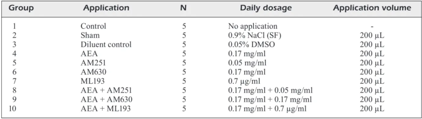
10 groups. Group 1 was the control group where no drug application was performed; groups 2 and 3 were sham and diluent control groups, respec-tively. Daily intraperitoneal medication was ap-plied to the subjects in groups 4-10 for one week. The application, sample number, daily dosage, and application volumes are provided in Table I. Dissection of Hippocampal Tissues
At the end of one week of applications de-scribed in Table I, the subjects were adminis-tered Isoflurane-USP 100 ml® (Adeka, Maslak,
Istanbul), an inhalation anesthetic, and then, decapitated under general anesthesia using a rodent decapitator (Decapitator Catalog No. 25303059999990000651400001, Remer, Ka-vacik, Istanbul). A light source (CL 6000 LED®, Zeiss™, Göteborg, Sweden) was used throughout the process. The subjects’ heads were taken to the operation site, and the skin and subcutaneous tissue were excised to uncover the osseous tissue. The anatomically uncovered osseous tissues were incised using iris scissors along the caudolateral border of the interparietal bone. Frontal, parietal, and interparietal bone tissues covering the dorsal surface of the cerebral tissue were excised. The cerebral tissue was carefully detached from the surrounding meninges. The cerebral tissue was extracted without damaging the adjacent osseous structures, and the excised cerebral tissue was immediately placed on a frosted.
The cerebellum, pons, and medulla were dis-sected from the cerebral cortex with a dissector, and the tissues were then detached from the cere-bral cortical structures with a surgical steel scal-pel (No. 15 blade). The interhemispheric fissure, also known as the medial longitudinal fissure, was incised with the scalpel. The two cerebral hemispheres were thus separated from each other. The left cerebral hemisphere was dissected using Table I. Group number, application, sample number, daily dosage, and application volume.
Group Application N Daily dosage Application volume
1 Control 5 No application
2 Sham 5 0.9% NaCl (SF) 200 µL
3 Diluent control 5 0.05% DMSO 200 µL
4 AEA 5 0.17 mg/ml 200 µL 5 AM251 5 0.05 mg/ml 200 µL 6 AM630 5 0.17 mg/ml 200 µL 7 ML193 5 0.7 µg/ml 200 µL 8 AEA + AM251 5 0.17 mg/ml + 0.05 mg/ml 200 µL 9 AEA + AM630 5 0.17 mg/ml + 0.17 mg/ml 200 µL 10 AEA + ML193 5 0.17 mg/ml + 0.7 µg/ml 200 µL
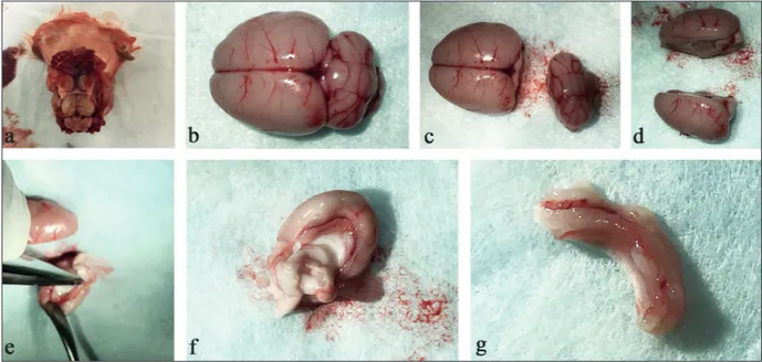
small curved forceps, a scalpel, and a dissector. The same procedure was performed on the right cerebral hemisphere, and bilateral hippocampal tissues were obtained24. The excised hippocampal
tissues were placed in tubes (Figure 1), and the extracted tissues were irrigated in 0.15 ml cold (+4°C) potassium chloride and dried with blotting paper.
Quantitative Real Time-Polymerase Chain Reaction (RT-PCR)
The tissues were then weighed and registered. Half of the extracted hippocampal tissue was used for RNA isolation and the other half for pro-tein isolation. The RNA was extracted from the tissues using a PureLink RNA Mini Kit (Thermo Fisher Scientific, Catalog No. 121830A, Waltham, MA, USA). The tissues were transferred to the lysis buffer (included in the kit) and fully disin-tegrated mechanically using a rotator. The RNA was isolated in accordance with the manufac-turer’s instructions, and the extracted RNA was reverse transcribed using a high capacity cDNA RT Kit (Thermo Fisher Scientific, Catalog No. 4368814, Waltham, MA, USA) to obtain cDNA. The reaction conditions were 10 min at 25°C, 120 min at 37°C, and 5 min at 85°C. The chang-es in MMP-9, NOTCH1, and PTEN exprchang-essions were determined with qPCR, which was
per-formed on an Applied Biosystems 7300/7500 Real-Time PCR system (Thermo Fisher Scien-tific, Waltham, MA, USA). To determine the gene expression profiles, all genes were amplified using TaqMan Gene Expression Assays MMP-9 (Cat#4331182, Mmp9. Rn00579162_m1, RefSeq NM_031055.1), NOTCH1 (Cat#4331182, Notch1. Rn01758633_m1, RefSeq NM_001105721.1), and PTEN (Cat#4331182, PTEN.Rn00477208_m1, RefSeq NM_031606.1) from Thermo Fisher Sci-entific, Waltham, MA USA. β-actin TaqMan Gene Expression Assay (Cat#4331182, Actb. Rn00667869_m1, RefSeq NM_031144.3, Thermo Fisher Scientific, Waltham, MA USA) was used for the quantification of gene expression.
Western Blotting
For the total protein isolation, the resected hippocampal tissues were weighed and placed in encoded Eppendorf tubes containing a Tri-ton X-100 (Sigma-Aldrich, Cat#10789704001, Sigma-Aldrich Chemie GmbH, Taufkirchen, Germany) protease inhibitor cocktail (Roche, Cat#04693159001, Taufkirchen, Germany). The tissues were mechanically degraded, and protein lysates were obtained.
The protein lysates were processed using a western blot test to reveal the expressions of MMP-9, NOTCH1, and PTEN. The total
pro-Figure 1. A, The cranial location of the cerebral tissue after the excision of the skin, subcutaneous tissue, and osseous tissue covering the cranium. B, Excised brain tissue on a frosted plate. C, Two cerebral hemispheres obtained through the excision of the cerebellum, pons, and medulla. D, and E, Medial surface of the right cerebral hemisphere extracted after the incision of the interhemispheric fissure and the dissection of the corpus callosum and hippocampal tissue in the right cerebral hemisphere.
tein amount was determined with a Bradford protein assay. Sodium dodecyl sulfate-polyacryl-amide gel electrophoresis (SDS-PAGE) was per-formed for the samples, each of which contained 100 mg of protein. Using iBlot, the samples were transferred to a polyvinylidene difluoride (PVDF) membrane (Thermo Fisher Scientific, Cat#IB401001, Waltham, MA, USA) to perform immunoblotting. The PVDF membranes were blotted with an MMP-9 monoclonal antibody (Thermo Fisher Scientific, Cat#MA5-15886, Waltham, MA, USA) at a dilution of 1:500, a NOTCH1 polyclonal antibody (Thermo Fisher Scientific, Cat#PA5-23181, Waltham, MA, USA) at a dilution of 5mg/ml final concentration, a phos-pho-PTEN (Ser380) polyclonal antibody (Thermo Fisher Scientific, Cat#PA5-17826, Waltham, MA, USA) at a dilution of 1:1000, and a monoclo-nal β-actin antibody (Thermo Fisher Scientific, Waltham, Cat#MA1-140) at a dilution of 1:5000. Immunoblotting was performed using a Western Breeze chemiluminescence kit (Thermo Fisher Scientific, Cat#WB7104, Waltham, MA, USA) according to the manufacturer’s instructions. The protein bands transferred to an X-ray film (Ther-mo Fisher Scientific, Cat#34090, Waltham, MA, USA) were analyzed using ImageJ software, and the specific amount of protein in each sample was determined.
Statistical Analysis
Non-parametric tests were used to analyze the data, as most of the variables did not meet the as-sumption of normality and the number of subjects per group (n = 5) was low. The relative quantifica-tion (RQ) and Western blot values in the experi-mental and control groups were compared using a Kruskal-Wallis test. A Mann-Whitney U test was used to show the differences between indepen-dent groups. Statistical analyses were performed using IBM SPSS 22 statistical software (IBM Inc., Armonk, NY, USA). The significance level was set at α = 0.01.
Results
RT-qPCR and Western blot assays were per-formed for all groups (i.e., 100 samples in total). MMP-9, PTEN, and NOTCH1 gene expressions were normalized using β-actin, an internal con-trol gene. Group 1 served as the concon-trol group, and changes in the gene expressions (RT-qPCR) and protein expressions (Western blot) of the
ex-perimental groups are presented as a fold change relative to group 1. For the RT-qPCR assays, the RQ value was set to one in group 1, and the gene expression level in this group was considered to be 100%, as shown in Table II.
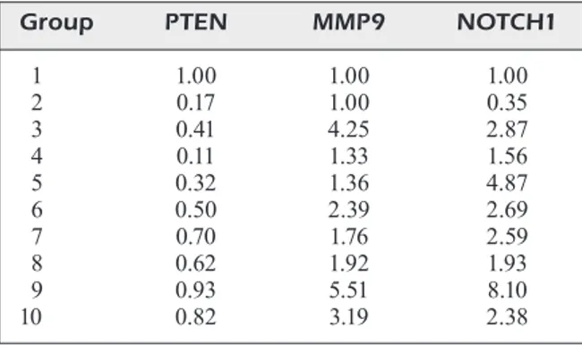
For the Western blot assays, the protein bands were quantified using ImageJ software, and the protein expression in group 1 was set to be 1 – that is, 100%, as shown in Table III.
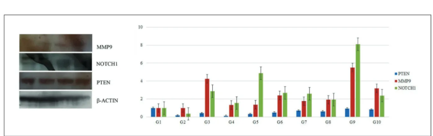
The Western blot tests were repeated at least three times for each sample. An illustrative West-ern blot figure from group 1 and a graph showing the protein expression fold change in all groups are shown in Figure 2. Illustrative Western blots for all groups are given in the Supplementary Data.
An analysis of the RQ values in the control and study groups revealed that there were no signifi-cant differences in the MMP-9 (χ2 = 10.848, p >
0.01.), NOTCH1 (χ2 = 8.771, p > 0.01), and PTEN
(χ2 = 7.727, p > 0.01) values. However, the Western
blot results revealed significant differences in the PTEN values (χ2 = 25.508, p < 0.01); the PTEN
values of group 1 were higher than those of groups 2, 3, and 4. Moreover, the values of group 4 were Table II. RQ values of the study groups.
Group MMP9 NOTCH1 PTEN
1 1 1 1 2 1.25 1.26 0.80 3 1.50 1.21 1.04 4 1.01 1.51 0.66 5 1.36 1.36 0.77 6 1.63 1.13 1.32 7 0.93 1.24 0.79 8 1.01 1.23 2.75 9 1.08 1.36 1.20 10 1.65 1.75 1.65
Table III. Protein expression values (r).
Group PTEN MMP9 NOTCH1 1 1.00 1.00 1.00 2 0.17 1.00 0.35 3 0.41 4.25 2.87 4 0.11 1.33 1.56 5 0.32 1.36 4.87 6 0.50 2.39 2.69 7 0.70 1.76 2.59 8 0.62 1.92 1.93 9 0.93 5.51 8.10 10 0.82 3.19 2.38
lower than those of groups 6, 8, and 9 (p < 0.01). There were also significant differences in NOTCH1 values (χ2 = 29.179, p < 0.01). The
NOTCH1 values of group 9 were higher than those of groups 1, 3, 4, 7, 8, and 10; the values of group 5 were higher than those of groups 1, 7, 8, and 10; and the values of group 2 were lower than those of groups 3, 4, 5, 9, and 10. No significant differences were observed in the MMP-9 values (χ2 = 15.472, p > 0.01).
Discussion
Clinical manifestations resulting from a life-time of neurodegeneration constitute the group of neurodegenerative diseases, such as Alzheimer’s, Parkinson’s, amyotrophic lateral sclerosis, and Huntington’s disease. Neurodegeneration can be defined as the progressive loss of structure or function of neurons.
While neurodegenerative diseases such as Alzheimer’s and Parkinson’s often occur due to aging, neurodegenerative diseases such as amy-otrophic lateral sclerosis and Huntington’s dis-ease can occur at young ages. In other words, neurodegeneration is associated with both aging and genetic factors25. Dementia, a symptom of
neurodegenerative diseases, is the destruction of mental and social abilities, especially memory, to the extent that it affects one’s daily life activities. Alzheimer’s disease is the most common disease in the primary degenerative dementia group with progressive cognitive disorders25,26.
In the macroscopic examination of the brains of Alzheimer’s patients, atrophy in the cerebral cortex and hippocampus, enlargement
of the sulcus, reduction in frontotemporal areas and para-hippocampal gyrus, and ventricular en-largement due to tissue loss are observations27.
Alzheimer’s disease can be classified as early, moderate, or severe. In the early stage, clinical symptoms include amnesia, aphasia, apraxia, and agnosia. Episodic memory impairment and dis-orientation are important main symptoms, and episodic memory is related to the deterioration of the hippocampus tissue28-30.
The hippocampus stores memories, and the amygdala functions by regulating emotional mem-ories31. The amygdala performs rapid and overall
processing while scanning whether the sensory stimulus matches an experience in an emotional memory. Therefore, not only hippocampus tissue damage but also amygdala damage may be import-ant in regulating emotional memories31.
Typical symptoms of Parkinson’s disease in-clude bradykinesia, postural instability, rigidity, and resting tremors. The main histopathological features of Parkinson’s disease are the loss of dopaminergic neurons from the substantia nigra to the basal ganglia and the accumulation of so-called Lewy bodies in the cytoplasm of surviving neurons30.
The eCBs plays a role in the regulation of brain development, the release of neurotrans-mitters, synaptic plasticity, and cytokine release from microglia by cell, tissue, organ, and organ-ism homeostasis. As a result, the eCBs plays a role in many neurological disorders, such as the aforementioned Alzheimer’s, Parkinson’s, amy-otrophic lateral sclerosis, and Huntington’s dis-eases32,33.
The microtubule-associated tau protein is a distinguishing feature of many neurodegenerative Figure 2. Western blot analysis. In the study, a western blot analysis was performed at least three times for each of 10 experimental groups, with five samples from each group. Illustrative Western blot figures are presented from group 1, and a graph showing the protein expression fold change in all groups is given. Each row shows the immunoblot results for MMP-9, NOTCH1, PTEN, and b-ACTIN.
diseases, such as Alzheimer’s disease and chronic traumatic encephalopathy34-37. It is a tauopathy
associated with epilepsy that contributes to the acceleration of cognitive regression in temporal lobe epilepsy and has diagnostic and therapeutic consequences34-37.
The most common adult epilepsy is mesial temporal sclerosis epilepsy, which is caused by hippocampal sclerosis and is associated with a high prevalence of cognitive impairment.
The potential efficacy of CB use in children with Dravet Syndrome and Lennox-Gastaut Syn-drome has been investigated regarding epilepsy, where a neurodegenerative disease relationship has been suggested35-38.
To reveal the mechanisms of occurrence of these diseases and to develop new treatments by creating experimental models that will make it possible to study these diseases in experimental animals.
Animal experiments and clinical studies have shown that cannabis-based therapy is beneficial in the treatment of different diseases, but further studies are still needed to investigate the efficacy and safety of such therapeutics39. The present
study is aimed at investigating changes in the gene and protein expression levels of MMP-9, PTEN, and NOTCH1 in rats treated with AEA, AM251, AM630, and ML193. Furthermore, the main role played of proteins involved in neurode-generative diseases was investigated.
LTP is effective in learning and memory, and the hippocampus plays an important role in trans-forming short-term memory into long-term mem-ory in the neocortex. In this process, the hippo-campus performs a critical function by providing the first inputs required for long-term memory, and then converts them into long-term memory by cre-ating and strengthening the synaptic connections necessary for sustainable long-term retention40.
The mechanism related to hippocampal memo-ry and the affecting factors have been investigated in studies on protective and therapeutic methods for neurodegenerative diseases41. Molecular and
pharmaco-molecular studies on gene expressions and proteins involved in hippocampal tissues have therefore gained popularity. The literature also indicates that neural progenitor cells can be affected by both extrinsic factors, such as growth factors, cytokines, and MMPs, and intrinsic fac-tors, such as transcription factors and regulators of signaling pathways42.
PTEN independently modulates the functional and structural features of hippocampal neurons
and plays a key role in the mechanisms of synap-tic plassynap-ticity. However, LTP is insufficient when PTEN is removed or downregulated in hippo-campal neurons, and behavioral abnormalities also arise – neurodegenerative pathologies have been reported to develop due to the suppression or down-regulation of PTEN in hippocampal neurons43.
PTEN, a bidirectional phosphatase enzyme that influences many proteins, negatively regu-lates PI3K and dephosphoryregu-lates phosphatidyli-nositol 3.4.5-triphosphate to phosphatidyliphosphatidyli-nositol 4.5-bisphosphate44. A loss of PTEN is directly
associated with an increase in Akt activity45. In
the present study, PTEN protein expression in-creased in the AM630-treated group (group 6), and ligands in CB receptor inhibitors increased in groups 8, 9, and 10. The increase in protein expression was statistically significant (p < 0.01).
The NOTCH1 signaling pathway – a highly conserved pathway that regulates cell fate – is a cell signaling system present in most multicellu-lar organisms. Mammals possess four different notch receptors (NOTCH1, NOTCH2, NOTCH3, and NOTCH4). The NOTCH receptor is a sin-gle-pass transmembrane receptor protein, and the NOTCH pathway is involved in the regulation of the characteristic functions of cells and in con-trolling the stem and precursor cell layers46. The
NOTCH1 signaling pathway is an evolutionarily conserved signaling mechanism that regulates development and is involved in plasticity-related processes, including changes in neurite struc-ture and the preservation of neural stem cells47.
NOTCH1 proteins have been reported to interact with presenilins and with β-amyloid precursor proteins, and may therefore play a role in familial and sporadic Alzheimer’s disease47. Null
het-erozygous mutations in NOTCH1 may provoke significant impairments in spatial learning and memory without affecting other forms of learn-ing, motor control, or exploratory activity47. The
authors have previously suggested that a decrease in NOTCH1 signaling may lead to neurodegen-eration and that abnormalities in NOTCH-de-pendent transcription may be associated with Alzheimer’s disease and Alagille and Cadasil syndromes47,48. In the present study, the NOTCH1
protein increased in the AM251-treated group (group 5) and the AEA + AM630-treated group (group 9) (p < 0.01).
MMP expression may increase due to the in-fluence of factors manipulating the mechanism of gene transcription during the remodeling of
tissues under various physiological and patholog-ical conditions49. Similarly, to the progression of
highly specific and potent orthosteric ligands, as well as the development of allosteric ligands, the eCBs has come to the forefront as a modulator of many physiological processes. As such, it has attracted interest from researchers who have investigated the role of the eCBs and medical cannabis in human physiology through phar-maceutical chemistry and pharmaco-molecular laboratories50. In the present study, the changes
in MMP-9, PTEN, and NOTCH1 gene protein expressions were evaluated in live mammalian subjects treated with AEA agonists and antago-nists. Salaga et al51 conducted research in which
a CB2-selective antagonist AM630 was used, and CB2R was distributed in the central and pe-ripheral tissues, including immunocytes. Fowler et al52 reported that there were no compounds
selectively inhibiting AEA synthesis and that the mechanisms obstructing the release and uptake of AEA were not fully elucidated. The authors emphasized that the selective agonists and antag-onists of CB1R and CB2R were well described. In their study, the modulation of the ECB system was stated to produce neuroprotective effects52.
Aguayo et al53 tested the hypothesis that MMP-9,
an enzyme that breaks down ECM components and synaptic proteins, such as β-dystroglycan (β-DG43), changed its activities and distribution in the rat hippocampus during an acute stress re-sponse. In that study, after 24 h of stress, MMP-9 net activity increased in the somatic area. That is to say, in the stratum pyramidal and granular cell layers and in the synaptic area, especially in the stratum radiatum and molecular layer of the hippocampus, MMP-9 enriched the hippocam-pal synapto-neurosome fractions without altering their potential enzymatic activity53.
Basavarajappa et al54 investigated the effects of
pharmaceuticals on neurodegenerative processes and reported that many exogenous and endoge-nous CBs, such as AEA and 2-AG, demonstrat-ed an important role in hippocampal memory processes in rodents. However, the mechanisms through which endogenous AEA regulates these processes remain unknown. The authors noted that the acute administration of URB597 in-creased the effects of AEA without changing the levels of 2-AG or CB1R in the hippocampus and neocortex and that, in hippocampal slices, URB597 damaged LTP in CB1R WT, but not in the knockout (KO) offspring. In that study, URB597 increased extracellular-signal-regulated
kinase (ERK) phosphorylation in WT without affecting ERK levels in WT or KO mice. The au-thors indicated that pharmacologically increased AEA damaged LTP, learning, and memory and inhibited Ca2+/calmodulin kinases-IV and cyclic
amp-response element binding protein phosphor-ylation through the activation of CB1Rs. The study showed the disruptive effects of the phar-macological elevation of AEA beyond normal concentrations on underlying physiological re-sponses54.
Terranova et al55 reported that delta
9-tet-rahydrocannabinol and the synthetic CB ago-nist HU-210(-)-11-OH-delta8-tetrahydrocannabi-nol-dimethylheptyl prohibited LTP induction in rat hippocampal slices.
Wiera et al14 noted that LTP was commonly
understood to be a memory substrate, and, in the hippocampal CA3-CA1 pathway, different forms of LTP hinged on NMDA receptors (nmdaLTP) or L-type voltage-gated calcium channels (vdc-cLTP). In their study, the regulation of different LTP forms by different MMPs in mice hippocam-pal slices was investigated. The authors indicated that MMP-3 inhibition or KO damaged late-phase LTP in the CA3-CA1 pathway, as well as the MMP-9 blockade, reduced late-phase LTP14.
Both early- and late-phase LTP were observed to be damaged when MMP-3 and MMP-9 were disrupted14. Immunoblotting, in situ
zymogra-phy, and immunofluorescence results revealed that LTP induction was correlated with an in-crease in MMP-3 expression and activity in the CA1 stratum radiatum14.
The findings of the study showed that the acti-vation of perisynaptic MMP-3 supported L-type channel-dependent LTP in the CA1 region, while NMDA LTP hinged on MMP-9. The authors stated that the underlying molecular signaling pathways in the hippocampal tissue were not well understood. The obtained results showed that dif-ferent MMPs may behave as molecular switches for some types of LTP14.
Szepezi et al56 reported that chemically
in-duced LTP (cLTP) induction in dissolved hippo-campal cultures increased MMP-9 activity, which controls the formation of spine head protrusions (SHP). The authors indicated that auto-active recombinant MMP-9 promoted the formation of SHPs in organotypic hippocampal slices and that blocking MMP activity or microtubule dynamics eliminated the appearance of SHPs56. The results
of the study demonstrated that MMP-9 plays a strong functional role in the formation of SHPs
and in the control of postsynaptic receptor distri-bution on the cLTP.
Dziembowska et al57 reported that MMP-9 had
a significant role in the regulation of synaptic plasticity and that MMP-9 mRNA was carried to dendrites for local translation and protein re-lease. The authors suggested that locally secreted MMP-9 may contribute to the structural and functional plasticity of the activated synapses57.
NOTCH receptors transduce extracellular sig-nals at the cell surface and change the gene ex-pression pattern of cells. NOTCH1 is one of the receptors that regulates PTEN expression and the activity of the PI3K-AKT signaling pathway58.
The relationship between MMP-9 and NOTCH1 signaling has been demonstrated, and it is well known that an increase in MMP-9 expression results in the strong activation of NOTCH1 signal-ing59. Moreover, PTEN down-regulates MMP-9 in
response to TNF-alpha through the transcription factor NF-kappaB and activation protein-160.
Wiera et al15 indicated that exogenous protease
could repair LTP in mice, whereas, in the wild-type, excess MMP-9 damaged LTP. The authors suggested that LTP maintenance in the MF-CA3 pathway may result in altered MMP-9 levels that may be harmful for cognitive processes, as ob-served in some neuropathologies15.
Brzdąk et al61 noted that MMP-NMDAR
correla-tions were not fully elucidated. Their study in-vestigated the involvement of MMP subtypes in E-S plasticity and NMDAR function in mouse hippocampal acute brain slices. The authors em-phasized that the temporal necessity for MMP-3/ NMDAR activity in E-S potentiation in the CA1 region mostly coincided, and MMP-3, but not MMP-2/9, activity was vital for the functional ac-quisition of NMDARs following LTP induction61.
Tsilibary et al62 suggested that there were
in-dications implicating MMPs in major neuropsy-chiatric disorders, probably by generating syn-aptic aberrations. MMP-9, NOTCH1, and PTEN signaling are intersecting signal pathways that are associated with brain development and the hippocampus and have been the subject of many studies that have investigated drug dependence or neurological diseases63. In this study, both the
agonist and antagonist drugs that were applied to the subjects resulted in changes in MMP-9 gene expression, but the changes observed were not statistically significant (p > 0.01). The pres-ent study is the first in vivo research seeking to investigate the effects of anandamide and its antagonists, which have been shown to cross the
blood-brain barrier64 on MMP-9, NOTCH1, and
PTEN gene and protein expressions.
Group 1 served as the control group during the Western blot and RT-qPCR assays and included untreated samples. Groups 2 and 3 were treated with the drug diluents that did not provoke any significant changes in gene and protein expres-sion. Changes in MMP-9, NOTCH1, and PTEN gene expressions were evaluated with RT-qPCR assays, and the observed changes were not con-sidered statistically significant. The Western blot results revealed that the changes in MMP expres-sion were not statistically significant and that the NOTCH1 protein increased in group 5, which was treated with AM251, and in group 9, which was treated with AEA + AM630. The increase in PTEN protein expression was statistically signif-icant in group 6, which was treated with AM630, and in groups 8, 9, and 10, all of which were treat-ed with ligands and CBR inhibitors.
A week of drug application did not change gene expression but did cause an acute increase in the amount of protein expression, and the hip-pocampal cells swiftly responded to the applied drugs. The applications did not change the MMP-9 gene and protein levels, yet the expression of NOTCH1-a signal pathway activated by MMP-9-increased, and the observed NOTCH1 activation was independent of MMP-9. The increase in PTEN expression in groups 6, 8, 9, and 10 was expected to suppress MMP-9 expression in the same groups, but MMP-9 expression remained unchanged.
The present study used rat hippocampal tis-sue for the analyses, and assays were performed using a very limited number of samples. The literature indicates that the sensitivity of human and animal tissues is different. Accordingly, the results obtained in these studies may differ from the results obtained with human samples and may be misleading, which is the primary limitation of this study.
Conclusions
This is the first study in which AEA, AM251, AM630, and ML193 were evaluated together in rat hippocampal tissues. The observed changes in MMP-9 gene expression following the adminis-tration of AEA – a potent endogenous agonist of CB1R and CB2R – and its antagonists were statis-tically insignificant. PTEN expression levels were increased with the administration of only AM630,
a potent and selective inverse agonist for CB2R in rat hippocampal tissue. PTEN expression levels were also increased with the combined applica-tion of AEA; AM251, an inverse agonist at the CB1R; AM630, a CB2R antagonist; and ML193, a GPR55 antagonist. An increase in NOTCH1 protein was observed following the administration of only AM251, an inverse agonist at the CB1R, and the concomitant administration of AEA and AM630, a CB2R antagonist. These results suggest that systemic manipulations of eCBs may alter neurodegenerative disorders associated with mem-ory loss. This is an important aspect of the current research. Medication that can act on the CB system should be developed for the treatment of diseases in hippocampal tissue, and further researches that include many more subjects should be performed to provide further insight into the efficacy of these pharmaceuticals on MMP-9, PTEN, and NOTCH1 signaling pathways.
Conflict of Interest
The Authors declare that they have no conflict of interests. Acknowledgements
The authors thank the Medical Research Center laborato-ry (Istanbul Medipol University) employees for all support and assistance
Availability of Data
The data and materials generated/analyzed in the present study are available from the corresponding author upon request. Ethical Approval
The entire experimental procedure was approved by the Is-tanbul Medipol University Local Ethics Committee for An-imals (Permission of Live Mammal Usage for Experiments No. 38828770-604.01.01-E.10835).
Authors’ Contribution
I.Y., N.K., and D.Y.S. designed the study and the experi-ments. I.Y. and N.K. performed the intraperitoneal injec-tions on rats. I.Y. and N.K. resected hippocampal tissues. I.Y., N.K., and D.Y.S. worked on the experiments and the molecular analysis of the tissues. I.Y. collected the data. I.Y., N.K., and H.O. carried out the statistical analysis. All the authors read and approved the final manuscript.
References
1) Zhang JC, Zhang WJ, Zhao Q, Chen Bn, Wang X,
Luo YP, Zhang FX. Adiponectin improves
isoflu-rane-induced cognitive dysfunction in elderly rats
via inhibiting p38-MAPK signal pathway in hippo-campus. Eur Rev Med Pharmacol Sci 2019; 23: 171-176.
2) Joshi n, onaivi es. Endocannabinoid system
com-ponents: overview and tissue distribution. Adv Exp Med Biol 2019; 1162: 1-12.
3) sZCZePanska-sadoWska e, CudnoCh-JedrZeJeWska a,
uFnaL M, Zera T. Brain and cardiovascular
diseas-es: common neurogenic background of cardio-vascular, metabolic and inflammatory diseases. J Physiol Pharmacol 2010; 61: 509-521.
4) PisCiTeLLi F, siLvesTri C. Role of the
endocannabinoi-dome in human and mouse atherosclerosis. Curr Pharm Des 2019; 25: 3147-3164.
5) BaZWinskY-WuTsChke i, ZiPPriCh a, dehghani F.
Endo-cannabinoid system in hepatic glucose metabo-lism, fatty liver disease, and cirrhosis. Int J Mol Sci 2019; 20: 2516.
6) eLLerT-MikLasZeWska a, CieChoMska ia, kaMinska B.
Cannabinoid Signaling in Glioma Cells. Adv Exp Med Biol 2020; 1202: 223-241.
7) garCía-Baos a, aLegre-Zurano L, CanTaCorPs L,
MarTín-sánCheZ a, vaLverde o. Role of
canna-binoids in alcohol-induced neuroinflammation. Prog Neuropsychopharmacol Biol Psychiatry 2021; 104: 110054.
8) horn h, BöhMe B, dieTriCh L, koCh M.
Endocanna-binoids in body weight control. Pharmaceuticals (Basel) 2018; 11: 55.
9) MaurYa n, veLMurugan Bk. Therapeutic
applica-tions of cannabinoids. Chem Biol Interact 2018; 293: 77-88.
10) Çakir M, Tekin s, doğanYiğiT Z, erden Y, soYTürk M,
ÇiğreMiş Y, sandaL s. Cannabinoid type 2
recep-tor agonist JWH-133, attenuates Okadaic acid in-duced spatial memory impairment and neurode-generation in rats. Life Sci 2019; 217: 25-33. 11) aLMeida v, Levin r, Peres FF, suiaMa Ma, vendraMini
aM, sanTos CM, siLva nd, Zuardi aW, haLLak JeC,
CriPPa Ja, aBíLio vC. Role of the endocannabinoid
and endovanilloid systems in an animal model of schizophrenia-related emotional processing/cog-nitive deficit. Neuropharmacology 2019; 155: 44-53.
12) PadaMseY Z, MCguinness L, Bardo sJ, reinharT M,
Tong r, hedegaard a, harT ML, eMPTage nJ.
Activ-ity-dependent exocytosis of lysosomes regulates the structural plasticity of dendritic spines. Neu-ron 2017; 93: 132-146.
13) Xiong L, duan L, Xu W, Wang Z. Nerve growth
fac-tor metabolic dysfunction contributes to sevoflu-rane-induced cholinergic degeneration and cogni-tive impairments. Brain Res 2019; 1707: 107-116. 14) Wiera g, noWak d, van hove i, dZiegieL P, Moons
L, MoZrZYMas JW. Mechanisms of NMDA receptor-
and voltage-gated L-Type calcium channel-de-pendent hippocampal LTP critically rely on prote-olysis that is mediated by distinct metalloprotein-ases. J Neurosci 2017; 37: 1240-1256.
15) Wiera g, WoZniak g, BaJor M, kaCZMarek L, MoZrZY -Mas JW. Maintenance of long-term potentiation in
hippocampal mossy fiber—CA3 pathway requires fine-tuned MMP-9 proteolytic activity. Hippocam-pus 2013; 23: 529-543.
16) uLugoL a. The endocannabinoid system as a
po-tential therapeutic target for pain modulation. Bal-kan Med J 2014; 31: 115-120.
17) BiLir ka, anLi g, oZkan e, gunduZ o, uLugoL a.
In-volvement of spinal cannabinoid receptors in the antipruritic effects of WIN 55,212-2, a cannabi-noid receptor agonist. Clin Exp Dermatol 2018; 43: 553-558.
18) CoLangeLi r, PieruCCi M, Benigno a, CaMPiani g, Bu -Tini s, di giovanni g. The FAAH inhibitor URB597
suppresses hippocampal maximal dentate after discharges and restores seizure-induced impair-ment of short- and long-term synaptic plasticity. Sci Rep 2017; 7: 11152.
19) siLva-CruZ a, CarLsTroM M, riBeiro Ja, seBasTião aM.
Dual influence of endocannabinoids on long-term potentiation of synaptic transmission. Front Phar-macol 2017; 8: 921.
20) WaLenTinY dM, gaMage TF, Warner Ja, nguYen Tk,
grainger dB, WiLeY JL, vann re. The endogenous
cannabinoid anandamide shares discriminative stimulus effects with Δ9-tetrahydrocannabinol in fatty acid amide hydrolase knockout mice. Eur J Pharmacol 2011; 656: 63-67.
21) BiaLuk i, WinniCka MM. AM251, cannabinoids
re-ceptors ligand, improves recognition memory in rats. Pharmacol Rep 2011; 63: 670-679.
22) ross ra, BroCkie hC, sTevenson La, MurPhY vL,
TeMPLeTon F, MakriYannis a, PerTWee rg.
Agonist-in-verse agonist characterization at CB1 and CB2 cannabinoid receptors of L759633, L759656 and AM630. Br J Pharmacol 1999; 126: 665-672. 23) rahiMi a, haJiZadeh MoghaddaM a, roohBakhsh a.
Central administration of GPR55 receptor agonist and antagonist modulates anxiety related behaviors in rats. Fundam Clin Pharmacol 2015; 29: 185-190. 24) YiLMaZ i, karaarsLan n, oZBek h. Practical
perfor-mance of hippocampal tissue resection in rats in pharmacomolecular research. Turk Neurosurg 2020 Sep 29. doi: 10.5137/1019-5149.JTN.31730-20.1 [Online Publishing].
25) hussain r, ZuBair h, PurseLL s, shahaB M.
Neu-rodegenerative diseases: regenerative mecha-nisms and novel therapeutic approaches. Brain Sci 2018; 8: 177.
26) duong s, PaTeL T, Chang F. Dementia: what
pharma-cists need to know. Can Pharm J 2017; 150: 118-129. 27) aLves L, Correia as, MigueL r, aLegria P, BugaLho P.
Alzheimer’s disease: a clinical practice-oriented review. Front Neurol 2012; 3: 63.
28) erkkinen Mg, kiM Mo, gesChWind Md. Clinical
neu-rology and epidemiology of the major neurode-generative diseases. Cold Spring Harb Perspect Biol. 2018; 10: a033118.
29) aPosToLova Lg. Alzheimer disease. Continuum
(Minneap Minn) 2016; 22: 419-434.
30) Wang XX, Zhang B, Xia r, Jia QY. Inflammation,
apoptosis and autophagy as critical players in
vascular dementia. Eur Rev Med Pharmacol Sci 2020; 24: 9601-9614.
31) BeChara a, TraneL d, daMasio h, adoLPhs r, roCk -Land C, daMasio ar. Double dissociation of
condi-tioning and declarative knowledge relative to the amygdala and hippocampus in humans. Science 1995; 269: 1115-1118.
32) MhYre Tr, BoYd JT, haMiLL rW, Maguire-Zeiss ka.
Parkinson’s disease. Subcell Biochem 2012; 65: 389-455.
33) esTrada Ja, ConTreras i. Endocannabinoid
recep-tors in the CNS: Potential drug targets for the pre-vention and treatment of neurologic and psychi-atric disorders. Curr Neuropharmacol 2020; 18: 769-787.
34) CrisTino L, Bisogno T, di MarZo v. Cannabinoids
and the expanded endocannabinoid system in neurological disorders. Nat Rev Neurol 2020; 16: 9-29.
35) Tai XY, koePP M, dunCan Js, FoX n, ThoMPson P, BaX -endaLe s, Liu JY, reeves C, MiChaLak Z, ThoM M.
Hy-perphosphorylated tau in patients with refractory epilepsy correlates with cognitive decline: a study of temporal lobe resections. Brain 2016; 139: 2441-2455.
36) BernasConi n. Is epilepsy a curable
neurodegener-ative disease? Brain 2016; 139: 2336-2337. 37) gasTon Te, sZaFLarski JP. Cannabis for the
treat-ment of epilepsy: an update. Curr Neurol Neuros-ci Rep 2018; 18: 73.
38) Morano a, FaneLLa M, aLBini M, CiFeLLi P, PaLMa e,
giaLLonardo aT, di BonavenTura C. Cannabinoids
in the treatment of epilepsy: current status and future prospects. Neuropsychiatr Dis Treat 2020; 16: 381-396.
39) Barrie n, kuruPPu v, ManoLios e, aLi M, Moghadd -aM M, ManoLios n. Endocannabinoids in arthritis:
current views and perspective. Int J Rheum Dis 2017; 20: 789-797.
40) MosCareLLo JM, Maren s. Flexibility in the face or
fear: Hippocampal-prefrontal regulation of fear and avoidance. Curr Opin Behav Sci 2018; 19: 44-49.
41) Zhao C, deng W, gage Fh. Mechanisms and
func-tional implications of adult neurogenesis. Cell 2008; 132: 645-660.
42) gouignard n, andrieu C, Theveneau e. Neural crest
delamination and migration: looking forward to the next 150 years. Genesis 2018; 56: e23107. 43) ogino M, iChiMura M, nakano n, MinaMi a, kiTagi
-shi Y, MaTsuda s. Roles of PTEN with DNA repair
in Parkinson’s Disease. Int J Mol Sci 2016; 17: 954.
44) TaMura M, gu J, MaTsuMoTo k, aoTa s, Parsons r,
YaMada kM. Inhibition of cell migration, spreading,
and focal adhesions by tumor suppressor PTEN. Science 1998; 280: 1614-1617.
45) sTaMBoLiC v, suZuki a, de La PoMPa JL, BroThers gM,
MirTsos C, sasaki T, ruLand J, Penninger JM, siderovs -ki dP, Mak TW. Negative regulation of
PKB/Akt-de-pendent cell survival by the tumor suppressor PTEN. Cell 1998; 95: 29-39.
46) ChiLLakuri Cr, shePPard d, Lea sM, handFord Pa.
Notch receptor-ligand binding and activation: in-sights from molecular studies. Semin Cell Dev Bi-ol 2012; 23: 421-428.
47) CosTa rM, honJo T, siLva aJ. Learning and
memo-ry deficits in Notch mutant mice. Curr Biol 2003; 13: 1348-1354.
48) sui Y, Zhang Y, dong C, Xu B, sun X. The small
mo-lecular CCR3 antagonist YM344031 attenuates neurodegenerative pathologies and improves learning and memory performance in a mouse model of Alzheimer’s disease. Brain Res 2019; 1719: 1-10.
49) karaarsLan n, gurBuZ Ms, CaLiskan T, aYan e, ak -er Fv, BerkMan MZ. The effect of matrix
metallo-proteinase-3 on the prognosis and biological be-haviour of meningiomas. Turk Neurosurg 2016; 26: 678-683.
50) ConsoLe-BraM LM, Zhao P, aBood Me. Protocols
and good operating practices in the study of can-nabinoid receptors. Methods Enzymol 2017; 593: 23-42.
51) saLaga M, ZaTorski h, ZieLişska M, Mosinska P, TiM -MerMans JP, kordek r, sTorr M, FiChna J. Highly
se-lective CB2 receptor agonist A836339 has gas-troprotective effect on experimentally induced gastric ulcers in mice. Naunyn-Schmiedeberg’s Arch Pharmacol 2017; 390: 1015-1027.
52) FoWLer CJ, hoLT s, niLsson o, Jonsson ko, Tiger
g, JaCoBsson so. The endocannabinoid
signal-ing system: pharmacological and therapeutic as-pects. Pharmacol Biochem Behav 2005; 81: 248-262.
53) aguaYo Fi, PaCheCo aa, garCía-roJo gJ, PiZarro-Bau -erLe Ja, doBerTi av, TeJos M, garCía-PéreZ Ma, ro -Jas Ps, FiedLer JL. Matrix metalloproteinase 9
dis-plays a particular time response to acute stress: cariation in its levels and activity distribution in rat hippocampus. ACS Chem Neurosci 2018; 9: 945-956.
54) Basavara JaPPa Bs, nagre nn, Xie s, suBBanna s.
Elevation of endogenous anandamide impairs LTP, learning, and memory through CB1 recep-tor signaling in mice. Hippocampus 2014; 24: 808-818.
55) Terranova JP, MiChaud JC, Le Fur g, souBrié
P. Inhibition of long-term potentiation in rat hippocampal slices by anandamide and
WIN55212-2: reversal by SR141716 A, a se-lective antagonist of CB1 cannabinoid recep-tors. Naunyn-Schmiedeberg’s Arch Pharmacol 1995; 352: 576-579.
56) sZePesi Z, BiJaTa M, rusZCZYCki B, kaCZMarek L,
WLodarCZYk J. Matrix metalloproteinases regulate
the formation of dendritic spine head protrusions during chemically induced long-term potentiation. PLOS One 2013; 8: e63314.
57) dZieMBoWska M, MiLek J, JanusZ a, reJMak e, ro -ManoWska e, gorkieWiCZ T, Tiron a, BraMhaM Cr,
kaCZMarek L. Activity-dependent local translation
of matrix metalloproteinase-9. J Neurosci 2012; 32: 14538-14547.
58) graBher C, von BoehMer h, Look aT. Notch 1
ac-tivation in the molecular pathogenesis of T-cell acute lymphoblastic leukaemia. Nat Rev Cancer 2006; 6: 347-359.
59) FaZio C, PiaZZi g, viTagLione P, FogLiano v, Munari -ni a, ProssoMariTi a, MiLaZZo M, d’angeLo L, naPoL -iTano M, ChieCo P, BeLLuZZi a, BaZZoLi F, riCCiardieL -Lo L. Inflammation increases NOTCH1 activity via
MMP9 and is counteracted by eicosapentaenoic acid-free fatty acid in colon cancer cells. Sci Rep 2016; 6: 20670.
60) Moon sk, kiM hM, kiM Ch. PTEN induces G1
cell cycle arrest and inhibits MMP-9 expression via the regulation of NF-kB and AP-1 in vascu-lar smooth muscle cells. Arch Biochem Biophys 2004; 421: 267-276.
61) BrZdak P, WLodarCZYk J, MoZrZYMas JW, WoJToWiCZ
T. Matrix metalloprotease 3 activity supports hip-pocampal EPSP-to-spike plasticity following pat-terned neuronal activity via the regulation of NMDAR function and calcium flux. Mol Neurobiol 2017; 54: 804-816.
62) TsiLiBarY e, TZinia a, radenoviC L, sTaMenkoviC v, LeBiT -ko T, MuCha M, PaWLak r, FrisChkneChT r, kaCZMarek
L. Neural ECM proteases in learning and synap-tic plassynap-ticity. Prog Brain Res 2014; 214: 135-157. 63) Tanveer r, goWran a, noonan J, keaTing se, BoW
-ie ag, CaMPBeLL va. The endocannabinoid,
anan-damide, augments Notch-1 signaling in cultured cortical neurons exposed to amyloid-β and in the cortex of aged rats. J Biol Chem 2012; 287: 34709-34721.
64) CaLaPai F, Cardia L, sorBara ee, navarra M, gangeMi
s, CaLaPai g, MannuCCi C. cannabinoids,
blood-brain barrier, and blood-brain disposition. Pharmaceu-tics 2020; 12: 265.