ENZYMATIC DEGRADATION OF
SELF-ASSEMBLED PEPTIDE NANOFIBER
GELS
a thesis submitted to
the graduate school of engineering and science
of bilkent university
in partial fulfillment of the requirements for
the degree of
master of science
in
material science and nanotechnology
By
Ayg¨
ul Zengin
ENZYMATIC DEGRADATION OF SELF-ASSEMBLED PEPTIDE NANOFIBER GELS
By Ayg¨ul Zengin February 2016
We certify that we have read this thesis and that in our opinion it is fully adequate, in scope and in quality, as a thesis for the degree of Master of Science.
Mustafa ¨ozg¨ur G¨uler(Advisor)
Ay¸se Beg¨um Tekinay
Erhan Bat
Approved for the Graduate School of Engineering and Science:
Levent Onural
ABSTRACT
ENZYMATIC DEGRADATION OF SELF-ASSEMBLED
PEPTIDE NANOFIBER GELS
Ayg¨ul Zengin
M.Sc. in Material Science And Nanotechnology Advisor: Mustafa ¨ozg¨ur G¨uler
February 2016
The self-assembled peptide nanofiber gels have received enormous attention because of their inherent biocompatible, biodegradable and functional proper-ties. They provide a smart platform for a range of applications such as tissue engineering, drug delivery and wound healing.
These gels are formed through noncovalent interactions such as hydrogen bond-ing, hydrophobic interactions and electrostatic interactions among the peptide amphiphile molecules at physiological conditions. In order to understand the stability of these gels in the presence of proteases in natural conditions, we stud-ied degradation behavior of the gels with proteinase K, which is a non-specific protease cleaving the peptide bonds. Degradation process was studied by mea-suring weight measurement and TEM imaging. In addition, sustained release of Rhodamine B from these gels was also studied in the presence of proteases. The results clearly demonstrated that presence of D- amino acids in the peptide nanofiber network significantly improves their stability against enzymatic degra-dation and change the release profile of the encapsulated molecules in the gels. These findings are interesting for biomedical applications of these materials due to their tunable degradation and controlled release behavior.
D-¨
OZET
KEND˙IL˙I ˘
G˙INDEN B˙IR ARAYA GELEN PEPT˙IT
NANOF˙IBERLERDEN OLUS
¸AN JELLER˙IN
ENZ˙IMAT˙IK DEGREDASYONU
Ayg¨ul Zengin
Malzeme Bilmi ve Nanoteknoloji, Y¨uksek Lisans Tez Danı¸smanı: Mustafa ¨ozg¨ur G¨uler
¸subat 2016
Kendili˘ginden bir araya gelen peptit nanofiber jeller, doalar gerei biy-ouyumlu, biyobozunur ve biyofonksiyonel ¨ozelliklerinden dolayı son derece ilgi ¸cekmektedirler. Bu yapılar, doku m¨uhendisli˘gi, ila¸c salımı ve yara iyile¸smesi gibi ¸ce¸sitli uygulamalar i¸cin akıllı bir platform olu¸stururlar.
Bu jeller, fizyolojik ko¸sullarda peptit amfifil molek¨ulleri arasındaki hidrojen ba˘gı, hidrofobik ve elektrostatik etkile¸simler gibi kovalent olmayan etkile¸simler aracılı˘gıyla olu¸sturulmu¸stur. Bu jellerin, proteaz i¸ceren do˘gal ko¸sullardaki stabilitesini anlamak i¸cin jellerin degredasyon davranı¸sı,spesifik olmayan bir ¸sekilde peptit ba˘glarını kıran bir proteaz olan proteinaz K ile ¸calı¸sılmı¸stır. De-gredasyon prosesi, a˘gırlıktaki azalmayı ¨ol¸cerek ve TEM g¨or¨unt¨ulemesi kullanarak yapılmı¸stır. Ek olarak, proteinaz K varlı˘gında, rodamin B'nin bu jellerinden s¨urekli salımı ¸calı¸sılmı¸stır. Sonu¸clar, D- peptit molek¨ullerin jel ve nanofiber a˘gında bulunmasıyla, bu yapıların enzimatik degredasyona kar¸sı kararlılıklarının arttı˘gını ve jel i¸cinde hapsedilen molek¨ullerin salım profillerinin de˘gi¸sti˘gini a¸cık¸ca g¨ostermi¸stir. Elde edilen bu bulgular, ayarlanabilir degredasyon ve kontroll¨u salım davranı¸sları a¸cısından bu malzemelerin biyomedikal uygulamaları i¸cin ¨
onemlidir.
Anahtar s¨ozc¨ukler : Peptit amfifil, kendili˘ginden bir araya gelme, supramolek¨uler jel, nanofiber, D-amino asitler, kontroll¨u salım, enzimatik degredasyon.
Acknowledgement
I would like to express my appreciations to my advisor Prof. Mustafa ¨ozg¨ur G¨uler for his guidance and support during the course of this thesis. I would like to express my special thanks to Prof. Ay¸se Beg¨um Tekinay for sharing her knowledge.
I would like to thank to my colleagues and my senior peers Dr. Ruslan Gari-fullin, Dr. ¨Ozlem Erol, G¨oksu C¸ inar, G¨ulcihan G¨ulseren, G¨ulistan Tansık, Melis S¸¸ardan and Mohammad Aref Khalily for helpful discussions.
I would like to express most sincere thanks to friends; Meryem Hatip, T¨urkan Bayrak, Seda Kizir. Their support always motivated me. In addition, special thanks to my officemates, Hepi Hari Susapto, Zeynep Ayta¸c, Aslı ¸celebio˘glu, Yelda Erta¸s, Fatma Kayacı, Zehra ˙Irem G¨urb¨uz. They helped me to work in such a warm environment.
I would like to special thank to my best buddies, Ali Torun, Benan ˙Inan, Hatice K¨ose, Belma Nural and Sinem Yakars¨onmez. We shared unforgettable memories together and I always felt their support including my high school and undergraduate years. I would like to thank my flat mates, G¨ok¸ce Aydo˘gan and Pelin G¨ulizar Ersan. I would like to thank Ula¸s Kudu for his support and help.
I would like to thank to Mr. Mustafa G¨uler and Ms.Zeynep Erdo˘gan for technical assistance and helpful discussion.
I would like to thank T ¨UB˙ITAK (The Scientific and Technological Research Council of Turkey) for grant numbers 114Z728, 214Z214 and B˙IDED-2210C fel-lowship.
Finally, I would like to express my most sincere gratitude to my family. I am eternally grateful for supporting and allowing me to pursue my ambitions
vi
Contents
1 Introduction 1
1.1 Self-Assembled Peptide Nanostructures . . . 2
1.1.1 Solid Phase Peptide Synthesis . . . 4
1.1.2 Characterization Techniques for Self-Assembled Peptide Molecules . . . 7
1.1.3 Applications of Self-Assembled Peptide Nanostructures . . 9
1.2 Self-Assembling Peptide Amphiphile Molecules . . . 13
1.3 Gels for Biomedical Applications . . . 19
1.4 Drug Delivery Application of Peptide Gels . . . 20
1.5 Controlled Release from Peptide Gels . . . 22
1.6 Enzymatic Degradation of Peptide Gels . . . 24
2 Enzymatic Degradation of Self-Assembled Peptide Nanofiber
CONTENTS viii
2.2 Experimental Section . . . 33
2.2.1 Materials . . . 33
2.2.2 Synthesis of Peptide Amphiphiles . . . 33
2.2.3 Characterizations of Synthesized Peptide Amphiphiles . . . 34
2.2.4 Weight Based Degradation of Self-Assembled Peptide Gels 37
2.2.5 Degradation of Self-Assembled Peptide Nanofibers . . . 37
2.2.6 Rhodamine B release from self-assembled peptide gels . . . 38
2.3 Results and Discussion . . . 40
3 Conclusions and Future Perspectives 63
List of Figures
1.1 Illustration of main types of peptide secondary structures. (a) β-sheet, (b) β- hairpin, (c) α-helix and (d) coiled-coil (Reproduced from Ref [1] with permission of the PCCP Owner Societies 2013) 3
1.2 Various self-assembled peptide nanostructures (Reproduced from Ref [2] with permission of John Wiley and Sons 2015) . . . 4
1.3 Basic steps of solid phase peptide synthesis (Reproduced from ref [3]). . . 6
1.4 Circular dichroism patterns for peptide secondary structures. Solid line, α-helix; long dashed line, anti-parallel β-sheet; dotted line, type I β-turn; cross dashed line, extended α-helix or poly (Pro) II helix; short dashed line, irregular structure. (Reproduced from Ref [4] with permission of Elsevier 2005). . . 8
1.5 Self-assembled peptide nanomaterials for a wide range of applica-tions (Reproduced from Ref [5] with permission of John Wiley and Sons 2015). . . 10
LIST OF FIGURES x
1.6 Molecular structure of PA nanofibers (a) Chemical structure of PA nanofiber formation, (b) SEM of 3D network IKVAV-PA nanofiber, (c) gel formation by adding cell culture media, and (d) molecular graphics illustration of self-assembly into high-aspect-ratio nanofibers (Reproduced with permission of Ref [6] with permission of The Royal Society of Chemistry). . . 15
1.7 Phase diagram of PA self-assembly behavior. (a) free molecules, (b) spherical micelles, (c) micelles with β-sheet, (d) long cylindrical fibers, (e) parallel sheet stacks, (f) single β-sheets and (g) amor-phous aggregate phase (reproduced from Ref [7] with permission of American Chemical Society 2008). . . 16
1.8 Molecular model of d-EAK16 (a) and l-EAK16 (B) (Produced from Ref [8] with permission of Creative Commons Attribution License 2008). . . 18
1.9 PTX release from RADA16-PTX hydrogel according to different concentrations of peptide in vitro. A) % release of drug from hy-drogel during 120 h, b) hyhy-drogel photographs which belongs to 30 min and 48 h after release (Reproduced from ref [9] with permis-sion of 2011 Dove Medical Press). . . 24
1.10 10-Hydroxycamptothecin release from D-amino acids containing nanofibers (a) Chemical Structures of GFFYGRGD, Nap-GDFDFD YGRGD, and 10-hydroxycamptothecin (HCPT); b) sta-bility of L-fiber and D-fiber against proteinase K digestion in PBS buffer solution (pH 7.4) (Reproduced from ref [10] with permission of 2014 American Chemical Society). . . 26
1.11 Illustration of projected degradation behaviors of PNF degrada-tion. (Reproduced from Ref [11] with permission of The Royal Society of Chemistry). . . 27
LIST OF FIGURES xi
1.12 Observation of degradation of peptide nanofibers by TEM a) TEM images of peptide nanofibers which is incubated with 0.5% trypsin-EDTA, b) Image J analysis of length of nanofibers, c) TEM images of peptide nanofibers which are incubated with cell culture media (Reproduced from ref [11] with permission of The Royal Society of Chemistry). . . 29
2.1 Schematic illustration of Rhodamine B release experimental setup 39
2.2 Chemical structures of peptide amphiphiles (a) L-K3PA, (b)
D-K3PA, (c) L-E3PA, and (d) D-E3PA . . . 42
2.3 Characterization of D-E3PA by using LC-MS. Liquid
chro-matogram of D-E3PA by the absorbance at 220 nm (top). mass
spectrum of D-E3PA (bottom). MS: (m/z) calculated 914.05,
[M-H]-observed 912.5169, [M-2H]-/2 observed 455.7582, [M-3H]-/3 ob-served 303.5042 . . . 43
2.4 Characterization of L-E3PA by using LC-MS. Liquid
chro-matogram of L-E3PA by the absorbance at 220 nm (top), Mass
Spectrum of E3PA (bottom). MS: (m/z) calculated 914.05, [M-H]
-observed 912.5164, [M-2H]-/2 observed 455.7581, [M-3H]-/3 ob-served 303.5041 . . . 44
2.5 Characterization of L-K3PA by using LC-MS. Liquid
chro-matogram of L-K3PA by the absorbance at 220 nm (top).
mass spectrum of K3PA (bottom). MS: (m/z) calculated
910.24, [M+H]+ observed 910.4739 [M+2H]+/2 observed 455.6983
LIST OF FIGURES xii
2.6 Characterization of D-K3PA by using LC-MS. Liquid
chro-matogram of D-K3PA by the absorbance at 220 nm (top). mass
spectrum of K3PA (bottom). MS: (m/z) calculated 910.24,
[M+H]+ observed 910.69166, [M+2H]+/2 observed 455.85202,
[M+3H]+/3 observed 304.23917 . . . 46
2.7 Circular dichroism spectra of synthesized peptide amphiphiles . . 47
2.8 TEM images of peptide amphiphile gels a) D-E3/K3 stained with
uranyl acetate, (b) L-E3/K3 stained with uranyl acetate, (c) DL
-E3/K3 stained with uranyl acetate . . . 48
2.9 SEM imaging of peptide amphiphile gels a) 1% (w/v) D-E3/K3;
(b) 1% (w/v) L-E3/K3; (c) 1% (w/v) DL-E3/K3 . . . 49
2.10 Photograph of the gels of (a) D-E3/K3 (1.0 wt%, pH 7.4); (b)
L-E3/K3 (1.0 wt%, pH 7.4); (c) DL-E3/K3 (1.0 wt%, pH 7.4) . . . 51
2.11 Rheology results of self-assembled peptide gels. Time sweep test (top), Amplitude sweep test (bottom) . . . 53
2.12 Weight based enzymatic degradation of peptide amphiphile gels . 54
2.13 Photograph of self-assembled peptide nanofiber gel degradation after 29 day incubation with proteinase K. (a) L-E3/K3 incubated
with proteinase K; (b) D-E3/K3 incubated with proteinase K; (c)
DL-E3/K3 incubated with proteinase K; (d) D-E3/K3 incubated
with Tris buffer; (e) L-E3/K3 incubated with Tris buffer ; (f)
DL-E3/K3 incubated with Tris buffer . . . 55
2.14 Degradation of L-E3/K3 peptide nanofibers visualized by TEM.
TEM images of L-E3/K3 nanofibers incubated with 1 mg/mL
pro-teinase K (top). TEM images of KRK peptide nanofibers incu-bated with Tris buffer (control). All scale bars are 100 nm. . . 58
LIST OF FIGURES xiii
2.15 Degradation of DL-E3/K3 peptide nanofibers visualized by TEM.
TEM images of DL-E3/K3 nanofibres incubated with 1 mg/mL
proteinase K (top). TEM images of KRK peptide nanofibers incu-bated with Tris buffer (bottom). All scale bars are 100 nm . . . . 59
2.16 Degradation of D-E3/K3 peptide nanofibers visualized by TEM.
TEM images of D-E3/K3 nanofibers incubated with 1 mg/mL
pro-teinase K (top). TEM images of KRK peptide nanofibers incu-bated with Tris buffer (bottom). All scale bars are 100 nm. . . 60
2.17 Rhodamine B release from self-assembled peptide gels (L-E3/K3, D-E3/K3 and DL-E3/K3) during 96 h . . . 62
List of Tables
1.1 Self-assembled peptides as drug delivery vehicles (Reproduced from Ref [12] with permission of American Chemical Society 2016) 12
2.1 peptide amphiphile molecules (*theoretical net charge at pH 7.4) . 35
Chapter 1
1.1
Self-Assembled Peptide Nanostructures
Molecular self-assembly is a powerful bottom-up approach for creating highly ordered structures. Weak non-covalent bonds including hydrogen bonds, ionic bonds, hydrophobic interactions, and van der Waals interactions have significant roles in mediating the structural conformation of molecules and controlling their interaction with other molecules. Nature has already utilized this path to generate hierarchically functional materials from a wide range of building blocks such as proteins, lipids, or DNA. The greatest advantage of this approach is creating nanostructures at multiple length scales and controlling their final architecture precisely by altering their molecular chemistry through modifying environmental conditions such as temperature, pH, and ionic strength. [13, 14]
Peptides are versatile building blocks for fabricating a wide range of self-assembling nanostructures for various applications due to their intrinsic biocom-patible, biodegradable and biofunctional properties. The variety in the nanostruc-tures which are formed by self-assembled peptides comes from the capability of the peptides of making use of different molecular interactions such as hydrophobic interactions, hydrogen bonding, electrostatic interactions and aromatic interac-tions. [15]
Secondary structures of peptides have major role in their organization into highly ordered, supramolecular architectures. The main types of secondary struc-tures are the α-helix, β-sheet, β-hairpins and coiled coils. (Figure 1.1)α-Helix is the most common protein secondary structure where amino acids are prone to form hydrogen bonds between the carbonyl groups and amide groups for promot-ing peptide backbone stabilization. Compared to β-sheet conformations, α-helix based assemblies have a tendency to be reversible. However, they can form more stable conformations by coming together with other α-helix structures. Until today, numerous α-helical based nanostructures were developed for various pur-poses. [1, 12, 16]
Figure 1.1: Illustration of main types of peptide secondary structures. (a) β-sheet, (b) β- hairpin, (c) α-helix and (d) coiled-coil (Reproduced from Ref [1] with permission of the PCCP Owner Societies 2013)
contain 2-5 helices which are wrapped around each other. Basically, coiled-coil nanostructure formation is based on repeated heptad units, -abcdefg-, in the peptide sequence. The amino acids in positions at a and d promote hydrophobic interactions and e and g provide electrostatic interactions in order to obtain the secondary structure formation. Such peptide sequences can assemble into a vari-ety of nanostructures such as nanofibrils or nanoparticles with the contribution of hydrophobic and electrostatic interactions. [1, 12, 16]
The β-sheet is the second widely studied secondary structure form which arises from laterally connected β-stands through hydrogen bonds. Hydrogen bonding between the carbonyl and amine groups and hydrophobicity stabilizes this struc-ture. They can be either parallel, which means two- β-sheet components aligned in the same direction, or antiparallel aligned in the opposite direction. Peptides have a tendency to form β-sheets and self-assemble into various supramolecular architectures such as nanofibers, nanovesicles, nanotubes. [1, 12, 16]
The β-hairpin is another secondary structure motif which arises as the short loop segments between antiparallel hydrogen bonded β-stands. They are typically
Figure 1.2: Various self-assembled peptide nanostructures (Reproduced from Ref [2] with permission of John Wiley and Sons 2015)
length and sequence. [1, 12, 16]
With the help of synthetic methods, it is possible to create a wide range of peptide based supramolecular nanostructures with different morphologies includ-ing fibers, tubes, vesicles, tapes and ribbons. (Figure 1.2) These specific nanos-tructures can arise from several factors such as chemical snanos-tructures and order of building blocks, molecular packaging properties, and environmental conditions such as pH, temperature, ionic strength and aging time. [2]
1.1.1
Solid Phase Peptide Synthesis
Solid phase peptide synthesis (SPPS) is a commonly used approach to readily synthesize peptides by coupling the carboxyl group of one amino acid to the amino group of another with high purity and yield. Pioneering work of Merrifield related to solid-phase peptide synthesis caused radical change within the peptide synthesis society in 1963. [17]
conditions. Particularly, it is useful for synthesis and design of unnatural amino acids containing peptides or natural complex peptides which are expressed in bac-teria. However, chain length and sequence of amino acids (particularly amyloid peptide synthesis) influence the yield of this method. [18]
The main logic behind the SPPS is a stepwise elongation an amino acid se-quence on a solid polymer bead through sequential steps of coupling and de-protection of protected amino acids. At the end of this process, elongated peptide is removed from these polymer beads which are called resin. These small beads provide a suitable environment which holds the peptide chain ro-bustly until cleaved by a reagent, for instance trifluoroacetic acid. Widely used solid polymer supports for peptide synthesis based on Fmoc chemistry are 4-hydroxymethylphenyloxymethyl polystyrene resin developed by Wang, 4-hydroxymethylphenoxyacetyl-poly (dimethylactylamide) resin developed by Atherton, and 2-chlorotritylchloride (CTC) resin developed by K. Barlos. Figure 1.3 gives an overview of solid phase peptide synthesis principles. [19–21]
There are two major strategies of SPPS: Fmoc/tBu and Boc/Bzl. [22–24] Recently, Fmoc- chemistry has become more favorable than Boc-chemistry be-cause of its safe and mild environmental conditions. 9-Fluorenylmethoxycarbonyl (Fmoc) represents the Fmoc protecting group. In order to get rid of the Fmoc group from a growing peptide sequence, basic conditions which are generally 20% piperidine in DMF are employed. Incubation with trifluoroacetic acid (TFA) pro-motes removing side-chain protecting groups and peptide chain from the polymer bead. [25, 26]
Figure 1.3: Basic steps of solid phase peptide synthesis (Reproduced from ref [3]).
1.1.2
Characterization
Techniques
for
Self-Assembled
Peptide Molecules
Compositional analysis of synthesized peptide molecules can be done by liquid chromatography and mass spectrometry (LC-MS). MS is a very rapid and sensi-tive technique for analysis of peptide/proteins. Matrix assisted laser desorption and ionization (MALDI) and electrospray ionization (ESI) are the two generally used ionization methods for MS analysis. These techniques can be merged with various mass analyzers (quadrupole, TOF). [27]
Structural analysis of self-assembled peptide molecules can be done by employ-ing circular dichroism (CD) and Fourier transform infrared (FTIR) spectroscopy. CD is used to determine the secondary structure of self-assembled peptides. α-helical, β-sheet, β-turn and random-coil conformations have distinctive dichroic signatures which are easily detected. (Figure 1.4)
Notably, CD offers a great opportunity to monitor secondary structure changes in response to various environmental stimuli such as pH, temperature, and ionic strength. [27]
Fourier transform infrared (FT-IR) spectroscopy is another suitable technique to analyze the secondary structure of self-assembled peptides. Similar to CD, α-helical, β-sheet and random-coil structures have specific absorption peaks. Major advantage of this technique over CD is being less sensitive to light scattering. This may enable to study higher concentrations of peptides. However, TFA salts of peptides and water limits this method because of their strong absorption in the amide I region. In order to achieve these limitations, excess TFA ions of peptides are usually removed by dissolving peptide in 0.1 M HCl solution. Then, lyophilization is employed to exchange water from the environment. [27]
Bulk mechanical properties of self-assembled peptide gels can be investigated by using oscillatory rheology. Mechanical rigidity of gels is typically evaluated by measuring the storage and loss modulus of gels against time, frequency, or
Figure 1.4: Circular dichroism patterns for peptide secondary structures. Solid line, α-helix; long dashed line, anti-parallel β-sheet; dotted line, type I β-turn; cross dashed line, extended α-helix or poly (Pro) II helix; short dashed line, irregular structure. (Reproduced from Ref [4] with permission of Elsevier 2005).
crosslinking, density may be obtained by using rheological measurements. [27]
Morphological characterizations of self-assembled peptide nanostructures can be achieved by using transmission electron microscopy (TEM), atomic force mi-croscopy (AFM) or scanning electron mimi-croscopy (SEM). TEM visualizes 2D self-assembled peptide nanostructures. It enables very high resolution by mea-suring absorption and diffraction of electrons. In general, dilute solutions of self-assembled peptide gels are dropped on copper grids and let dry. Contrast agents such as uranyl acetate are frequently employed to obtain more detailed nanostructures. The major limitation for this technique is hydration. Nanostruc-tures may change after drying and visualized in different from that in the original state. To overcome this potential drawback, cryo-TEM can be performed. [27]
In addition to TEM, atomic force microscopy (AFM) can be an essential in-strument in studying self-assembled peptide nanostructures. AFM relies on mea-surement of force between the cantilever tip and samples surface. It provides information about 3D surface topologies of self-assembled peptide materials. [27]
Scanning electron microscopy is a commonly used electron microscopy tech-nique. It gives valuable information about 2D surface properties of samples. This technique is based on the measurements of scattered electrons. [27]
1.1.3
Applications of Self-Assembled Peptide
Nanostruc-tures
Self- assembled peptide nanostructures are attractive candidates for various ap-plications such as regenerative medicine, drug delivery, bioimaging, biominer-alization and nanoelectronics. Particularly, biocompatible, biodegradable and biofunctional nature of these materials make them notable for rational design of bioinspired and biomimetic materials. [5, 28] (Figure 1.5)
Self-assembled peptide networks can be used as smart platforms for controlled delivery of drugs and oligonucleotides. In a study, slow release of ODNs was achieved from a nanofibrous platform which was formed by mixing the cationic PAs (C12-VVAGK-Am, Lys-Pa) with Bcl-2 antisense oligonucleotide (ODN) through electrostatic interactions. The results indicated that changing concentra-tions of PA-ODN mixtures affected the release behavior and PAs also influenced the uptake of ODNs by the cells. [29]In a different study, cell penetrating peptides (CPPs) were employed to encapsulate paclitaxel (PTX), which is a hydropho-bic anticancer drug in order to enhance the solubility of the drug and achieve multidrug resistance. As a conclusion, they reported that tat nanofibers may effectively carry encapsulated drug molecules into the cells by use of adsorptive-mediated endocytosis pathway. [30]
Figure 1.5: Self-assembled peptide nanomaterials for a wide range of applications (Reproduced from Ref [5] with permission of John Wiley and Sons 2015).
Self-assembled peptide nanostructures were also used to provide antimicro-bial activity. The principle behind this application was that naturally occur-ring antimicrobial peptides have capability to disrupt the bacterial membrane. One of the first examples towards antibacterial activity belongs to peptide nan-otubes. They were used to disrupt the bacterial membrane integrity by forming nanochannels inside and to kill bacteria by osmotic pressure. [31] In addition, it is reported that peptide nanotubes were also employed as a template for fabri-cation of silver nanowires for possible nanoelectronic applifabri-cations and electrodes for biosensing. [32, 33]
In the field of bioimaging, magnetic resonance imaging (MRI) is one of the most powerful diagnosis techniques which enables to visualization of 3D structure of the living tissue. The major drawback of this technique is lack of strong contrast agents which promotes prolonged in vivo imaging time. In a pioneering study, Stupp group developed an MR agent-conjugated PA system to enhance the relaxivity. They used a derivative of DOTA which is covalently attached to PAs. It was shown that DOTA conjugated Pas were self-assembled into both spheres and cylindrical nanofibers and the relaxivity of MR agent was increased. [34]
Self-assembled peptide nanostructures can be further used in the regeneration of soft tissues such as blood vessels and skin, and hard tissues such as bone and cartilage. [6, 35]
T able 1.1: Self-assem bled p eptides as drug deliv ery v ehicles (Repro duced from Ref [12] with p ermission of American Chemical So ciet y 2016) Comp osition/Sequences Supramolecular Structure Secondary Structure Cargo L-diphen ylalanine: N H2 -FF-COOH Microtub es – Rho damine B Fmo c-FF-COOH Hydrogel nanop erticles – Do xorucbucin MAX8:VKVKVKVK-V DPPTKVEVKVKV-NH 2 Fibrils β -hairpin Curcumin (A C-FLIVI) 2 KKKKK-CONH 2 Nano v esicles β -sheet 5(6)-carb o xyfluorescein RAD A16-I:Ac-(RAD A) 4 -CONH 2 Nanofib er h ydrogel β -sheet Pindolol, Quinine, Tim olol maleate RAD A16-I:Ac-(RAD A) 4 -CONH 2 Nanofib er h ydrogel – Human IgG RAD A16-I:Ac-(RAD A) 4 -CONH 2 Nanofib er h ydrogel β -sheet Lysozyme, trypsin inhibitor, IgG, BSA SA 2-7 : Ac-AA VVLLL W(E) 2-7 -COOH Nano v esicles PPI I Zinc-ph thalo cy anine (TT-ZnPcNH 2 RAD A16-I:Ac-(RAD A) 4 -CONH 2 Nanofib er h ydrogel – Cytokines: β F GF Ac-(RAD A) 4 -GGDGEA-CONH 2 – – BDNF Ac-(RAD A) 4 -GGPFSSTKT-CONH 2 – – VEGF NH 2 -PSF CFKFEP-COOH Nanofib ers β -sheet Pyrene MAX8:VKVKVKVK-V DPPTKVEVKVKV-NH 2 Fibrils β -hairpin Lysozyme, α -lactalbumin, m y oglobin, lactoferrin EAK16-I I:NH 2 -AEAEAKA-KAEAEKAK-COOH Nanofib er β -sheet Ellipticine EAK16-IV:AEAEAEAEA-KAKAKAK-COOH Globular nanostruc tures, short nanofib ers β -turn EAK16-I I: FEFEFKFKFE-FEFKFK-COO H Nanofib er not rep orted
1.2
Self-Assembling Peptide Amphiphile Molecules
Peptides are versatile building blocks for fabricating novel materials. Particularly, peptide amphiphiles (PAs) have received broad interest in creating nanoscale, functional biomaterials for various applications. [36, 37]
The PA molecules basically consist of four key elements: (1) a hydrophobic tail, (2) β-sheet forming sequence, (3) charged groups and (4) a bioactive epi-tope. The first region, hydrophobic tail, is commonly a long alkyl tail such as fatty acids which promotes aggregation through the hydrophobic collapse. It can be adjusted by using different alkyl chain lengths or hydrophobic moieties. This segment allows to present bioactive epitopes on the nanofiber surface. The second region is β-sheet forming sequence which is typically composed of hydrophobic amino acids with high hydrogen-bonding capability in the form of β-sheet. This leads to the formation of one-dimensional (1D) assemblies. The third region de-rives solubility in water through the charged amino acid residues. To be able to promote nanofiber growth by environmental stimuli such as changing pH or mak-ing use of ionic strength, mainly weak acids or bases are used as charged amino acids. The number of these amino acids should be enough to guarantee ade-quate solubility. Otherwise, electrostatic repulsion between the charged moieties can disrupt the self-assembly mechanism of the PAs. Lastly, the fourth region consists of a bioactive epitope which can be tuned for various purposes such as cell adhesion, cell migration, and differentiation. Since this region presents the opposite end of the hydrophobic tail, bioactive epitopes are easily displayed on the nanofiber surface. [37–40] In neural applications, laminin derived IKVAV se-quence is commonly used as an epitope incorporated in to PAs for promoting cell adhesion, cell migration and neurite growth. (Figure 1.6) Another frequently used bioactive epitope is RGDS, which is present in many extracellular matrix (ECM) proteins, and derives cell adhesion. Incorporation of such epitopes in to the PAs provides a biomimetic network resembling ECM. [41–44]
three major forces: Hydrophobic interactions of the alkyl tails, hydrogen bond-ing between the β-sheet formbond-ing sequence and electrostatic interactions among the charged amino acids. Interaction between these forces determines the struc-tural properties such as size, shape and interfacial curvature. [45–47] Stupp group used molecular simulations which are based on only hydrophobic interactions and hydrogen bonding in order to understand PA self-assembly behavior in water. According to this model, only hydrophobic interactions of PA molecules gener-ate micelles of finite size. On the contrary, only hydrogen bonding between the molecules leads to 1D β-sheet. Contribution of both hydrophobic interactions and hydrogen bonding influence the assembly kinetics thanks to changed strength of intermolecular interactions. (Figure 1.7) Characterization of PA assemblies by advance microscopy techniques such as transmission electron microscopy (TEM), scanning electron microscope (SEM), and atomic force microscopy (AFM) demon-strated that they have strong propensity to form cylindrical nanofibers with high-aspect-ratio instead of tapered shape as predicted before. [48, 49]
After the EAK16 peptide motif (AEAEAKAKAEAEAKAK) was found out from a yeast protein Zuotin in 1992, first computational estimations demon-strated that it has α-helix structure since the possible ionic bonds come from lysine and glutamic acid containing side chains. [50]When only the amino acid sequence was changed but not the composition, it was found that this peptide has remarkably steady β-sheet structure. However, later studies have shown that L-EAK16 peptide shows remarkably stable β-sheet structure rather than α-helix. [51,52]In addition, it was reported that this peptide can spontaneously form highly ordered nanofiber structures. Therefore, the first self-assembling peptides were explored. [53]
1.6: Molecular structure of P A nanofib ers (a) Chemical structure of IKV A V-P A nanofib er formation, (b) SEM of net w or k IKV A V-P A nanofib er, (c) gel formation b y adding cell culture media, and (d) molecular graphics illustration bly in to high-asp ect-ratio nanofib ers (Repro duced with p ermission of Ref [6] with p ermission of The Ro y al y of Chem istry).
Figure 1.7: Phase diagram of P A self-assem bly b eha vior. (a) free molecules, (b) spherical micelle s, (c) micelles with β -sheet, (d) long cylindrical fib ers, (e) p arallel sheet stac ks, (f ) single β -sheets and (g) amorphous aggregate phase (repro duced from Ref [7] with p ermission of American Chemical So ciet y 2008).
After their discovery, various types of self-assembling peptides containing L-amino acids with a variety of L-amino acid sequences, compositions and lengths have been researched in the past decades. In one work, Luo et al. investigated a 16-residue self-assembling peptide d-EAK16 which contains only D-amino acids to explore the change in its structural characteristics at different pH values, tem-peratures and in the presence of some denaturing chemicals. They reported that changes in the temperature and the pH value trigger the secondary structure transition of d-EAK16 between β-sheet and α-helical conformation. While the secondary structure was β-sheet at 25°C, it turned into α-helix structure when the temperature was raised above 80°C. On the other hand, d-EAK16 peptide demonstrated different secondary structure tendencies in different pH values. Al-though it displayed α- helix structure at pH 0.76 and pH 12, it formed typical β-sheet structure at neutral pH. Moreover, they reported that there was no sig-nificant influence of salt and denaturing agents such as urea, guanidine-HCl, and 0.1% SDS. [8]
Furthermore, Luo et al. synthesized hybrid chiral peptides by changing L-and D- amino acids in sequence, EA*K16 L-and E*AK*16. In this work, the self-assembly behavior was observed to be reduced. Additionally, d-EAK16, EA*K16 and E*AK*16 contributed to wound healing process showing that d-EAK16 was more advantageous in rapid hemostasis than the peptides containing alternating L- and D- amino acids. [54]
Figure 1.8: Molecular model of d-EAK16 (a) and l-EAK16 (B) (Produced from Ref [8] with permission of Creative Commons Attribution License 2008).
D-form self-assembled peptides are interesting due to their exclusive properties: (i) their stability against enzymatic degradation, (ii) their easily regulated self-assembly process by environmental stimuli, (iii) their ability to rapidly stop bleed-ing, (iv) and their contribution to sustained cell growth. In an inspiring work, D-form self-assembled peptides were used in 3D cell cultures to examine the re-lation between the nanofiber network and cell activities. Cell viability on D-form peptides was quite high and there are low-levels of apoptosis. Same behavior was observed in the case of L-peptides. Also, d-EAK nanofibers demonstrated good stability in the presence of different enzymes such as trypsin, pronase, and pepsin as well. On the other hand, rheological studies with L-EAK16 and D-EAK16 in salt solution indicated that d-EAK16 It was more responsive to ions compared to l-EAK16 and this enhances the mechanical properties of d-EAK16. [55] As a conclusion, using chiral D-form self-assembling peptides may trigger creation of new generation biomaterials and promote a variety of applications of D-form self-assembling peptides in the biomedical field.
1.3
Gels for Biomedical Applications
Hydrogels are three-dimensional (3D) polymeric networks that can absorb large amounts of water. They have immense inherent capacity for various tissue en-gineering and drug delivery applications thanks to their high water content, vis-coelastic properties and diffusion properties. Based on the cross linking prop-erties, hydrogels can be divided into two main classes: Chemical (covalent) or physical gels. Traditional synthesis approaches including crosslinking copolymer-ization, crosslinking of polymeric precursors and polymerpolymer reactions are applied for production of chemical gels. Although various types of hydrogels can be produced by using these methods, they have many disadvantages such as no precise control in the structure, possible toxicity of cross linking procedures and forming of strong irreversible structures as a result of the covalently bound network. In the case of physical hydrogels, molecular network chains are held together by non-covalent interactions such as hydrogen bonding, hydrophobic in-teractions, and electrostatic interactions. These interactions can be influenced by the environmental changes such as temperature, pH and ionic strength. As a result, non-covalently bound hydrogel matrices may be reversible. [56]
Hydrogels can be composed of a variety of synthetically derived (e.g. polyethy-lene oxide, polyethypolyethy-lene glycol, polyvinyl alcohol, polyacrylic acid, polypeptides) and naturally derived (e.g. agorase, chitosan, collagen, hyaluronic acid, alginate, fibrin, gelatin) materials. [57] All the materials which are used in hydrogel matri-ces have their own disadvantages and advantages. Synthetically derived hydrogels provide more controllable and reproducible network architecture and chemical composition. However, they exhibit lower bioactivity. On the other hand, natu-rally derived hydrogel materials offer great biocompatibility and biodegradability which result in harmless degradation products and inherent biological function-ality. But they may not provide adequate mechanical strength and may evoke immune responses. [58] Limited control over the gelation process, mechanical strength and degradation behavior of the natural material based hydrogels has accelerated the development of novel synthetic hydrogels. Recently, peptide based
hydrogel systems which are based on self-assembly of molecules into highly or-dered 3D structures in response to changes in ionic strength, temperature, pH or electrostatic interactions has been studied extensively. Owing to their inherent biocompatible and biodegradable characteristics and tunable functionality, they have an important place for various applications such as controlled drug delivery, tissue engineering, wound healing and biosensing. [59, 60](Table 1.2)
Self-assembling hydrogel systems are driven by weak non- covalent interactions such as hydrogen bonding, hydrophobic effect, van der Waals interactions and electrostatic interactions. Self-assembly is a highly specific and smart gelation mechanism as the toxic chemicals are eliminated, and it is possible to trigger cross linking via environmental stimulation and the material properties are more controllable due to reversible nature of the assembly and disassembly processes.45
Self-assembled peptide nanofiber hydrogels form a noteworthy class of peptide based hydrogels. They have many advantages compared to the traditional poly-meric hydrogels such as easy gel formation by changing pH, salt concentrations or using oppositely charged nanofibers, tunable mechanical strength, elimination of toxic crosslinking agents and in vivo injectability. There are many studies in the literature to prove that these hydrogels are responsive to external stimulation in promoting triggered assembly, disassembly, or degradation. [61–64]
1.4
Drug Delivery Application of Peptide Gels
Even though there are several approved biopharmaceutical products and ongo-ing clinical studies, there are still number of restrictions in the administrations of drugs. [65, 66] For instance, the propensity of proteins to be physically or chemically degraded may restrict the therapeutic efficacy, and decrease the drug half-life. [67]To overcome this limitation, hydrogel biomaterials have been uti-lized as delivery vehicles for protein based drugs. Encapsulation of therapeutics within the gel matrices may enhance the stability towards enzymatic degradation, prolong half-life of drugs and enable controlling over the release rate. [68, 69]
A potential strategy for the design of gel networks is the use of self-assembled molecules which are based on non-covalent interactions such as hydrogen bond-ing, hydrophobic interactions, electrostatic interactions, and generation of highly ordered nanostructures. Using peptides as molecular building blocks for self-assembly is promising thanks to tunable bulk properties of gels through the programmable amino acid sequence, and their biocompatible and biodegradable characteristics. For instance, with the help of solid phase synthesis method, de-sired material properties such as mesh size, hydrophilicity/hydrophobicity, and degradation rate may be achieved easily by changing amino acid residues. This enables a smart platform for controlling molecular diffusion by engineered pep-tide molecules. Additionally, concentration of the peppep-tides which are used for gel formation influences the mesh size of gels and release behavior directly. [70]
Doxorubicin (DOX), camptothecin (CPT) and cisplatin (CDDP) are com-monly used drugs for delivery applications. They are toxic for both cancer cells and healthy cells and use passive diffusion to enter the cells. [71]Working principle of these drugs is based on inhibition of DNA synthesis. There are two major ways to decrease the side effects and enhance the therapeutic efficacy by using peptide molecules. One of them is conjugation of peptides with these drugs via amide bonds, ester bonds, hydrozone bonds and enzymatically cleavable bonds. Soudy et al. have conjugated to peptides which have high selectivity for breast cancer cells to doxorubicin via ester and amide bonds. Cell uptake studies revealed that the drug-peptide conjugates containing ester bonds were 6-10 times more specific for the cancer cells. Although they showed similar toxicity to free dox against the cancer cells, they were approximately 40 times less toxic to the healthy cells. [72]
Another strategy is encapsulating the drug molecules within the peptide gel networks directly during self-assembly process. For example, Kim et al. built a bioresponsive cisplatin (CDDP) delivery platform by using MMP-2 specific self-assembling peptide amphiphile nanofiber gels in order to form a system of CDDP-PA gels, PA molecules and the drug. In order to form the system, the reagents were held at 37°C for 5 h. [73]
In another study, curcumin molecule, which is an inflammatory and anti-tumorigenic molecule, was encapsulated into a hydrogel formed by self-assembling peptide (MAX8) in order to overcome the major drawbacks such as insolubility in water, low bioavailability and not being able to localize the curcumin delivery. In vitro studies with medulla blastoma cells revealed that encapsulation of curcumin within the hydrogel network did not influence its bioactivity and curcumin release can be controlled by changing concentration of peptide. [74]
1.5
Controlled Release from Peptide Gels
Hydrogels have been widely used as smart vehicles in controlled drug-delivery systems. There are many key parameters related to drug-delivery studies in-cluding (i) enhancing therapeutic efficacy and retention time of the drugs; (ii) reducing possible side effects of drugs; (iii) controlling release behavior; (iv) cost of therapy, (v) simple clinical application and administration.
To be able to understand molecular release mechanisms in hydrogels, a va-riety of models have been developed. These models are basically classified as (i) diffusion controlled; (ii) swelling-controlled, and (iii) chemically-controlled. Diffusion-controlled is the most appropriate mechanism for understanding drug release from hydrogels. In order to model the release, Fick’s diffusion law is gen-erally employed. [75]Drug diffusivity is mostly defined by using free volume, and hydrodynamic, or obstruction-based theories. Swelling-controlled release arises while the diffusion rate of drug is faster than hydrogel swelling. Drug release from hydroxypropyl methylcellulose (HPMC) hydrogel tablets is usually modelled with this route. For instance, Methocel networks which are mixtures of methylcellu-lose and HPMC are commercially available for swelling controlled drug release design with a wide range of delivery periods. [76–79] Chemically-controlled release based on molecular release comes from the reactions such as enzymatic degrada-tion, or polymer chain cleavage happening within a delivery matrix. Commonly, degradation of hydrogel network will regulate the drug release properties. [80]
Self-assembled peptide nanofiber hydrogels are promising platforms for creat-ing controlled drug delivery systems. Peptides can spontaneously assemble into highly ordered nanofibers with a stable secondary structure and form hydrogels under physiological conditions. Compared with the traditional polymer mate-rials, they are more biocompatible, nontoxic, nonimmunogenic and tunable in terms of molecular design, and gelation behavior, and they can be functionalized according to the desired application. Also, their hydrophilic three-dimensional networks (99.5% w/v) may provide diffusion paths for various molecules.
The diversity of the amino acid sequence of these self-assembling peptides leads to controllable nanofiber properties at molecular level. This programmability may be advantageous for creating innovative delivery platforms as they provide diffusion of wide range of molecules. Tan and coworkers designed a drug delivery system by using self-assembling peptides RADAFI and RADAFII which contain the same amino acid compositions and different positions of one phenylalanine residue. These peptides show different physical morphology due to the alteration of Q
-Q
stacking while they are self-assembling into nanofibrillar network. Also, they encapsulated different sized guest molecules such as phenylalanine (Phe), tryptophan (Trp), and phenol red (PR) into these hydrogel networks and observed release kinetics of these molecules according to Fickian diffusion model. Results showed that release properties were related to both the peptide design and the encapsulated molecules. The release from RADAFI was governed by the guest size while the guest lipophilicity controlled the release of RADAFII. [81]
Injectable and in situ-forming characteristics of hydrogels give them a remark-able potential as delivery vehicles for various hydrophobic drugs. Liu et al. have developed an injectable in situ gel drug delivery system based on mixture of self-assembling RADA16 peptide and paclitaxel (PTX) which is a hydrophobic antitumor drug. RADA16-PTX solution was able to form hydrogel under physio-logical conditions and exhibited extended toxic effect on growth of breast cancer cells in vitro. In the figure 1.4, it can be seen that the controlled release properties of the system are related to the hydrogel elasticity which is dependent on pep-tide concentrations. Higher peppep-tide concentrations had prolonged drug release.
h release. [9] (Figure 1.9)
Figure 1.9: PTX release from RADA16-PTX hydrogel according to different con-centrations of peptide in vitro. A) % release of drug from hydrogel during 120 h, b) hydrogel photographs which belongs to 30 min and 48 h after release (Repro-duced from ref [9] with permission of 2011 Dove Medical Press).
1.6
Enzymatic Degradation of Peptide Gels
To entirely exploit a hydrogel in a biomedical framework, understanding of the degradation route is vital to design useful materials for desired applications. Degradation properties of a biomaterial give valuable information about the prob-able destiny in vivo. An understanding of the enzyme kinetics associated with these materials is important in order to design more resistant materials and pro-long their life in the body.
Among the hydrogel materials, self-assembled peptide hydrogels have attracted considerable research attention because of their biological relevance. Recently, their applications for controlled drug release, which require that the hydrogels resist digestive enzymes and possess long term stability, have been studied exten-sively.
Recently, unnatural amino acids containing hydrogels have gained significant momentum thanks to their resistance to hydrolysis. They propose durable sta-bility against the many proteolytic enzymes and show controlled release in vivo.
In one study, presence of D-amino acids in the peptide backbone dramatically enhanced the resistance of hydrogelator against proteinase K, which is a powerful proteolytic enzyme, however, still has its own limitations. For example, the ex-treme use of D-amino acids frequently causes immunogenic reactions and restricts their applications in vivo. [10](Figure 1.10)
There are many approaches for understanding degradation process of hydrogels in literature. Mazza et al. suggested two mechanisms for enzymatic interaction of peptide nanofibers with carboxipeptidase. [11]First mechanism proposes enzy-matic degradation which is based on the separation of nanofiber integrity into smaller fragments. Enzyme attack at many spots breaks the H-bonds and dis-turbs the assembly into nanofibers. Broken H-bonds can cause more disordered packing as it enhances the molecular mobility. Hence, amino acids may be cleaved effortlessly due to less sterically hindered structure. The second mechanism of-fers enzymatic degradation, which causes a decrease in nanofibers length and generation of aggregates. (Figure 1.11)
To be able to verify that they incubated KRK peptide nanofibers in 0.05% trypsin EDTA, they visualized them by using TEM at different time intervals. After 30 minutes of incubation, nanofibers were stable and protected their struc-tural integrity. There was a decrease in nanofiber length after 24 h of incubation. At day 14, peptide nanofibers were hardly observed by TEM. Also, they incu-bated peptide nanofibers with cell culture medium (MEM) which has no enzyme. Results showed that there was no significant degradation in MEM except for the eventual aggregation due to the buffering effect. (Figure 1.12) They concluded that enzymatic degradation of peptide nanofibers prefers the second mechanism due to the significant reduction in nanofiber length and structural degradation.
Figure 1.10: 10-Hydro xy camptothecin release from D-amino acids con taining nanofib ers (a) Chemical Structures of Nap-GFFYGR GD, Nap-GDFDFD YGR GD, and 10-h ydro xycamptothec in (HCPT); b) stabilit y of L-fi b er and D-fib er against proteinase K digestion in PBS buffer sol ution (pH 7.4) (Repro duced from ref [10] with p ermission of 2014 American Chemical So ciet y).
1.11: Illustratio n of p ro jected degradation b eha viors of PNF degradation. (Repro duced from Ref [11] with p ermis-of The Ro y al So ciet y of Chemistry).
In a different study, Hartgerink and coworkers demonstrated that the MMP-2 specific cleavage site containing peptide nanofiber gels were utterly degraded with collagenase IV in HEPES-buffered saline solution after one month. In order to analyze the degradation behavior, weight loss method and TEM imaging were employed (Figure 1.12) Findings indicated that the peptide nanofiber gels lost 50% of their weight in the presence of collagenase IV in one week. [82]
After three weeks, the nanofibers lost their uniform structure and transformed to egg shaped aggregates or twisted ribbons. The interesting point of this study was that there was no instant breaking of the fiber into two pieces at the enzyme specific cleavage site. In place of that, enzyme generates defects in the fiber packaging. Increasing the number of the defects changed the favored packing from cylindrical structures to smaller, non-uniform fragments, which were visualized by TEM.
Figure 1.12: Observation of degradation of peptide nanofibers by TEM a) TEM images of peptide nanofibers which is incubated with 0.5% trypsin-EDTA, b) Image J analysis of length of nanofibers, c) TEM images of peptide nanofibers which are incubated with cell culture media (Reproduced from ref [11] with permission of The Royal Society of Chemistry).
Chapter 2
Enzymatic Degradation of
Self-Assembled Peptide
2.1
Introduction
Peptide amphiphiles (PAs) constitute important class of self-assembled peptides. They consist of four segments: (i) long alkyl tail which promotes hydrophobic collapse; (ii) β-sheet forming sequence for generation of hydrogen bonding, (iii) charged groups in order to provide solubility, electrostatic intractions, reactions etc. and (iv) bioactive epitopes. They can self-assemble into highly ordered hierarchical nanostructures such as nanofibers, nanotubes and nanospheres as a result of pH change, concentration (monomer amount), salt addition or ionic strength. [83–85] The self-assembly of PA molecules allows the formation of high-aspect-ratio nanofibers, which can then arrange themselves into three-dimensional networks to generate gel matrices.
Recently, peptide based supramolecular gels, which are composed of L-amino acid sequences have received extensive interest for various biomedical applications such as tissue engineering scaffolds, drug delivery vehicles and wound healing cov-ering and biomineralization matrices. They display many advantages such as bio-compatibility, biodegradability and easy functionalization for specific purposes. In order to use these peptide based gels effectively in in vivo applications, these matrices should be stable against the enzymatic degradation during treatment. However, some studies revealed that small peptide-based hydrogelators may show inherent susceptibility to proteolytic degradation and this leads to faster degra-dation and shortened in vivo half life time. For example, in vivo stability of Nap-FFY hydrogels is shorter than 24 h. [86]
Current endeavors have focused to design and synthesize new types of peptide based molecules in order to promote prolonged biostability, bioavailability and biofunctionality. The use of unnatural amino acids such as D-amino acids for rational peptide design and engineering have became more attractive. [86] Li et al. integrated D-amino acids into peptide backbone to enhance the stability of peptide derivatives towards the enzymatic degradation. According to the results, D-amino acids containing hydrogelators exhibited increased biostability against
the presence of D-amino acids on the peptide backbone probably reduces the intermolecular interactions by changing the peptide conformation and disrupts the self-assembly. [87] In addition; it is noteworthy that the excessive use of unnatural amino acids causes immunogenic response frequently and obstructs in vivo applications.
In a comprehensive study, researchers determined in vivo stability, biodistri-bution and toxicity of self-assembling L-fibers (NapGFFYGRGD), and D-fibers (NapGDFDFDYGRGD). The results exhibited that the D-fibers are more stable
than L-fibers in vitro and in vivo. While L-fibers were completely degraded in plasma during 6 h, D-fibers preserved their overall structure in plasma for the pe-riod of 24 h. Biodistrubution studies revealed that the L-fibers mostly accumulate in stomach, while D-fibers prefer liver for accumulation. In a pioneering study, D-amino acids containing dipeptide hydrogelators were also used for controlled drug release applications in vivo. [88]
In this work, we visualized the proteolytic stability of D-E3/K3PA, L- E3/K3PA
and DL- E3/K3PA nanofibers in the presence of proteinase K for 14 days with the
help of TEM. Then, we determined the degradation percentage of peptide gels in both enzyme and no enzyme containing buffer solution by the weight loss method during 29 days. Finally, we investigated the connection between the degradation behavior and bulk release properties by measuring the absorbance of Rhodamine B dye, which was released from the PA gels with UV-Vis spectroscopy. This study will be helpful to understand the link between molecular structure and bulk release mechanism, for developing smart drug delivery systems.
2.2
Experimental Section
2.2.1
Materials
9-Fluorenylmethoxycarbonyl (Fmoc) and other protected amino acids, lauric acid, [4-[α-(2,4-dimethoxyphenyl) Fmoc-amino methyl]phenoxy] acetomidonorleucyl-MBHA resin (Rink amide acetomidonorleucyl-MBHA resin), and 2-(1H-Benzotriazol-1-yl)-1,1,3,3-tetramethyluronium hexafluorophosphate (HBTU), were purchased from Nov-aBiochem. All solvents were purchased from Sigma Aldrich.
2.2.2
Synthesis of Peptide Amphiphiles
Standard Fmoc solid phase peptide synthesis method was used to synthesize D-Lauryl-Val-Val-Ala-Gly-Glu-Glu-Glu-OH (D-E3PA),
L-Lauryl-Val-Val-Ala-Gly-Glu-Glu-Glu-OH (L-E3PA) and D-Lauryl-Val-Val-Val-Ala-Gly-Lys-Lys-Lys-Am
(D-K3PA), L-Lauryl-Val-Val-Val-Ala-Gly-Lys-Lys-Lys-Am. D-K3PA and
L-K3PA were constructed on MBHA Rink Amide resin; D-E3PA and L-E3PA were
constructed on Fmoc Glu(OtBu)-Wang resin. Amino acid couplings were per-formed with 2 equivalents of Fmoc-protected amino acid, 1.95 equivalents of HBTU and 3 equivalents of N,N-diisopropylethylamine (DIEA) and then the mix-ture was shaken for at least 2 hours. In order to remove the Fmoc group, 20% (v/v) piperidine/ N,N-Dimethylmethanamide (DMF) solution was used during a 20 min incubation period. 10% (v/v) acetic anhydride solution in DMF was used for blocking the remaining free amine groups after coupling for 30 min. After each step, the resin was washed with DMF, dichloromethane (DCM), and DMF (three times each). The PAs were cleaved from the resin by using TFA/TIS/H2O mixture (95:2.5:2.5 ratio) for 2 h. The excess TFA was removed by applying rotary evaporation method. The remaining of the peptide solution was dispersed in diethyl ether and left for overnight incubation at -20°C. Then, the peptides were removed from diethyl ether by centrifugation. After the drying session,
freeze-dried. (Table 2.1)
2.2.3
Characterizations
of
Synthesized
Peptide
Am-phiphiles
2.2.3.1 Liquid Chromatography Mass Spectrometry (LC-MS)
1 mL of water was used to dissolve 1 mg of the peptides, then the solutions were sonicated for 15 min. LC-MS measurements were taken with Agilent Tech-nologies 6530 Accurate-Mass QTOF system equipped with a Zorbox Extend-C18 column. 0.1% (v/v) ammonium hydroxide water (A) and 0.1% (v/v) ammo-nium hydroxide acetonitrile (B) were used as mobile phase for the analysis of the negatively charged peptides (D-E3PA and L-E3PA); 0.1% formic acid-water (A) and 1% formic acid- acetonitrile (B) were used as a mobile phase for the pos-itively charged peptides (D-K3PA and L-K3PA). The flow rate of mobile phase was 0.65 mL/min. For the first 2 min, flow composition was 98% A 2% B, then B increased to 98% between 2nd and 25th min and then turned back to 2% for the next two minutes. LC chromatogram was obtained at 220 nm.
2.2.3.2 Preparative High Performance Liquid Chromatography (Prep-HPLC)
An Agilent 1200 preparative reverse-phase HPLC system was used to purify the peptides. The mobile phase for the negatively charged peptides (D-E3PA and
L-E3PA) were 0.1% (v/v) ammonium hydroxide water (A) solution and 0.1%
(v/v) ammonium hydroxide acetonitrile (B) solution; for the positively charged peptides ((D-K3PA and L-K3PA), it was 0.1% (v/v) trifluoroacetic acid (TFA)
Table 2.1: peptide amphiphile molecules (*theoretical net charge at pH 7.4)
PA sequence Nomenclature Net charge
Lauryl-VVAGKKK L-K3PA +3
Lauryl-vvaGkkk D-K3PA +3
Lauryl-VVAGEEE L-E3PA -4
Lauryl-vvaGeee D-E3PA -4
2.2.3.3 Circular Dichroism (CD)
The secondary structure of peptide nanofibers were investigated by JASCO J815 CD spectrometer. Crude peptides were dissolved in double distilled water and sonicated to obtain a homogeneous solution as 1% (w/v). Final peptide solution concentrations were 10.94 mM for D-and L-E3PA and 10.98 mM for D- and
L-K3PA. Oppositely charged peptide solutions were neutralized by mixin at 4:3
(D:L) ratio to form self-assembled peptide nanofiber gels through the electrostatic interactions at neutral pH. In order to prepare 100 L of D- E3/K3 gel: 57 L
D-K3PA and 43 L D-E3PA ; for L- E3/K3gel: 57 L L-K3PA and 43 L L-E3PA; and for
DL- E3/K3 gel: 28.5 L D-K3PA, 28.5 L L-K3PA, 21.5 L D-E3PA, 21.5 L L-E3PA
solutions were mixed and left for overnight incubation. (Table 2.2) Peptide gels were diluted 30 times without sonication to form final concentrations of 0.033% (w/v). 300L of the diluted PA gel solutions were put into 1 mm thick quartz cells and measured from 300 nm to 190 nm, with data interval and data pitch of 0.1 nm, scanning speed of 100 nm/min, and three times of accumulations. The digital integration time (DIT) was selected as 1 s, the bandwidth as 1 nm, and the sensitivity as standard sensitivity. The measurements were taken at room temperature.
Table 2.2: Nomenclature and compositions of the PA gels
Nomenclature Gel/nanofiber compositon Volume mixing ratio D-E3/K3 D-E3PA/D-K3PA 3:4
L-E3/K3 L-E3PA/L-K3PA 3:4
DL-E3/K3 DL-E3PA/DL-K3PA 3:4
2.2.3.4 Oscillatory Rheology
Rheology measurements were performed with an Anton Paar Physica RM301 Rheometer equipped with a 25 mm parallel plate at 25°C. Previously prepared 1% (w/v) oppositely charged peptide solutions were mixed as with 250 µL total volume. Measuring distance was determined as 0.5 mm. Gelation kinetics of the gels was investigated with time-sweep test. During the test, angular frequency was 10 rad s-1 and strain was 0.01%. Amplitude sweep test was performed with
increasing strain amplitude from 0.01 to 1000% at constant angular frequency (10 rad s-1.
2.2.3.5 TEM Imaging
A FEI Tecnai G2F30 TEM instrument was used to visualize resulting nanostruc-tures of diluted gel samples. Briefly, overnight incubated 1% (w/v) PA gels were diluted in 1:30 ratio by addition of double distilled water. 10 µL of diluted gel samples were dropped onto TEM grid and waited for 10 min. After that, excess sample solution was separated from grid surface by micropipeting. 2% (w/v) uranyl acetate which is a negative stain was casted onto grid and waited for 5 min. Then, excess uranyl acetate was removed and the grid was left to dry.
2.2.3.6 SEM Imaging
To visualize 3D nanofiber network formation of self-assembled PA gels, scanning electron microscopy (SEM) was performed. 1% (w/v) PA gels were prepared on cleaned silicon wafer surfaces. Then, they were dehydrated in 20%, 40%, 60%, 80% and 100% ethanol solutions, respectively. The dehydrated PA nanofiber gels were dried with a Tourismis Autosamdri-815B critical-point-drier to maintain the 3D network arrangement. Samples were coated with 6 nm gold/palladium prior to imaging. A FEI Quanta 200 FEG scanning electron microscope was employed to image gel networks.
2.2.4
Weight Based Degradation of Self-Assembled
Pep-tide Gels
In order to determine degradation behavior of D-E3/K3, L-E3/K3 and DL-E3/K3,
the gels were prepared as described above. First, empty glass vials were weighted and then 300 µL of the gels were made. After 3 h passed from the attained equilibrium of the gels, their weights were measured. Then, prepared gels were separated into two groups, which were then incubated with (i) 900 µL Proteinase K solution at 1mg/mL in Tris buffer or Tris Buffer as a control. (n=3) The sample solutions were changed at definite time intervals including day 2, day 4, day 8, day 15, day 22 and day 29.
2.2.5
Degradation of Self-Assembled Peptide Nanofibers
1 mg of each peptide amphiphile molecule was dissolved in 100 µL double distilled water and sonicated to obtain homogenous peptide solutions with concentration of 1% (w/v). Then, the oppositely charged peptide amphiphile molecules were mixed to form self-assembled peptide gels and left for overnight incubation. In order to get peptide nanofiber solutions with concentration of 0.1% (w/v), the
solutions were mixed with 900 µL Tris buffer or proteinase K solution (1 mg/mL). TEM imaging was done at defined time intervals including 0 h, 24 h, 48 h, day 7, and day 14.
2.2.6
Rhodamine B release from self-assembled peptide
gels
In the release studies, 1 mg of each peptide amphiphile molecule was dissolved in 100 µL double distilled water to form peptide solutions at a concentration of 1% (w/v). For D-E3/K3 and L-E3/K3 gels, 1% (w/v) K3PA solution (160 µL)
was mixed with 750 µM Rhodamine B (20 µL) and then the oppositely charged 1% (w/v) E3PA solution (120 µL) was added on to the K3PA-Rhodamine B
mix-ture to form self-assembled peptide gel. In the case of DL-E3/K3 gels, 1% (w/v)
D-K3PA (80 µL) and L-K3PA (80 µL) solutions; 1% (w/v) D-E3PA (60 µL) and
L-E3PA (60 µL) solutions were mixed separately, vortexed and sonicated for 15
min. Then, 160 µL of D- and L- mixture of K3PAs were blended with 750 µM
Rhodamine B (20 µL) and then D- and L- mixture of E3PA solution (120 µL) was
added to promote gelation. (Figure 2.1) The samples were prepared in quartz cuvettes and left for 3 h of incubation to attain equilibrium of the gels. Further-more, 2.2 mL Proteinase K solution (1 mg/mL) or Tris Buffer solution (10mM) as control was added on top of the gels (n=3). Absorbance of the solutions were measured between 400 and 600 nm with Varian Cary 100 UV-Vis spectropho-tometer at definite time intervals including 0 h, 2 h, 8 h, 12 h, 24 h, 48 h, 72 h, 96 h at room temperature. (Figure 2.1)
Figure 2.1: Sc hematic illustration of Rho damine B release exp erimen tal setup
2.3
Results and Discussion
In this work, four different peptide amphiphile molecules, L-form Lauryl-VVAGEKKK-Am [L-K3PA], D-form Lauryl-VVAGEKKK-Am [D-K3PA], L-form
Lauryl-VVAGEEE-OH [L-E3PA], D-form Lauryl-VVAGEEE-OH [D-E3PA] were
designed and synthesized according to Fmoc-SPPS approach and followed by alkylation with lauric acid. Designed peptides are composed of three major seg-ments: (i) a hydrophobic alkyl group, (ii) β-sheet forming sequence, (iii) and charged groups. Lauric acid is used as hydrophobic alkyl tail to promote ag-gregation. The second segment (VVA) which is adjacent to alkyl tail is the β-sheet forming sequence. This region has the hydrophobic amino acids which have great propensity to form intermolecular hydrogen bonding, which gener-ally ends up forming β-sheets. The last region is the charged groups (positively charged, KKK; and negatively charged EEE) which is responsible for solubility of the peptides in water.
Basically, the negatively charged E3PA and the positively charged K3PA
molecules were mixed in order to achieve self-assembled peptide amhiphile nanofibers which are driven by electrostatic interactions at physiological pH. These nanofiber structures in the aqueous media transform into three dimen-sional PA gel network. (Figure 2.2)
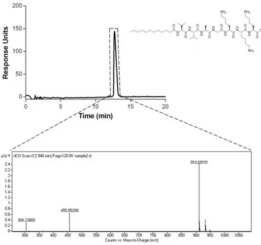
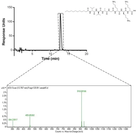
The peptide amphiphile molecules were purified using Prep-HPLC after the synthesis to separate the unintended materials from the mentioned peptides. The purified peptide amphiphiles were dissolved in double distilled water with con-centration of 1 mg/mL and then analyzed by using Q-TOF LC-MS to determine the final purity and the molecular weight of the peptides.
The chromatograms showed that the purity of both peptides were more than 95%. (Figure 2.3-6) According to the mass spectra results, the molecular weights of E3PA and K3PA were found as 912 g/mol and 914 g/mol, respectively, which
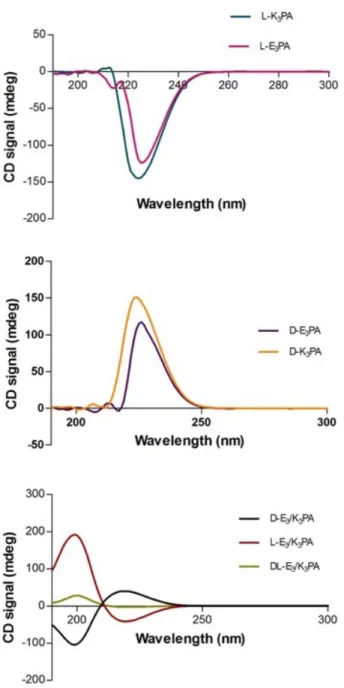
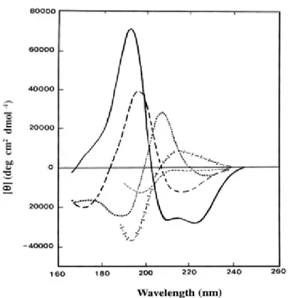
Circular dichroism (CD) spectroscopy was used for determination of the sec-ondary structures and the chiral conformations of the designed PAs. This tech-nique relies on the absorbance differences between right-handed polarized light and left-handed polarized light which comes from the molecule chirality in the far-UV region. CD spectra of the samples were red-shifted relative to typical β-sheets with a maximum at 195 nm and a minimum at 216 nm at neutral pH. The red-shifted β-sheet signals are associated with a twisted structure. 50% mixture of these chiral molecules decreased the intensity. This could be due to batch to batch variation in synthesis causing variable TFA content. TFA molecules which did not separate from lysine residues of the L- and D- K3PA may have role in
this difference. (Figure 2.7)
TEM imaging was employed to visualize the nanofibers and the network prop-erties. It revealed that the co-assembly of the oppositely charged peptide am-phiphiles self-assemble into high-aspect-ratio nanofiber structures with diameters of about 6-10 nm and lengths in terms of micrometers. (Figure 2.8)
SEM imaging was also performed to observe 3D network of self-assembled PA gels. Resulting images indicated that D-E3/K3, L-E3/K3 and DL- E3/K3 gel
Figure 2.2: Chemical structures of peptide amphiphiles (a) L-K3PA, (b) D-K3PA,
Figure 2.3: Characterization of D-E3PA by using LC-MS. Liquid chromatogram
of D-E3PA by the absorbance at 220 nm (top). mass spectrum of D-E3PA
(bot-tom). MS: (m/z) calculated 914.05, [M-H]- observed 912.5169, [M-2H]-/2
Figure 2.4: Characterization of L-E3PA by using LC-MS. Liquid chromatogram
of L-E3PA by the absorbance at 220 nm (top), Mass Spectrum of E3PA
(bot-tom). MS: (m/z) calculated 914.05, [M-H]- observed 912.5164, [M-2H]-/2 ob-served 455.7581, [M-3H]-/3 observed 303.5041
Figure 2.5: Characterization of L-K3PA by using LC-MS. Liquid chromatogram
of L-K3PA by the absorbance at 220 nm (top). mass spectrum of K3PA (bottom).
MS: (m/z) calculated 910.24, [M+H]+ observed 910.4739 [M+2H]+/2 observed
Figure 2.6: Characterization of D-K3PA by using LC-MS. Liquid chromatogram
of D-K3PA by the absorbance at 220 nm (top). mass spectrum of K3PA (bottom).
MS: (m/z) calculated 910.24, [M+H]+ observed 910.69166, [M+2H]+/2 observed
Figure 2.8: TEM images of p eptide amphiphile g els a) D-E 3 /K 3 stained with uran yl acetate, (b) L-E 3 /K 3 stained with uran yl acetate, (c) DL -E 3 /K 3 stained with uran yl acetate
2 .9: SEM imaging of p eptide amphiphile gels a) 1% (w/v) D-E 3 /K 3 ; (b) 1% (w/v) L-E 3 /K 3 ; (c ) 1% (w/v) DL-E 3 /K 3
par After the synthesis and characterizations of the peptide amphiphile molecules, gel formation properties were investigated. The D-K3PA and the
L-K3PA had good solubility in water and their final pH values were around 5.
Solubilities of the D-, and the L- E3PA in water were enhanced by adding NaOH
(1 M) into the peptide solutions and their final pH values were around 7.4. When the oppositely charged PAs were mixed, they formed milky white gels which can be observed by naked eye. (Figure 2.10) These three-dimensional networks con-tain extended nanofibers which can bundle up and trap the water molecules inside and result in gel formation.
2.10: Photograph of the gels of (a) D-E 3 /K 3 (1.0 wt%, pH 7.4); (b) L-E 3 /K 3 (1.0 wt%, pH 7.4); (c) DL-E 3 /K 3 (1.0 pH 7.4)
![Figure 1.5: Self-assembled peptide nanomaterials for a wide range of applications (Reproduced from Ref [5] with permission of John Wiley and Sons 2015).](https://thumb-eu.123doks.com/thumbv2/9libnet/5733878.115138/24.918.177.790.173.434/figure-assembled-peptide-nanomaterials-applications-reproduced-permission-wiley.webp)
![Figure 1.8: Molecular model of d-EAK16 (a) and l-EAK16 (B) (Produced from Ref [8] with permission of Creative Commons Attribution License 2008).](https://thumb-eu.123doks.com/thumbv2/9libnet/5733878.115138/32.918.267.686.168.499/figure-molecular-produced-permission-creative-commons-attribution-license.webp)