SELECTIVE FLUORESCENCE SENSING OF BIOLOGICAL
THIOLS USING A BODIPY BASED BIFUNCTIONAL PROBE
AND
THE CATALYTIC ACTIVITY OF SHORT PEPTIDE
AMPHIPHILE NANOSTRUCTURES: IMPLICATIONS ON THE
ORIGIN OF LIFE
A THESIS
SUBMITTED TO THE DEPARTMENT OF CHEMISTRY
AND THE GRADUATE SCHOOL OF ENGINEERING AND SCIENCE OF BİLKENT UNIVERSITY
IN PARTIAL FULFILLMENT OF THE REQUIREMENTS FOR THE DEGREE OF
MASTER OF SCIENCE
By YİĞİT ALTAY
I certify that I have read this thesis and that in my opinion it is fully adequate, in scope and in quality, as a thesis for the degree of Master of Science.
………. Prof. Dr. Engin U. Akkaya (Advisor)
I certify that I have read this thesis and that in my opinion it is fully adequate, in scope and in quality, as a thesis for the degree of Master of Science.
………. Assoc. Prof. Dr. Dönüş Tuncel
I certify that I have read this thesis and that in my opinion it is fully adequate, in scope and in quality, as a thesis for the degree of Master of Science.
………. Asst. Prof. Dr. Salih Özçubukçu
Approved for the Graduate School of Engineering and Science:
………. Prof. Dr. Levent Onural Director of the Graduate School
i
ABSTRACT
SELECTIVE FLUORESCENCE SENSING OF BIOLOGICAL THIOLS USING A BODIPY BASED BIFUNCTIONAL PROBE AND THE CATALYTIC ACTIVITY OF SHORT PEPTIDE AMPHIPHILE NANOSTRUCTURES: IMPLICATIONS ON
THE ORIGIN OF LIFE Yiğit Altay
M.S. in Department of Chemistry Supervisor: Prof. Dr. Engin U. Akkaya
July, 2013
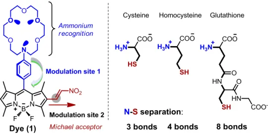
Chemosensor development is an attractive field of modern chemistry and there exist large amount of contribution from all over the world. The biological importance of thiols triggered the development of sensors to differentiate especially cysteine (Cys), homocysteine (Hcy) and glutathione (GSH) which play key roles in biological systems. Concentration of those thiols results in number of diseases and their structural similarity complicates the differentiation. Optical probes especially fluorescent ones are widely employed for that purpose since it offers simplicity, sensitivity and low detection limits as well as real time analysis. BODIPY core is decorated with a Michael acceptor nitro-styrene group to covalent incorporation of thiols and with an aza-crown moiety to recognition of N-terminus of them. The work in this thesis is the first example in which one of them is separated from others or three of them separated from each other’s by chain length difference using fluorescence spectrometry.
Formation of short peptides (2-4 aa residues) is considered to be likely under primordial conditions, following a number of scenarios. In this work, it is constructed a short peptide library limiting our choice of amino acids to those believed to be available at larger concentrations such as Gly, Ala, Asp and Cys. It is demonstrated that when acylated at the N-terminus, nanostructures of varying size and shapes were formed. Investigations on the catalytic activity of these nanostructures under different conditions are presented. The findings on the correlation of peptide structure and nanostructure formation and/or catalytic activity are presented.
ii
ÖZET
BODIPY TABANLI BİFONKSİYONEL SENSOR İLE BİYOLOJİK TİYOLLERİN FLORESANS SEÇİCİ TAYİNİ VE YAŞAMIN KAYNAĞI ARAYIŞINDA KISA
PEPTİT AMFİFİL NANOYAPILARIN KATALİTİK AKTİVİTELERİNİN İNCELENMESİ
Yiğit Altay Kimya, Yüksek Lisans
Tez Yöneticisi: Prof. Dr. Engin U. Akkaya Temmuz, 2013
Kemosensör geliştirme çalışmaları modern kimyanın ilgi çeken alanlarından birisidir ve bu alana tüm dünyadan büyük katkılar bulunmaktadır. Tiyollerin biyolojik sistemlerdeki önemi büyüktür. Biyolojik öneme sahip tiyollerden sistin (Cys), homosistin (Hcy) ve glutatiyon (GSH) kilit rol oynamaktadır. Benzer yapılara sahip bu tiyolerin hücre içi değerlerinin değişmesi durumunda sedef hastalığı, karaciğer yetmezliği, kanser, lökosit kaybı gibi birçok hastalık meydana gelmektedir. Bu sebeplerden ötürü, tiyol tayini çalışmaları büyük öneme sahiptir. Optik problar ve özellikle floresans olan optik problar kolay, hassas ve düşük algılama limitleri ve gerçek zamanlı analiz imkânları nedeniyle sıklıkla bu amaca yönelik kullanılmaktadır. Bu çalışmada BODIPY tiyollerin kovalent bağlanması için Michael alıcısı nitro-stiren grubu ile ve tiyollerin N-uçlarının tanınmasını sağlayan aza-taç eter grubu ile modifiye edilmiştir. İlk defa bu çalışma ile bu üç önemli tiyol, zincir uzunlukları arasındaki farklardan yararlanarak floresans spektroskopisi ile birbirinden ayırt edilmiştir.
Birçok senaryoya göre ilksel koşullar altında kısa peptitlerin (2-4 aminoasit) oluşabildiği bilinmektedir. Bu çalışmada, ilkel koşullarda daha yüksek derişimlerde bulunduğu bilinen Gly, Ala, Asp ve Cys aminoasitleri ile kısıtlanarak kısa peptit kütüphanesi oluşturulmuştur. Asetillenmiş N-ucuna sahip peptitlerin, değişik boyut ve şekillerde nanoyapılar oluşturduğu gözlenmiş ve bu yapıların değişik koşullar altındaki katalitik etkinlikleri incelenmiştir. Bu çalışmada peptit yapıları ve oluşan nanoyapılar ve/veya katalitik etkinlik ilişkileri üzerindeki bulgular sunulmuştur.
iii
Dedicated to my beloved mother and father
iv
ACKNOWLEDGEMENTS
I would like to express my sincere appreciation to my research supervisor Prof. Engin Umut Akkaya. It would have been impossible for me to accomplish the work without his guidance, support and encouragement. I consider being his student as a great privilege and I would like to take this opportunity to offer my deepest gratitude for everything he has done for me. It has been an honor being a member of his research group.
I would like to thank Salih Özçubukçu whom I have learnt what I know about peptide chemistry. The days spent in his lab were not only contributed to my scientific knowledge but the discussions with him provided me a better understanding of science and its philosophy.
I also would like to express my appreciation to all of those with whom I have had the pleasure to work within the past four years. I am sincerely grateful to my colleagues Safacan Kölemen, Ruslan Guliyev, Tuğba Özdemir, Sündüs Erbaş, Onur Büyükçakır for their support and helpful discussions during the course of this research project. It has been a pleasure working with group members of Akkaya Lab. I thank them all for helpful discussions and collaborations as well as for their friendship.
I have received great support, encouragement and companionship from Can Aykanat, Pınar Kınay, Tuğçe Durgut and Tuba Yaşar during the past few years. I thank them all from the bottom of my heart for their valuable friendship.
I am grateful for the privilege of meeting Meniz Tezcan who has significantly important contributions on the work presented in this thesis. Other than being an outstanding collaborator, she is one of the brightest minds that I have ever met.
Last but not the least; I would like to thank my family for their unconditional love, continuous support and understanding for what I am doing and what I want to do. I owe them a lot for their support throughout my life.
v
LIST OF ABBREVIATIONS
BODIPY :BoradiazaindaceneCD : Circular Dichroism
Cys : Cystein
DIEA : Diisopropylethyl amine
DMF : Dimetylformamide
EtOH : Ethanol
FL : Fluorophore
FMOC : Fluorenylmethyloxycarbonyl FTIR : Fourier Transform Infra-Red
GSH : Glutathione
HBTU : O-(Benzotriazol-1-yl)-N,N,N′,N′tetramethyluronium
hexafluorophosphate
Hcy : Homocystein
HRMS : High Resolution Mass Spectroscopy ICT : Internal Charge Transfer
MeOH : Methanol
NMR : Nuclear Magnetic Resonance PeT : Photoinduced Electron Transfer pNPA : p-nitrophenylacetate
PS : Photosensitizer
SEM : Scanning Electron Microscopy TFA : Trifluoroacetic acid
vi
TABLE OF CONTENTS
CHAPTER 1: INTRODUCTION ... 1
CHAPTER 2: BACKGROUND ... 3
2.1 What is Supramolecular Chemistry? ... 3
2.2 Molecular Recognition ... 4
2.2.1 Recognition, Information and Complementarity ... 4
2.2.2 Design Principles of Molecular Receptors ... 6
2.2.3 Anion Recognition ... 7
2.2.4 Cation Recognition ... 8
2.3 Photophysics of Light Absorption and Emission ... 10
2.4 Energy and Electron Transfer Processes ... 13
2.4.1 Intramolecular Charge Transfer (ICT) ... 13
2.4.2 Photoinduced Electron Transfer (PeT) ... 15
2.5 Fluorescent Chemosensors and BODIPY Dyes ... 18
2.6 What is Life? ... 21
2.7 History of the Search into the Origin of Life ... 22
2.7.1 Historical Outlook ... 22
2.7.2 Spontaneous Generation ... 24
2.7.3 Pasteur and Darwin ... 25
2.8 Primordial Soup Hypothesis ... 26
2.9 Complex Biological Molecules and Protocells ... 27
2.10 Early Conditions ... 27
2.11 Origins of Homochirality ... 29
2.12 Self-Organization and Replication ... 30
vii
2.14 Current Models ... 33
2.14.1 The “Prebiotic” RNA World ... 33
2.14.2 Autocatalysis ... 34
2.14.3 Panspermia and Exogenesis ... 34
2.14.4 Extraterrestrial Organic Molecules ... 35
2.15 Peptide Nanostructures ... 36
2.16 Solid State Peptide Synthesis ... 38
2.16.1 Mechanism ... 41
CHAPTER 3: Designing a Gluthathione Selective Fluorescent Sensor: The Use of Multiple Modulators in Signal Transduction ... 44
3.1 Introduction ... 44
3.2 Design.. ... 44
3.3 Results and Discussion ... 46
3.4 Experimental Details ... 56
3.4.1 General ... 56
3.4.2 Synthesis Scheme of Dye 1: ... 57
3.4.3 Synthesis of Dye 3 ... 61
3.4.4 Absorbance and Fluorescence Spectra ... 64
3.4.5 Absorbance and Fluorescence Spectra ... 66
3.4.6 Kinetic Studies ... 72
3.4.7 Cell Culture and Confocal Microscopy ... 73
CHAPTER 4: The Catalytic Activity of Short Peptide Amphiphile Nanostructures: Implications on the Origin of Life ... 74
4.1 Introduction ... 74
4.2 Design… ... 75
4.3 Results and Discussion ... 76
viii
4.4.1 General ... 89
4.4.2 General Procedure for Solid State Peptide Synthesis ... 89
4.4.3 Scanning Electron Microscopy ... 91
4.4.4 Circular Dichroism ... 91 4.4.5 LC-MS ... 91 CHAPTER 5: CONCLUSION ... 93 BIBLIOGRAPHY ... 94 APPENDIX A ... 105 APPENDIX B ... 115 APPENDIX C ... 126 APPENDIX D ... 129 APPENDIX E ... 131
ix
LIST OF FIGURES
Figure 1. Structures of Cys, Hcy and GSH. ... 1
Figure 2. Schematic representation of hydrolysis reaction by peptide nanostructures. 2 Figure 3. From molecular to supramolecular chemistry ... 3
Figure 4. Valinomycin (1) and enniatin (2). ... 9
Figure 5. Crown ether and its derivatives: 18-crown-6... 9
Figure 6. A cryptand. ... 9
Figure 7. Structures of typical fluorescent substances. ... 11
Figure 8. Distribution of dyes in the visible region. ... 11
Figure 9. Perrin-Jablonski diagram and illustrations of the relative positions ... 12
Figure 10. Spectral shifts of ICT type sensors. ... 14
Figure 11. Crown containing ICT sensors. ... 14
Figure 12. Molecular orbital representation of electron transfers. ... 15
Figure 13. Molecular action of a fluorescent PeT potassium cation sensor. ... 16
Figure 14. Contributions to fluorescent PeT sensors from all over the world.. ... 17
Figure 15. PeT based sensors. ... 18
Figure 16. Schematic representations for the types of fluoroionophores... 19
Figure 17. Structures and numbering of dipyrromethane, dipyrrin and Bodipy ... 19
Figure 18. Important contributions to chemical modifications of the BODIPY core.20 Figure 19. Mono-, di-, tri- and tetra-styryl BODIPY derivatives. ... 20
Figure 20. A scheme of the origin of life theories. ... 22
Figure 21. The Apparatus of Miller-Urey experiment. ... 32
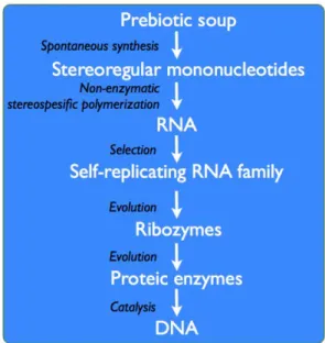
Figure 22. Origin of life in the “prebiotic” RNA world. ... 34
Figure 23. Possible peptide nanostructure formation routes via stacking ... 37
Figure 24. Synthetic cycle for solid state peptide synthesis (Fmoc/tBu approach). .. 39
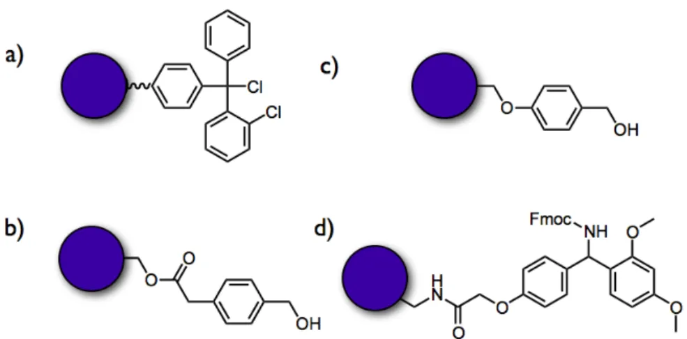
Figure 25. SSPS resins. ... 41
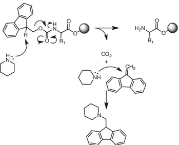
Figure 26. N-Fmoc deprotection mechanism. ... 42
Figure 27. Mechanism of HBTU-DIEA activation of C-terminus. ... 43
Figure 28. The structure and the signal modulation sites of the target probe. ... 46
Figure 29. Partial 1H-NMR (in CD3OD) spectra depicting the changes on GSH ... 48
Figure 30. Emission response to biological thiols at two different pH values. ... 49
x
Figure 32. The structures of control Bodipy dyes 2 and 3.. ... 51
Figure 33. pH titration of GSH adduct in buffered solutions. ... 52
Figure 34. Structures of control molecules Dye 4 and 5. ... 53
Figure 35. Absorption spectra of Dye 2 and 4 and emission spectra of Dye 4. ... 54
Figure 36. Absorption spectra of Dye 5 and emission spectra of Dye 2 and 5. ... 54
Figure 37. Time lapse (a-c) confocal microscopy pictures ... 55
Figure 38. Structures of Dye 1,2 and 3. ... 56
Figure 39. Synthesis scheme of Dye 1. ... 57
Figure 40. Synthesis scheme of Dye 3. ... 61
Figure 41. HPLC spectrum of dye 1. ... 65
Figure 42. Absorbance spectra of Dye 1 (8.0×10⁻⁶ M) and Dye 1 + Thiols ... 66
Figure 43. Absorbance spectra of Dye 1 (8.0×10⁻⁶ M) and Dye 1 + Thiols ... 66
Figure 44. Absorbance spectra of Dye 1 (8.0×10⁻⁶ M) and BODIPY 1 + Thiols. ... 67
Figure 45. Absorbance spectra of Dye 2 (8.0×10⁻⁶ M) at pH 6.0 ... 67
Figure 46. Absorbance spectra of Dye 3(8.0×10⁻⁶ M) and BODIPY 3 + Thiols ... 68
Figure 47. Absorbance spectra of Dye 3 (8.0×10⁻⁶ M) and BODIPY 3 + Thiols ... 68
Figure 48. Emission spectra of Dye 1 (8.0×10⁻⁶ M) and Dye 1 + Thiols ... 69
Figure 49. Emission spectra of Dye (8.0×10⁻⁶ M) and BODIPY 1 + Thiols ... 69
Figure 50. Emission spectra of Dye 1 (8.0×10⁻⁶ M) and Dye 1 + Thiols ... 70
Figure 51. Emission spectra of Dye 1 (8.0×10⁻⁶ M), BODIPY 1 + Thiols ... 70
Figure 52. Emission spectra of Dye 2 (8.0×10⁻⁶ M) at pH 6.0 ... 71
Figure 53. Emission spectra of Dye 3 (8.0×10⁻⁶ M) and Dye 3 + Thiols ... 71
Figure 54. Emission spectra of Dye 3 (8.0×10⁻⁶ M) and Dye 3 + Thiols ... 72
Figure 55. Fluorescence kinetic studies of Dye 1 + Thiols. ... 72
Figure 56.Schematic representation of the mechanism of our model. ... 74
Figure 57.Peptide amphiphiles synthesized in this study. ... 75
Figure 58. Scanning electron microscope (SEM) micrograph of peptides ... 77
Figure 59. SEM images of peptide Decanoyl-GDCA-COOH ... 78
Figure 60. SEM images of peptide Decanoyl-AGCD-COOH ... 78
Figure 61. SEM images of peptide Decanoyl-CADG-COOH ... 78
Figure 62. SEM images of peptide Decanoyl-ACGD-COOH ... 79
Figure 63.Maximum absorption wavelengths of products of pNPA hydrolysis. ... 80
Figure 64.Hydrolysis of pNPA in phosphate buffer pH 7.0 ... 81
xi
Figure 66.Plot of the absorption maximum at 400nm vs time for TRIS buffer ... 82
Figure 67. Plot of the absorption maximum at 400nm vs time for pNPA ... 83
Figure 68. Normalized relative rates of nanostructure forming peptides... 84
Figure 69. Plot of the absorption maximum at 400 nm vs time for pNPA ... 85
Figure 70. Plot of the absorption maximum at 400 nm vs time for pNPA ... 85
Figure 71. Plot of the absorption maximum at 400nm vs time for pNPA ... 86
Figure 72. Plot of the absorption maximum at 400 nm vs time for pNPA ... 86
Figure 73. Normalized relative rates of nanostructure and peptide catalysis ... 87
Figure 74.Chaotic combinatorial synthesis scheme. ... 88
Figure 75. Possible products formed by the chaotic synthesis... 88
Figure 76. LC-MS profile of peptide Decanoyl-DAGC-COOH. ... 92
Figure 77. QTOF-ESI-MS spectrum of peptide Decanoyl-DAGC-COOH. ... 92
Figure 78. SEM images of peptide Ethanoyl-ADGC-COOH ... 105
Figure 79. SEM images of peptide Butanoyl-ADGC-COOH ... 105
Figure 80. SEM images of peptide Hexanoyl-ADGC-COOH ... 106
Figure 81. SEM images of peptide Octanoyl-ADGC-COOH ... 106
Figure 82. SEM images of peptide Decanoyl-ADGC-COOH ... 106
Figure 83. SEM images of peptide Decanoyl-CADG-COOH ... 107
Figure 84. SEM images of peptide Decanoyl-DGAC-COOH ... 107
Figure 85. SEM images of peptide Decanoyl-ACGD-COOH ... 108
Figure 86. SEM images of peptide Decanoyl-ADCG-COOH ... 108
Figure 87. SEM images of peptide Decanoyl-AGCD-COOH ... 109
Figure 88. SEM images of peptide Decanoyl-AGDC-COOH ... 109
Figure 89. SEM images of peptide Decanoyl-CDAG-COOH ... 110
Figure 90. SEM images of peptide Decanoyl-CGDA-COOH ... 110
Figure 91. SEM images of peptide Decanoyl-CDGA-COOH ... 111
Figure 92. SEM images of peptide Decanoyl-ACDG-COOH. ... 111
Figure 93. SEM images of peptide Decanoyl-DCGA-COOH ... 112
Figure 94. SEM images of peptide Decanoyl-DGCA-COOH ... 112
Figure 95. SEM images of peptide Decanoyl-GACD-COOH ... 113
Figure 96. SEM images of peptide Decanoyl-GADC-COOH ... 113
Figure 97. SEM images of peptide Decanoyl-GCDA-COOH ... 114
Figure 98. SEM images of peptide Decanoyl-GDCA-COOH ... 114
xii
Figure 100. Circular Dichroism spectrum of peptide Butanoyl-ADGC-COOH... 115
Figure 101. Circular Dichroism spectrum of peptide Hexanoyl-ADGC-COOH ... 116
Figure 102. Circular Dichroism spectrum of peptide Octanoyl-ADGC-COOH... 116
Figure 103. Circular Dichroism spectrum of peptide Decanoyl-ADGC-COOH ... 117
Figure 104. Circular Dichroism spectrum of peptide Decanoyl-CADG-COOH ... 117
Figure 105. Circular Dichroism spectrum of peptide Decanoyl-CGDA-COOH ... 118
Figure 106. Circular Dichroism spectrum of peptide Decanoyl-ACDG-COOH ... 118
Figure 107. Circular Dichroism spectrum of peptide Decanoyl-ACGD-COOH ... 119
Figure 108. Circular Dichroism spectrum of peptide Decanoyl-ADCG-COOH ... 119
Figure 109. Circular Dichroism spectrum of peptide Decanoyl-AGCD-COOH ... 120
Figure 110. Circular Dichroism spectrum of peptide Decanoyl-AGDC-COOH ... 120
Figure 111. Circular Dichroism spectrum of peptide Decanoyl-CDAG-COOH ... 121
Figure 112. Circular Dichroism spectrum of peptide Decanoyl-CDGA-COOH ... 121
Figure 113. Circular Dichroism spectrum of peptide Decanoyl-DCGA-COOH ... 122
Figure 114. Circular Dichroism spectrum of peptide Decanoyl-DGCA-COOH ... 122
Figure 115. Circular Dichroism spectrum of peptide Decanoyl-DGAC-COOH ... 123
Figure 116. Circular Dichroism spectrum of peptide Decanoyl-GACD-COOH ... 123
Figure 117. Circular Dichroism spectrum of peptide Decanoyl-GADC-COOH ... 124
Figure 118. Circular Dichroism spectrum of peptide Decanoyl-GCDA-COOH ... 124
Figure 119. Circular Dichroism spectrum of peptide Decanoyl-GDCA-COOH ... 125
Figure 120. 1H NMR spectrum of Dye 2. ... 126
Figure 121. 13C NMR spectrum of Dye 2 ... 126
Figure 122. 1H NMR spectrum of compound 3 ... 127
Figure 123. 13C NMR spectrum of compound 3 ... 127
Figure 124. 1H NMR spectrum of Dye 1 ... 128
Figure 125. 13C NMR spectrum of Dye 1 ... 128
Figure 126. TOF-ESI-MS spectrum of compound 3 ... 129
Figure 127. TOF-ESI-MS spectrum of Dye 2 ... 129
Figure 128. TOF-ESI-MS spectrum of Dye 1 ... 129
Figure 129. TOF-ESI-MS spectrum of compound 4 ... 130
Figure 130. TOF-ESI-MS spectrum of compund 5 ... 130
xiii
LIST OF TABLES
Table 1. Impact of the electromagnetic radiations on molecular structures. ... 10
Table 2. Characteristic times for transitions between electronic states. ... 13
Table 3. Concentrations of organic compounds found in Murchison meteorite. ... 36
Table 4. Selected photophysical parameters for the dye 1. ... 47
Table 5. Exponential fit of the kinetic studies of Dye 1 + Thiols (200 equivalents). 73 Table 6. Characteristics of peptide amphiphiles. ... 79
Table 7. Exponential fit equations of Decanoyl- DACG-COOH ... 82
Table 8. Exponential fit equations of Decanoyl- DACG-COOH in TRIS buffer ... 82
Table 9. Examples of Enzymatic Rate Acceleration ... 83
Table 10. Exponential fit equations of the nanostructure forming peptides ... 84
Table 11. Time required to reach complete hydrolysis and calculated rates. ... 84
Table 12. Exponential fit equations of the nanostructure forming peptides ... 86
1
CHAPTER 1
INTRODUCTION
Thiols have a great importance in biological systems. Among possible biologically relevant thiols three of them play key roles; namely, cysteine (Cys), homocysteine (Hcy) and glutathione (GSH) (Figure 1). Those thiols have similar structures and generally results in number of diseases such as psoriasis, liver damage, cancer, and leucocyte loss if levels of cellular thiols have altered. In more detail, Cys deficiency causes slow growth of children, liver damage, loss of muscle, and hair depigmentation. Elevated level of Hcy in plasma triggers Alzheimer diseases. On the other hand, GSH which is a non-protein-thiol takes place in cellular functions such as intracellular signal transduction, gene regulation, and intracellular redox activities. Apart from above three, thiophenols are known to be a highly toxic and environmentally unfriendly. Thus, the detection of thiols has become a very important task. Optical probes especially fluorescent ones are widely employed for that purpose. This is apparent because fluorescence detection offers simplicity, sensitivity and low detection limits as well as real time analysis.
Figure 1. Structures of Cys, Hcy and GSH.
Probe design for thiol sensing mostly utilizes thiols strong nucleophilicity and high binding affinity towards metal ions. Based on these features, fluorescence sensing of thiols involves characteristics reactions between probe and thiol such as Michael addition, cyclization, and cleavage of disulfide bond in the presence of thiols. One can find many literature examples with these approaches. In most of them Cys, Hcy and GSH were detected together or only two of those were separated from the other. There is no example in which one of them is separated from others or three of them separated from each other’s by fluorescence spectra.
2
The molecular origin(s) of life is one of the most essential questions in modern biology and chemistry. The RNA world hypothesis could give satisfactory explanations for the evolvement of the biochemical networks but it cannot explain the connection between the primeval molecules and first RNA molecules. It is obvious and practically impossible that molecules with high complexity cannot be evolved spontaneously. Formation of tubular, vesicular and fibrilar structures via self-assembly of peptides as simple as dipeptides has been demonstrated recently. In addition to that, dipeptides can be used as template for synthesis of other peptides and can act as catalysts. In contrast to complex RNA molecules, it is very likely that functional short peptides can be synthesized under primordial earth conditions.
Figure 2. Schematic representation of hydrolysis reaction by peptide nanostructures.
In this thesis, a novel mechanism for the origin of life is proposed using the ability of short peptides to form well-ordered nanostructures and catalysis of chemical reactions. This model may help to explain early processes that led to the evolution of current biochemical systems that allow the functioning of living systems. For that purpose four natural amino acids (glycine, alanine, aspartic acid and cysteine) are selected because of their simplicity, primordial occurrence and known catalytic activity. A peptide library is constructed with alternating the sequence of amino acids. In each sequence, each amino acid used once and N-terminus of the peptides are acetylated with ten-carbon-long hydrocarbon chains to make peptides amphiphilic. Under same conditions, some of the peptides formed nanostructures adopting β-sheet conformation. Catalytic properties of these peptides are studied on the hydrolysis of p-nitrophenyl acetate benefiting from the fact of microscopic reversibility principle.[1]
3
CHAPTER 2
BACKGROUND
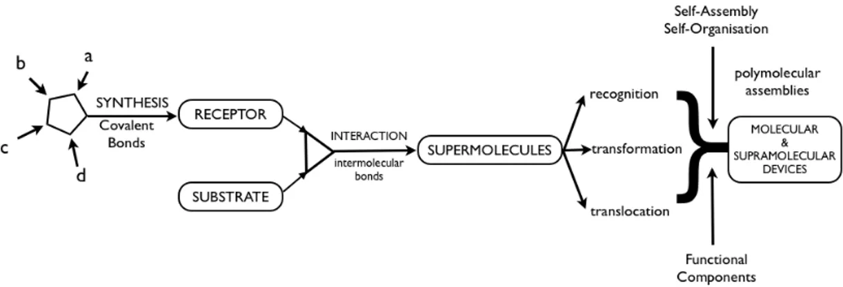
2.1 What is Supramolecular Chemistry?Physics declared its reign with the beginning of everything, the Big Bang. Then chemistry came along as the temperature decrease; elementary particles formed small atoms, and they united to make more and more complex molecules. These molecules formed aggregates, aggregates formed membranes, defining the protocells of which eventually give rise to emergence of life. Life is the highest expression of chemistry.
Molecular chemistry focuses on the covalent bond. Starting from Wöhler’s[2] urea synthesis to the synthesis of Vitamin B12 by Woodward[3] and Eschenmoser,[4] molecular chemistry is strengthened more and more. But there was a gap for the non-covalent interactions waiting to be filled. And the supramolecular chemistry aimed to gain control over non-covalent intermolecular interactions.
Supramolecular chemistry is a highly interdisciplinary field of science focusing on the chemical, physical and biological characteristics of molecular systems that are held together by the non-covalent intermolecular interactions rather than the molecule itself. Intermolecular forces are weaker with respect to covalent bonds. This is the reason why supramolecular species are thermodynamically less stable, kinetically more labile and dynamically more flexible.
Figure 3. From molecular to supramolecular chemistry; molecules, supermolecules, molecular and
4
2.2 Molecular Recognition
Molecular recognition can be defined by the information and the energy carried by the selection of substrate by a certain receptor molecule. It may also involve a specific function.[5] Recognition can be considered as binding with a purpose. Receptors are such an example. Structurally well-defined set of intermolecular interactions undergoes through recognition processes. Complex or the supermolecule is formed upon binding of guest (analyte) to the host (receptor). Characterization can be done by its thermodynamic and kinetic stability and selectivity. It means that the amount of energy and information brought into operation are characteristic for such processes.
2.2.1 Recognition, Information and Complementarity
Molecular information can be stored molecularly and can be read out supramolecularly through molecular recognition. At the beginning of 1970’s, studies on selective complexation of metal ions facilitated the use of notions of recognition and information which were used in relation with biological systems.[6] Since then molecular recognition has become a very frequently used term and a major field of research in chemistry.
Architecture of the receptor is capable of storing information in its binding sites; and the rate of formation and dissociation of the supermolecule provides a read out. Conformation, chirality and dynamics also come into play during the receptor characterization in addition to its size and the shape. In addition to eventual reactivity that may allow the coupling of complexation with other processes (protonation, deprotonation, oxidation and reduction); features such as size, shape, charge, polarity, polarisability, van der Waals interactions, number and the arrangement on the receptor architecture carries information for the characterization of binding sites. Thickness, hydrophilicity or hydrophobicity and overall polarity plays important role in the action of ligand layer. In addition, the mutual balance between solvation of guest by host and the complexation affects the stability and selectivity.
5
The most fundamental and the general notion of supramolecular chemistry is the information. In this respect, supramolecular chemistry could be considered as molecular informatics that concerns molecular storage and read out and processing the information via molecules or supermolecules.[7,8]
Interactional and geometrical complementarity between associating species is implied by recognition. So that the double complementarity principle extend over energetics in addition to geometrical ones such as the lock and key and steric fit concepts introduced by Emil Fischer.[9]
In order to get high recognition by a receptor molecule ρ, there should be a large difference between the binding free energies of a given substrate σ and the other substrates. Deviation from the statistical distribution is the ultimate result. There are several factors that should be taken into consideration to achieve large differences in affinity:
1) Complementarity between ρ and σ in terms of size and shape 2) Interactional complementarity.
3) Large contact areas between ρ and σ.
4) Multiple interaction sites to compensate the relative weakness of non-covalent interaction.
5) Strong overall binding.
In addition, two partners (ρ and σ) should have overlapping hydrophobic/hydrophobic or hydrophilic/hydrophilic domains to overcome the medium effects that play an important role in interaction of solvent molecules with ρ and σ.
Biological molecular recognition represents the most complex expression of molecular recognition leading to highly selective binding, reaction, transport, regulation etc. It provides study cases, illustration and inspiration for the unraveling of basic principles and for the design of model systems as well as abiotic receptors.
6
2.2.2 Design Principles of Molecular Receptors
Molecular receptors are defined by their characteristic property of binding selectively to ionic or molecular substrates (or both) by means of various intermolecular interactions, leading to an assembly o two or more species, a supermolecule. The principles of molecular recognition should be expressed in the design of a molecular receptor. Receptor chemistry represents a generalized version of coordination chemistry. But it is not limited to transition-metal ions and extending to all type of substrates: cationic, anionic, or neutral specie of organic, inorganic or biological nature. [10]
In order to achieve high recognition, every factor should be taken into account in the design of the receptor. In particular, complementarity depends on a well-defined three dimensional architecture with the correct arrangement of binding sites. Furthermore, ρ and σ will be in contact over a large area, if ρ is able to wrap around its guest so as to establish numerous non-covalent binding interactions and sense its molecular size, shape, and architecture. These are important especially in design of receptor molecules containing intramolecular cavities, clefts or pockets into which the substrate may fit.
Macrocyclic structures are of special interest for designing artificial receptors. They are large and may therefore contain cavities of appropriate size and shape. They possess numerous branches, bridges, and connections that allow the construction of a given architecture having specific dynamic features. Binding of a substrate into the cavity will yield an inclusion complex, a cryptate. [11] In addition to maximizing the contact area, inclusion also leads to almost complete solvent exclusion from the receptor site. Thus the displacement of solvent molecules by the substrate on binding is minimized.
The balance between rigidity and flexibility is important especially for binding and the dynamic properties of ρ and σ. On one hand, rigid receptors are expected to have highly efficient recognition since they are highly stabile and highly selective. On the other hand, substrates bind to flexible receptors by an induced fit.[12] This may present high selectivity but lower stability since a part of the binding energy is used
7
in the conformational change. The designed dynamics are more difficult to control than rigidity. So receptor design covers both static and dynamic aspects of macrocyclic structures.
Chelating and macrocyclic ligands are frequently employed due to high thermodynamic stability of their complexes. The chelate effects refers to the enhanced stability of complex containing chelate rings as compared to a similar system containing fewer or no rings. Starting from the five membered chelate rings, chelate effect decreases with increasing ring size. Longer chains have higher configurational entropy and thus ring formation becomes increasingly improbable.
The macrocyclic effect is related to the chelate effect and refers to the increased thermodynamic stability of macrocyclic systems compared to their acyclic analogues. Macrocyclic hosts are less heavily solvated than their acyclic analogues and therefore less energy is required for desolvation (coordination is more enthalpically favorable). Macrocyclic ligands are less flexible and consequently have less disorder to lose on complexation than their acyclic analogues (in other words, coordination is more entropically favorable because of the relative rigidity of the receptor).
The enhanced binding of guest species provided by chelating or macrocyclic hosts has been employed in the design of many receptors operating through a variety of intermolecular forces.
2.2.3 Anion Recognition
Anionic species play crucial roles in chemistry and in biology; however their binding characteristics have not extensively studied. The coordination chemistry of anions is expected to have significant outcomes in novel structures and chemical and biological properties. In the recent years, research in this field is increasingly active and slowly builds its path in the area of coordination chemistry. [13,14,15]
8
Anionic substrates possess a wide range of geometries: spherical (F-, Cl-, Br-, I-), linear (N3-, OCN-), planar (NO3-, R-CO2-), tetrahedral (SO4-, ClO4-) and octahedral (M(CN)6n-).
Polyammonium macrocycles and macropolycycles have been studied most extensively as anion receptor molecules. They bind to inorganic anions, carboxylates and phosphates selectively with electrostatic and structural effects.
2.2.4 Cation Recognition
Cation detection is of great interest to many scientists from different fields including chemists, biologists, geologists and environmentalists. In many biological activities cations plays an important role such as enzyme activation (Zn2+, Mg2+, Mn2+) , transmission of nerve impulses (Na+, Mg2+ and Ca2+), regulation of cell activity (Zn2+, Mg2+ and Ca2+) and muscle contraction (Na+, K+ and Ca2+). Moreover, some metal ions are key component of metalloenzymes. In the treatment of high blood pressure and manic depression potassium and lithium are important serum parameters, respectively. Additionally, early detection of environmentally important toxic heavy metals such as mercury, cadmium and lead has great importance. For the cation detection, several methods are available: flame photometry, atomic absorption spectrometry, ion sensitive electrodes, electron microprobe analysis, neutron activation analysis, etc. These techniques are rather expensive and requires large amount of sample and continuous monitoring cannot be carried out.
Fluorescent sensors on the other hand provide real time analysis opportunities with high accuracy and selectivity. The design consists of a fluorophore connected to an ionophore. High stability and selectivity, metallic affinity, kinetically rapid sensitization and ease of target delivery are some of the criteria for good chemosensors. In such designs, signaling moiety converts the recognition into optical signal in the form of emission or shift in the absorption spectrum. These photophysical changes are due to perturbation of photoinduced processes.
9
Cyclic polyethers are one of the commonly used in recognition processes of positively charged metal ions (alkali, alkaline-earth and lanthanide cations). Three main classes may be distinguished:
1) natural macrocycles having antibiotic properties such as valinomycin or the enniatins;[16,17]
Figure 4. Valinomycin (1) and enniatin (2).
2) synthetic macrocyclic polyethers, the crown ethers,[18] their derivatives[19] and spherands.[20]
Figure 5. Crown ether and its derivatives: 18-crown-6 (3), dibenzo-18-crown-6 (4), diaza-18-crown-6
(5),
3) synthetic macropolycyclic ligands, the cryptands[21,22] and cryptospherands.[23,24]
10
There are numerous reports and studies on these supramolecules. For instance, valinomycin gives a strong and selective complex in which a K+ ion is included in the macrocyclic cavity. Similar inclusion takes place in the complexes of crown ethers such as the complex of Rb+ ion with dibenzo-18-crown-6.
2.3 Photophysics of Light Absorption and Emission
“Light plays an essential role in our lives: it is an integral part of the majority of our activities. The ancient Greeks, who for ‘to die’ said ‘to lose the light’, were already well aware of this” said Lois de Broglie in 1941. Light gives the color and brilliance to all works of art and nature. This brilliance is realized by absorption of light by the molecules.
An electronic transition occurs upon the absorption of an electron in which one electron from an orbital of a molecule in the ground state HOMO (or HOMO-1 etc.) to an unoccupied orbital LUMO (or LUMO+1 etc.). Photons being part of the UV-visible region have enough energy to induce such transitions. Photons being part of other regions may result in a wide variety of changes in the molecule upon excitation. These changes are summarized in Table 1.
Radiation Wavelength Photon Energy Results of Absorption
Gamma-rays < 0.01 nm > 1 MeV Nuclear reactions
X-rays 0.01-10 nm 124 eV-120 keV Transitions of inner shell electrons Ultra violet 10-400 nm 3.1-124 eV Transitions of outer shell electrons Visible 400-750 nm 1.7-3.1 eV Transitions of outer shell electrons Infrared 750nm-15 µm 80meV-1.7 eV Molecular vibrations
Far IR 15µm-1 mm 1.2meV-80meV Molecular rotations Radar 1mm-1 m 1.2µeV-1.2meV Oscillation of mobile electrons
Table 1. Impact of the electromagnetic radiations on molecular structures.
Fluorescence is defined as the emission of photons accompanying the S1-S0 relaxation. Since emission occurs from S, its characteristics do not depend on the excitation wavelength. Fluorescence and absorption have same 0-0 transitions however, fluorescence occurs at higher wavelengths (lower energy) than the
11
absorption since some of the excited state energy is lost due to vibrational relaxations.
Figure 7. Structures of typical fluorescent substances.
In general, ground and excited states have similar vibrational level differences, so that the fluorescence spectrum often resemble the first absorption band and the gap between the maximum of the first absorption band and the maximum of fluorescence is called the Stokes Shift. So the main cause of Stokes shift is the rapid decay to the lowest vibrational level of S1. In addition to this effect, fluorescent molecules can display further Stokes’ shift due to solvent effects, excited-state reactions, complex formation and energy transfer.
12
2.3.1 Characteristics of Fluorescence Emission
There are several possible transitions between electronic states. Photon absorption, internal conversion, fluorescence, inter-system crossing, phosphorescence, delayed fluorescence and triplet-triplet transitions are the possible processes that are used to describe radiative and non-radiative transitions between electronic states. In the Perrin-Jablonski diagram (Figure 9) singlet electronic states are depicted as S0, S1, S2,... and the triplet states T1, T2, T3,... . Characteristic times of these processes can be found in the Table 2.
Figure 9. Perrin-Jablonski diagram and illustrations of the relative positions of absorption,
fluorescence and phosphorescence processes.
Most elementary particles are in their ground state at room temperature. When these particles are irradiated by photons with proper energies, the electrons move to a higher energy state, which can also be termed as excited state. Once a molecule is excited by absorption of a photon, it can return to the ground state with emission of fluorescence, but many other pathways for de-excitation are also possible. These are internal conversion, intersystem crossing, intramolecular charge transfer and conformational change. Moreover, interactions in the excited state with other molecules such as electron transfer, proton transfer, energy transfer, excimer
13
formation, exciplex formation and photochemical transformations may compete with de-excitation.
Characteristic Times Absorption 10-15 s
Vibrational Relaxation 10-12 – 10-10 s
Lifetime of the excited state S1 10-10 – 10-7 s (Fluorescence)
Intersystem Crossing 10-10 – 10-8 s Internal Conversion 10-11 – 10-9 s
Lifetime of the excited state T1 10-6 – 1 s (Phosphorescence) Table 2. Characteristic times for transitions between electronic states.
2.4 Energy and Electron Transfer Processes
Light absorption significantly affects the electronic properties of molecules. It may induce intra- or intermolecular electron transfer processes leading to electron-hole separation.
2.4.1 Intramolecular Charge Transfer (ICT)
Intramolecular charge transfer (ICT) is a process that leads to blue or red shift in the emission spectrum of a fluorescent molecule and very common mechanism used in signaling. In the design of ICT-type chemosensors, fluorophore and the receptor units are directly bound to each other. The receptor is conjugated to the π-system of the fluorophore where it acts as an electron donor or electron acceptor according to the state of the fluorophore.
The electron density is redistributed when conjugated receptor-fluorophore system is excited by the absorption of a photon. This redistribution forms a dipole and internal charge transfer from donor to acceptor is thus triggered. When analyte bind to the system, it interacts with this excited state dipole. Interaction leads to significant changes in the both emission and absorption spectra.[25]
14
Figure 10. Spectral shifts of ICT type sensors: a) Interaction with the donor group, b) Interaction with
the acceptor group.
If the receptor is electron rich, interaction with the cation leads to reduction of e-donation ability of that group which results in the weakening of conjugation. This interaction destabilizes the excited state more and results in the increase in the E gap between ground state and the excited state and thus a blue shift is observed in the absorption spectrum. In the reverse case scenario, if the receptor is electron poor, interaction with the cation leads to increase the electron withdrawing ability of that group. The interaction stabilizes the excited state more than ground state and so the E gap between ground state and excited state and thus red shift in the absorption spectrum is observed (Figure 10).
In literature, ICT is a frequently used mechanism in the design of fluorescent sensors. Compounds 1[26] exhibit blue shift in both absorption and emission spectra upon cation binding and 2[27] produce Ca2+-induced red-shifts in the emission spectra. (Figure 11)
15
In addition to chemosensing applications, ICT systems are also widely used in optoelectronics, such as organic light emitting devices,[28,29] nonlinear optical devices,[30] and solar cell materials.[31]
2.4.2 Photoinduced Electron Transfer (PeT)
The photogeneration of charge-seperated species by photoinduced electron transfer (PeT) is an important phenomenon for initiating photocatalytic reactions and for the transfer of photosignal.
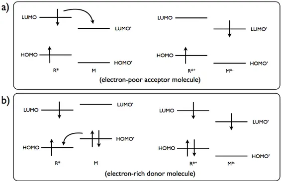
Photoinduced electron transfer (PeT) corresponds to the primary photochemical processes of the excited-state species, R*→I, where R* can be an electron donor (therefore oxidised, R* + M → R*+ + M*-) or electron acceptor (therefore reduced, R* + M → R*- + M*+) when reacting with another molecule, M. In, an electron is transfered between excited species and ground-state species. Since the electron transfers occur by electron-exchange interactions, orbital overlap is required.
Figure 12. Molecular orbital representation of a) oxidative and b)reductive electron transfer.
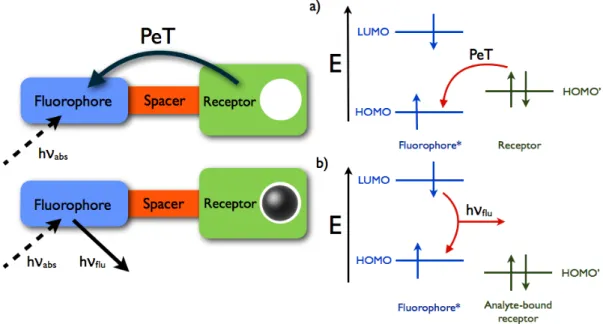
Probe molecules used for PeT consist of three major components: fluorophore (fluorescent chromophore), spacer and receptor. Spacers, as small as methylene
16
bridges, are used to cut off the conjugation between fluorophore and the receptor to eliminate other possible pathways. Cation receptor group contains electron donating group and PeT occurs from the receptor to fluorophore and results in quenching of the latter. Upon binding of cation or H+, fluorescence properties of the fluorophore restored as the electron transfer is inhibited. An electron is promoted from the HOMO to the LUMO when the fluorophore is excited. Thus PeT occurs from the HOMO of the receptor to that of the fluorophore. On binding, the redox potential of the electron-donating receptor is raised so that its HOMO becomes lower in energy than that of fluorophore. Thus, since PeT is not possible, fluorescence is observed.
Figure 13. Molecular action of a fluorescent PeT potassium cation sensor as a molecular switch using
a macrocyclic electron donor and anthracene fluorophore.
In literature, there are many examples of fluorescent probes for cations, including H+, that are normally nonluminescent, but become fluorescent upon binding of cation.
17
Figure 14. Contributions to fluorescent PeT sensors from all over the world. [A. P. de Silva, T. S.
Moody, G. D. Wright, Analyst, no. 134, pp. 2385–2393,2009. <http://
http://pubs.rsc.org/en/Content/ArticleLanding/2009/AN/b912527m>] - Reproduced by permission of The Royal Society of Chemistry.
PeT is the other widely used mechanism in the design of molecular probes. Molecule
3[32] is the first molecule used in a molecular logic operation, namely used as a AND
gate operator. There are two PeT pathways, one from electron rich tertiary amino and the other from crown ether group, blocking the fluorescence of anthracene moiety. Upon protonation of the amino group only does not recover the fluorescence since the PeT is still active through the crown ether moiety. Same thing is applicable for the binding of a Na+ ion to the 18-crown-6 group. However, when both of the analytes, H+ and Na+, present PeT is blocked and the fluorescence emission of anthracene is recovered.
18
Figure 15. PeT based sensors.
Molecule 4[33] is another molecular logic gate operator acting as an OR gate operator. It has poor chemoselectivity for transition metals, however shows significant emission enhancement upon addition of either Pb2+ or Eu3+ /Tb3+ ions or both of the ions.
2.5 Fluorescent Chemosensors and BODIPY Dyes
Fluorescent detection is a highly sensitive, easily processible, simple and cheap method to detect molecules or ions of interest. So that fluorescent chemosensors draw great attention. In the design of chemosensors, recognition and signaling processes have significant importance. In signaling, fluorescent core itself used as the signaling unit and a synthetic or biological receptor unit is incorporated to the system. Receptor set the limits of binding and selectivity. Because of that reason, a receptor should strongly and selectively bind to the target analyte. The fluorophore transforms information into optical signal as emitting light. As seen in the previous sections, binding of an analyte to the receptor may lead to significant changes in the photophysical properties of the fluorophore. With the help of these changes, presence and/or concentration of the analyte can be determined easily. What changes in the photophysical properties after the chemosensor undergoes complexation can be adjusted as desired in the design of the chemosensor. It can be in the form of enhancement or quenching of fluorescence signal via PeT mechanism, or shift in the emission wavelength via ICT mechanism.
There are several design models for fluorescent probes in accordance with the desired signaling process. Fluorophore-spacer-receptor or integrated systems are
19
examples of such designs (Figure 16). In fluorophore-spacer-receptor design, spacer cuts off the conjugation between receptor and the fluorophore whereas in the integrated systems receptor is a part of π-electron system of the fluorophore.
Figure 16. Schematic representations for the types of fluoroionophores.
Fluorescent dyes are used in a broad field in chemistry including supramolecular chemistry, photochemistry, biochemistry and physical chemistry. Among the large variety of fluorescent dyes known, boradiazaindacenes (BODIPY) have drawn increasing attention due to its high molar absorptivity, high fluorescence quantum yield, small Stoke’s shift, high photostability and ease of synthesis.
Numbering of the BODIPY core skeleton shows differences with its precursor dipyrromethane. The central carbon is named as meso position which comes from the porphyrin nomenclature.
Figure 17. Structures and numbering of dipyrromethane, dipyrrin and Bodipy
BODIPYs are stable under a wide range of pH values but start to decompose in strongly acidic or basic conditions. Also solvent polarity does not have significant
20
role in their characteristics. In addition to these properties, one of the reasons that draw attention is the ease of functionalization and tunability. Today it is well known that all positions (1–8) on the BODIPY core are open to chemical modifications (Figure 18). Making structural modifications brings out new members of BODIPY family with some shifted photophysical properties.
Figure 18. Important contributions to chemical modifications of the BODIPY core.
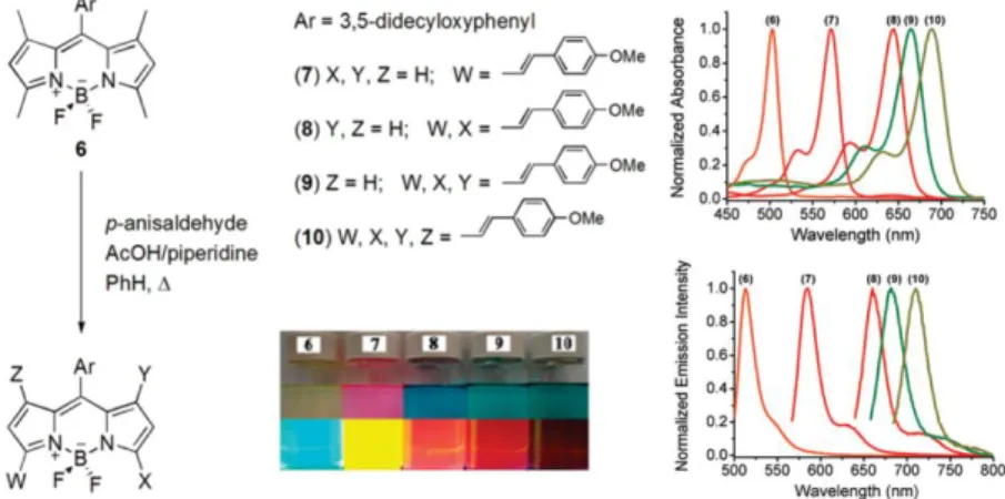
Akkaya et al. have important contributions to BODIPY chemistry especially in modification of the core in addition to their contributions to its applications in energy transfer cassettes, light harvesting systems, solar sensitizers, photodynamic therapy agents, fluorescent chemosensors and molecular logic gates. In the year 2009, they first reported the first member of the tetrasytryl-BODIPY dyes (molecule 10). Styryl groups are introduced to the BODIPY via Knoevenagel condensation on 1,3,5, and 7 position. Starting from unsubstituted core (abs. 500 nm/ ems. 525 nm); mono, di, tri and tetra substituted derivatives successfully managed to cover almost all the visible region extending to the near IR region with modifications on styryls.
Figure 19. Mono-, di-, tri- and tetra-styryl BODIPY derivatives. Adapted with permission from (O.
Buyukcakir, O. A. Bozdemir, S. Kolemen, S. Erbas, and E. U. Akkaya, “Tetrastyryl-Bodipy Dyes: Convenient Synthesis and Characterization of Elusive Near IR Fluorophores”, Org. Lett., vol. 11 , no.
21
2.6 What is Life?
Is it possible to define “life”?
Obviously, one needs an operational definition to describe what life is and how can it be characterized and, at the same time, to have a historic description relating life as we know it to the history. Our inability to give a complete definition of life was discussed by Küppers.[34] However, in spite of this conclusion, as well as similar conclusions reached earlier by other researchers, the scientific literature is abundant with attempts to define life (see Appendix E).
Historically there always been huge division between what people define living and non-living systems. As a consequence, we consider beautiful and complex crystals as non-living systems and rather beautiful and more complex animals as living. Although in the last hundred and fifty years science blurred the sharp distinction between the two, now it is certain that there is a continuum between living and non-living systems. For instance, viruses are the natural systems that cannot actually satisfy all the properties of living systems such that it needs another host to reproduce and evolve.
The scientific approaches to the characterization of life may be divided according to two aspects of life: One view focuses on the molecular level, whereas the second is cell-centered view. Most of the characterizations and definitions in Appendix E can be attributed to the first point of view. The second view is presented by the notion of
autopoiesis (Greek word for “self-producing”), according to which life cannot be
characterized by means of just one component or attribute of its complex pattern.
Then what are the characteristics of life? First of all, life has a body. It differentiates the self from its surroundings. Second, life should have a metabolism. This is a process of life uses to convert resources into building blocks so that it can build and maintain itself. Third, life has a kind of inheritable information. Human beings reserve this information as DNA in their genome and inherit to its offspring. If the first two, body and metabolism, are coupled, moving and reproducing systems may
22
be obtained. With the addition of inheritable information, system would be more lifelike and would even capable of undergoing evolution.
2.7 History of the Search into the Origin of Life
A scheme of the major landmarks in the history of the search into the origin of life can be seen in Figure 20. Starting with Thales and ending with some of the names of the most recent theories.
Figure 20. A scheme of the origin of life theories.
2.7.1 Historical Outlook
The scientific methodology was originated by the Greek philosophers, some 2.600 years ago. Curiosity was not forbidden by Greek traditions and beliefs. Moreover,
23
their philosophers explained the world in rational terms of mechanisms and natural processes and entities, not by divine actions. Two of the trivial questions they asked were what differentiates living from nonliving entities, and how plants and animals were formed. The Greek philosophers developed different ideas and proposals with regards to the problems they studied.
Thales, scholar from Miletus, is believed to have been the first Greek philosopher. He tried to discover the laws of nature by reasoning and understanding nature and observing the latter. According to him, magnet is a living entity since it causes iron bodies to move. Amber is also a living entity, since it can move other substances. Moreover, water is the substance of which the world is made; it is the ultimate substance out of which all things are generated and eventually perishes.[35]
A pupil of Thales, Anaximander first presented the elements of Darwinian evolution theory. He thought that adaptation to different conditions was needed in order to survive. According to him, life was formed spontaneously and abiogenetically, in the sea. Another supporter of abiogenesis was Xenophanes who was proposed that life is formed from nonliving substance.
The origin of life is explained by Empedocles as the association of soil and humidity under the influence of heat. Plants were the first formed out of these combinations. These were followed by the formation of limbs of animals which were seperated from each other but striving to combine with each other. Empedocles’ proposition seems to contain elements of selection and struggle but not of evolution.
Leucippus is believed to have been the first philosopher who developed the concept of atoms. According to his school, there are four kinds of atoms: stone atoms, dry and heavy; water atoms, wet and heavy; air atoms, light and cold; fire atoms, light and hot. Combinations of these kinds of atoms make up all known and unknown materials. On the other hand Democritus, the pupil of Leucippus, argued that there is an infinite number of worlds like ours. Since there is infinite number of atoms, there should be an infinite number of worlds made up of these atoms. Epicurus was the most prominent follower of Democritus’ atomic teaching. According to him, notion of motion of the atoms was a built-in feature, independent of any divine forces.
24
Aristotle was the most prominent opponent of the Democritus’ atomic world. Randomness and lack of theology was not overlapping with the Aristotle’s teachings. But he was apparently the first philosopher who described the graduality of living creatures and addressed the gradual transition between nonliving matter and plants and between plants and animals.
The Greek philosophers developed the school of thought that later became the mainstream of Western thinking regarding the origin of life, namely the spontaneous generation theory.
2.7.2 Spontaneous Generation
According to spontaneous generation theory, all living forms were formed by the generation power of the nature. Moreover, many of these living forms are still being formed today under certain favorable conditions, by spontaneous generation, not by means of seeds or parents.
The spontaneous generation theory was adopted and modified by the Stoic philosophers and accepted by the scholars of Rome and Alexandria. It was also adopted by the Christian church in both the East and West and become a dominant school in the Christian world for almost 2.000 years. It was considered almost natural in Europe because of the long tradition of belief. However, strictly religious arguments raised in time since God created all living creatures during the first days of creation.
Other and more rational doubts came from scientists. Ample observations supported the dictum of William Harvey, the discoverer of the mechanical principles governing the blood circulation: “Omne vivum ex ovo” (All living come from the egg). With regard to the spontaneous generation, Jan Baptistat van Helmont quoted if one puts a soiled shirt into the mouth of a jar containing grains of wheat, the ferment released from the soiled shirt, combining with the odor of the grain, transmutes the mixture into mice in about 21 days.[36] His ideas about the spontaneous generation were reflecting the contemporary thinking.
25
The initiation of critical experiments related to the spontaneous generation theory was carried out by Redi. Redi was interested in the problem of the deterioration of meat. Butchers of his time were covering fresh meat with a cloth or muslin to protect it from flies. He conducted experiments with meats and cloths and control experiments. And the observed the appearance of maggots in the meat. The presence of maggots in the uncovered meats only was connected to the source of maggots, namely flies. Thus it was the flies that caused the appearance of worms in the meat, not the spontaneous generation. Redi was also one of those experimentalists who thought that without experimental demonstration, belief is useless.
A technological breakthrough is happened in 1590 with the invention of optical microscope by Dutch brothers Francis and Zachary Janssen. It has magnification power of one order of magnitude larger than a magnifying lens had a decisive power on the assessment of the validity of spontaneous generation theory. This microscope developed by Antoni van Leeuwenhoek in 1676 had a magnification power of 275, just enough to discover microorganisms. Thus during the 17th century, the spontaneous generation theory was re-examined more critically than in earlier generations.
2.7.3 Pasteur and Darwin
During the 200 years between the classical experiments of Redi and Pastuer, spontaneous generation theory was tested over and over again experimentally and gradually shifted its focus to smaller organisms. Pastuer designed an experimental system based on a glass flask of special design that a cotton plug prevents the entrance of bacteria into the bottle. The flask and the content is boiled for some time for sterilization and observed that no bacteria developed after sterilization. Thus he proved that bacteria are not formed by spontaneous generation.
Although Pasteur’s work ended 200 year of controversy, it raised new questions without answers regarding the origin of life. Almost at the same time that Pasteur started his famous experiments regarding the spontaneous generation of bacteria, Darwin’s book On the Origin of Species was published. Before Darwin, controversy
26
was on the origin of present day organisms; after Darwin, controversy also involved the origin of life.
Charles Darwin suggested that the original ignition of life may have begun in a pond of ammonia, phosphoric salts, electricity and heat. And the first compounds formed were chemically ready to undergo more complex changes. However, the sterile conditions of the to-day laboratories may affect the origin of life studies since at the present day such matter would be instantly absorbed which would not have been the case before living creatures were formed.
2.8 Primordial Soup Hypothesis
The biochemist Alexandre Ivanovitch Oparin adopted evolution as a central theme and integrated various scientific disciplines, mainly organic chemistry, biochemistry, geochemistry and astrophysics to develop a coherent and partially testable scenario in his first book entitled Vozniknovenie Zhizny na Zemle (The Origin of Life on Earth).
Based on available studies of his time, Oparin suggested that the prebiotic earth had a reducing atmosphere. He reasoned that the synthesis of certain organic compounds that are necessary building blocks for the evolution of life is possible only under an atmosphere containing no molecular or atomic oxygen. Organic compounds formed are thus gradually become more and more complex. And these complex molecules aggregated to form coacervates that are held together by hydrophobic forces from a surrounding liquid. Coacervates are capable of absorption and assimilation of other organic molecules from its environment. This can be speculated as mimicking of metabolism and coacervates served as the first chemical entities capable of undergoing evolutionary process, which eventually leads to the emergence of primitive living forms. These hypothetical creatures are named as eubiont and would probably be considered as an extreme anaerobic heterotrophic prokaryote.
27
2.9 Complex Biological Molecules and Protocells
Sidney W. Fox is experimented the primordial soup theory and the abiogenesis between 1964 and 1988. One of the most common sources for condensation reactions in the prebiotic environment is likely to have heat followed by dehydration. In one of the experiments, Fox heated mixture of amino acids with excess aspartic acid and glutamic acid at 150ºC to 180ºC for a few hours. And he observed formation of peptides having cross-linked, thread-like, submicroscopic architectures by dry heating. These peptides are called "proteinoids".
n amino acids( with excess asp and glu) polypeptide(proteinoid) + n H2O
The most successful attempts to synthesize polynucleic acids under plausible prebiotic conditions were carried out by Ferris and his coworkers. In of these studies, a montmorillonite clay mineral was used as an adsorbent and catalyst. The nucleotide monomers were activated by imidazole.
Polymerization reactions on the surface of iron(III) hydroxide oxide are another successful candidate for the protocell formation. A major problem in the polymerization reactions carried out in the presence of water, such as condensation of polynucleotides, is the destruction of the condensing agents and reactive intermediates by water, thus preventing the production of large polymers. Based on earlier works, Weber used iron(III) hydroxide oxide(Fe(OH)O) in the oxidative polymerization of 2,3-dimercapto-1-propanol. In addition to prebiotic plausibility of the reaction under study, it was noted by Weber that “redox reactions could have provided the energy for the earliest type of polymer synthesis involved in the origin of life.” Weber’s experiment plus theoretical considerations suggest that polysulfides could have had important functions in prebiotic reactions.
2.10 Early Conditions
The earth is about 4.57 billion years old. At the Hadean area (starting from the formation of planets till 3.8 billion years ago from now) its surface is was about 1500K according to the recent models and so surface was molten. In the process
28
known as “iron catastrophe”, melted iron-group elements (Fe, Ni and Co) passed through the lighter silicate molten rocks down to the core.[37] As the accretion input decreased, surface gradually cooled down and solid rocks started to emerge. A steam atmosphere began to condense and rain down to form primordial oceans. Surface temperatures below 100 ºC could have developed 4.4 billion years ago.[38] There is no geological evidence for prebiotic organic chemical processes prior to 3.8-3.9 billion years old terrestrial sediments.
Seawater composition during Hadean and Archean eras are not exactly known. Bu the most important feature of the early oceans were the oxidation-reduction reactions. In the circulating seawater, soluble Fe2+ was extracted from hot igneous rocks to serve as the major reductant of this water. Model calculations suggested that the redox capacity of the prebiotic oceans serve as the major source of reducing power needed for prebiotic synthesis.[28]
Banded iron formations (also known as banded ironstone formations or BIFs) are distinctive units of sedimentary rock that are almost always of Precambrian age. Banded iron formation is an important process in relation with the prebiotic atmosphere. The formation of these bands was a result of the photo-oxidation of soluble Fe2+ in the oceans, with the generation of ferric ions and molecular hydrogen.
2 3
2
½ Fe HhFe H
The importance of this reaction is that it could have supplied the reduced raw materials such as H2, CH4, and HCN to the primitive atmosphere. Banded iron formation also served as a sink for the oxygen produced by the photolysis of water vapor in the atmosphere.
The properties of early atmosphere provide deeper understanding of the origin of life processes, whether life originated near surface environments, in the hydro-thermal vents or somewhere else in space, then being brought to Earth by extraterrestrial bodies such as asteroids or comets.