MIRNA BASED IDENTIFICATION OF PROSTATE
CANCER BY INVESTIGATION OF URINARY
EXOSOMES
A THESIS SUBMITTED TO
THE GRADUATE SCHOOL OF ENGINEERING AND SCIENCE OF BILKENT UNIVERSITY
IN PARTIAL FULFILLMENT OF THE REQUIREMENTS FOR THE DEGREE OF
MASTER OF SCIENCE IN
MOLECULAR BIOLOGY AND GENETICS
By Naz Bozbeyoğlu
i
MIRNA BASED IDENTIFICATION OF PROSTATE CANCER BY INVESTIGATION OF URINARY EXOSOMES
By Naz Bozbeyoğlu August 2020
We certify that we have read this thesis and that in our opinion it is fully adequate, in scope and in quality, as a thesis for the degree of Master of Science.
_________________________ İhsan Gürsel (Advisor)
_________________________
Serkan İsmail Göktuna
_________________________ Erdem Koç
Approved for the Graduate School of Engineering and Science:
________________________ Ezhan Karaşan
ii
ABSTRACT
MIRNA BASED IDENTIFICATION OF PROSTATE CANCER BY INVESTIGATION OF URINARY EXOSOMES
Naz Bozbeyoğlu
M.Sc. in Molecular Biology and Genetics Advisor: İhsan Gürsel
August 2020
Prostate cancer is one of the most incident cancer subtypes with high mortality rate. Currently, diagnosis of prostate cancer is based on rectal examination, Prostate Specific Antigen (PSA) testing and biopsy. Normal range of PSA is defined as 0-4 ng/ml and individuals with higher PSA levels are considered as potential prostate cancer patients. However, PSA fluctuates as a result of many factors and it is shown to increase with age. Thus, PSA testing causes significantly high false positive results and many healthy men have biopsy unnecessarily due to high PSA levels or they even get overtreated. This situation has huge psychological as well as financial effects on these people. Herein, we investigated the diagnostic potential of urinary exosomal microRNAs (miRNA) in prostate cancer. Rather than investigation of cellular miRNAs, we focused on exosomal miRNAs because of high integrity of exosomes and their abundance in many biofluids including urine. Development of a sensitive diagnostic method from urine would be advantageous because accurate diagnosis of prostate cancer would be possible by a non-invasive procedure. miRNAs are the small non-coding RNAs and they suppress expression of target genes via degradation of mRNA or post-translational regulation. miRNAs have high potential as cancer biomarkers because they can act as tumor suppressor and repress oncogenic gene expression or function as oncogenic miRNA and suppress tumor suppressor gene expression. For this reason, we identified several candidate exosomal tumor suppressor and oncogenic miRNAs and continued our study with the most potent ones. At the beginning of the study, we validated that we efficiently isolated exosomes via several techniques such as flow cytometry, Dynamic Light Scattering (DLS), Nanoparticle Tracking Analysis (NTA) and Transmission Electron Microscopy (TEM). Then, we studied differential expression of candidate miRNAs in prostate cancer PC-3 cell line. Similar to our literature search findings, we found that expression levels of miR-107, miR-139, miR-145 and miR-204 were significantly lower in PC-3 exosomes in comparison to healthy urinary exosomes. On the other hand, oncomiRs; miR-21-5p, miR-375-5p and
miR-574-iii
3p were upregulated in PC-3 cell line exosomes. Of note, expression of another candidate miRNA; miR-30a was almost the same in PC-3 exosomes with healthy controls. We used these results as preliminary data and collected urine specimens from 17 prostate cancer patients. We firstly analyzed their PSA level-age and PSA Level- Gleason Score correlations. Our analyses revealed that PSA level was increasing with age and PSA was not correlated with Gleason Score. We concluded that PSA was being affected by several reasons in addition to tumor formation and it was not correlated with disease progression. Again, this was a finding which supported that a more sensitive diagnosis method than PSA testing was necessary for prostate cancer. When we detected expression levels of candidate miRNAs in urinary exosomes of prostate cancer patients and healthy controls, we observed that miR-107, miR-139, miR-145 and miR-204 were downregulated whereas miR-375-5p was upregulated in patients’ urinary exosomes. For miR-21, there was a very slight upregulation and miR-30a-5p and miR-574-3p levels were almost the same with healthy controls. Further, we wondered the diagnostic potential of these miRNAs in our patient cohort and we performed Receiver Operator Characteristic (ROC) Curve analysis for them. When they were used as combination, our miRNAs had 77% (AUC=0.7731) accuracy in distinguishing patients from healthy controls. After that, we examined miRNA dysregulations in patients with different PSA levels with the hypothesis that they may have different miRNA expression profiles. We saw that expression of miRNAs in exosomes patients with PSA<10 ng/ml and PSA=10-15 ng/ml were very similar with our expectations; downregulation of tumor suppressor and upregulation in oncogenic miRNAs whereas patients with PSA>15 ng/ml had a unique profile and all miRNAs were downregulated. Due to sample size limitations, we only tested diagnostic potential of candidate miRNAs in exosomes of patients with PSA<10 ng/ml and observed that AUC was 0.8500 so we could discriminate patients and healthy controls with 85% accuracy by using our candidate miRNA panel in testing. Taken together, our findings indicated that candidate urinary exosomal tumor suppressor and oncogenic miRNAs which we suggested in thesis are very potent in diagnosis of prostate cancer with differential expressions in prostate cancer patients with different PSA levels and this study opens the way for development of a non-invasive prostate cancer diagnosis method.
iv
ÖZET
ÜRİNER EKSOZOMLARIN İNCELENMESİ İLE PROSTAT KANSERİNİN MİRNA TABANLI TANIMLANMASI
Naz Bozbeyoğlu
Moleküler Biyoloji ve Genetik, Yüksek Lisans Tez Danışmanı: İhsan Gürsel
Ağustos 2020
Prostat kanseri, görülme sıklığı ve ölüm oranı en yüksek olan kanser tiplerinden biridir. Günümüzde prostat kanseri teşhisi, rektal muayene, Prostat Spesifik Antijen (PSA) testi ve biyopsiye dayanmaktadır. Normal PSA aralığı 0-4 ng / ml olarak tanımlanır ve bundan daha yüksek PSA seviyeleri olan kişiler potansiyel prostat kanseri hastaları olarak kabul edilir. Ancak PSA seviyesi birçok faktörün bir sonucu olarak değişkenlik gösterebilir ve yaşla birlikte arttığı gösterilmiştir. Bu nedenle, PSA testi, önemli ölçüde yanlış pozitif sonuçlara neden olur ve birçok sağlıklı erkek, yüksek PSA seviyeleri nedeniyle gerekmediği halde biyopsi yaptırır ve hatta tedavi görür. Bu durumun bu insanlar üzerinde çok büyük psikolojik etkilerinin yanı sıra finansal etkileri de vardır. Biz bu çalışmada, prostat kanserinde üriner eksozomal mikroRNA'ların (miRNA) tanısal potansiyelini araştırdık. Hücresel miRNA'ları araştırmak yerine, eksozomların yüksek bütünlüğü ve idrar dâhil birçok biyolojik sıvı içerisindeki bolluğu nedeniyle eksozomal miRNA'lara odaklandık. İdrardan hassas bir teşhis yönteminin geliştirilmesinin avantajlarından biri prostat kanserinin doğru teşhisinin invazif olmayan bir yöntemle mümkün olabilmesidir. miRNA'lar küçük kodlamayan RNA'lardır ve mRNA degradasyonu veya post-translasyonel düzenleme yoluyla hedef genlerin ifadesini bastırırlar. miRNA'lar, kanser biyobelirteçleri olarak yüksek potansiyele sahiptir çünkü tümör baskılayıcı olarak hareket edip onkojenik gen ifadesini bastırabilirler veya onkojenik miRNA olarak davranıp tümör baskılayıcı gen ifadesini baskılayabilirler. Biz bu bilgilerden yola çıkarak, birkaç aday eksozomal tümör baskılayıcı ve onkojenik miRNA belirledik ve çalışmamıza en güçlü miRNA adaylarıyla devam ettik. Çalışmanın başında, eksozomları verimli bir şekilde izole ettiğimizi akış sitometrisi, Dinamik Işık Saçılımı Spektrometresi (DLS), Nanopartikül İzleme Analizi (NTA) ve Transmisyon Elektron Mikroskobu (TEM) gibi çeşitli tekniklerle doğruladık. Daha sonra prostat kanseri PC-3 hücre hattında aday miRNA'ların ekspresyonunu inceledik. Literatür araştırma bulgularımızla uyumlu olarak, miR-107, miR-139, miR-145 ve miR-204 ekspresyon seviyelerinin, sağlıklı üriner
v
ekzsozomlara kıyasla PC-3 eksozomlarında anlamlı derecede düşük olduğunu bulduk. Diğer yandan miR-21-5p, miR-375-5p ve miR-574-3p PC-3 hücre hattında yukarı regüle edildi. Diğer bir aday miRNA olan miR-30a seviyesi için PC-3 eksozomları ve sağlıklı eksozomlarda neredeyse hiçbir bir fark gözlenmedi. Bu önbulguların ardından miRNA seviyelerinin prostat kanseri hastalarındaki değişimlerini görmek için 17 prostat kanseri hastasından idrar örnekleri topladık. İlk olarak bu hastaların PSA düzeyi-yaş ve PSA düzeyi-Gleason Skor korelasyonlarını analiz ettik. Analizlerimiz PSA düzeyinin yaşla arttığını ve PSA'nın Gleason Skoru ile korele olmadığını ortaya koydu. PSA'nın tümör oluşumuna ek olarak farklı çeşitli nedenlerden etkilendiği ve hastalığın ilerlemesi ile ilişkili olmadığı sonucuna vardık. Yine bu, prostat kanseri için PSA testinden daha duyarlı bir tanı yönteminin gerekli olduğunu destekleyen bir bulguydu. Prostat kanseri hastalarının ve sağlıklı kişilerin idrar ekzozomlarında aday miRNA'ların ekspresyon seviyelerini tespit ettiğimizde miR-107, miR-139, miR-145 ve miR-204'ün azaldığını gözlemledik, miR-375-5p ise hastaların üriner ekzozomlarında yukarı regüle edildi. miR-21 seviyelerinde çok az bir artış vardı ve miR-30a-5p ve miR-574-3p seviyeleri sağlıklı ve hastada neredeyse aynıydı. Ayrıca, bu miRNA'ların hasta grubumuzdaki tanısal potansiyelini merak ettik ve bunlar için Alıcı İşletim Karakteristiği (ROC) analizi gerçekleştirdik. miRNA'larımız kombinasyon olarak kullanıldıklarında hastaları sağlıklılardan ayırt etmede % 77 (AUC = 0.7731) doğruluğa sahipti. Daha sonra, farklı PSA seviyelerine sahip hastaların farklı miRNA ekspresyon profillerine sahip olabileceği hipotezi ile miRNA düzensizliklerini inceledik. PSA<10 ng/ml ve PSA=10-15 ng/ml olan hastaların eksozomlarında miRNA ekspresyonunun beklentilerimizle çok benzer olduğunu gördük; çoğu tümör baskılayıcı miRNA’nın aşağı regülasyonu ve onkojenik miRNA'nın yukarı regülasyonu söz konusuydu. PSA>15 ng/ml olan hastalar kendilerine özgü bir profile sahipti ve tüm miRNA'ların seviyeleri hastalara göre düşüktü. Örnek sayısının az olması nedeniyle, aday miRNA'ların tanısal potansiyelini yalnızca PSA<10 ng/ml olan hastaların eksozomlarında test ettik ve AUC'nin 0.8500 olduğunu gözlemledik. Bu, aday miRNA'larımızı kullanarak hastaları ve sağlıklıları % 85 doğrulukla ayırt edebildiğimiz anlamına gelmektedir. Sonuç olarak, bu çalışmadan elde edilen veriler, tezde önerdiğimiz aday üriner eksozomal tümör baskılayıcı ve onkojenik miRNA’ların, prostat kanseri tanısında potansiyellerinin çok yüksek olduğunu ve buna ek olarak farklı PSA düzeylerine sahip hastalarda regülasyonlarının farklı olduğunu göstermiş ve bu çalışma invazif olmayan prostat kanseri tanı yöntemi geliştirilmesinin yolunu açmıştır.
vi
Acknowledgements
First of all, I would like to express my gratitude to my advisor Prof. İhsan Gürsel for his guidance, patience and support throughout my Master’s studies. He has encouraged me about everything related to life and he has been a great mentor. I am grateful that he gave me the chance and privilege for working in his lab.
I appreciate the members of my thesis jury: Asst. Prof. Serkan Göktuna for his invaluable suggestions and Asst. Prof. Erdem Koç not for only his continuous support and contribution but also for the collaboration and for providing the urine samples of prostate cancer patients.
I would like to thank Tamer Kahraman for patiently sharing his knowledge with me and for help and support he provided me throughout my Master’s studies. I want to give many thanks to all past and present THORLAB members: Muzaffer Yıldırım, Tuğçe Yıldırım, Banu Bayyurt, Göksu Gökberk Kaya, Havva Özgen Kılgöz, Bilgehan İbibik, Aslı Gülce Bartan, Tuğçe Bildik, Nilsu Turay, Artun Bülbül, Seda Sabah for their friendship and understanding in the lab during my studies. We have so many good memories together that I will never forget. I would like to thank İhsan Cihan Ayanoğlu from Troll Lab for his help in statistical analyses. I want to express my gratitude for lab technicians Abdullah Ünnü and Okan Erşahan, our lab managers Pelin Makas and Seda Şengül Birkan for their invaluable help and support and to Yavuz Ceylan for his conversations.
I am so grateful to Gizem Kılıç and Özlem Bulut for their precious friendship, support and for providing a perfect place to work in. I would love to express my deepest thanks to my dearest labmates İrem Evcili and Berfu Saraydar for always being there when I need them and for all the help and support they gave me. Long-lasting lab hours would not be bearable without İrem and I will miss working so close to Berfu. They are everlasting friends for me. I feel so lucky to have friends like İbrahim Oğuzhan Tarman and Ege Dedeoğlu. They have always cheered me up and supported me. The perfect coffee breaks, lunches, dinners and time we spent together were unforgettable. I would like to thank Oğuzhan also for his help and support about technological issues in my Master’s studies.
vii
One of my biggest gratitude is for Andaç Bahadır Özdemir. He has always encouraged and supported me in my Master’s studies as well as in my personal life. I am so glad that he was with me in times of stress as well as joy. His love, support and understanding is invaluable.
My greatest thanks are for my family; my mother Sibel, my father Cengiz and my sister Eda. I always feel their confidence and endless support with me everywhere, about everything. I am deeply grateful for what they have done for me throughout my life and I would like to thank them for giving me the opportunity to live in a house full of love. Their love and support makes my achievements possible.
viii
ix
Contents
ABSTRACT ... ii ÖZET ... iv Acknowledgements ... vi Contents ... ixList of Figures ... xii
List of Tables ... xiv
Abbreviations ... xv
Chapter 1 ... 1
Introduction ... 1
1.1 Cancer ... 1
1.1.1 Prostate Cancer... 1
1.1.1.1 Diagnosis of Prostate Cancer ... 2
1.1.1.1.1 Prostate-Specific Antigen (PSA) ... 3
1.1.1.1.2 Biopsy and Gleason Score ... 3
1.1.1.2 Therapy of Prostate Cancer ... 4
1.2. Extracellular Vesicles... 5
1.2.1 Biogenesis of Exosomes ... 5
1.2.2 Exosomes in Health and Disease ... 7
1.2.3 Diagnostic Applications of Exosomes in Cancer ... 8
1.2.4 Therapeutic Applications of Exosomes in Cancer ... 9
1.3 microRNAs ... 9
1.3.1 Biogenesis of microRNAs ... 10
1.3.2 microRNAs and Cancer ... 11
1.3.2.1 microRNAs as Cancer Biomarkers ... 13
1.3.2.2 microRNAs in Prostate Cancer Diagnosis ... 14
1.3.2.2.1 Tumor Suppressor microRNAs ... 15
1.3.2.2.1.1 hsa-miR-30a-5p ... 15 1.3.2.2.1.2 hsa-miR-107 ... 16 1.3.2.2.1.3 hsa-miR-139-5p ... 16 1.3.2.2.1.4 hsa-miR-145-5p ... 17 1.3.2.2.1.5 hsa-miR-204-5p ... 18 1.3.2.2.2 Oncogenic microRNAs ... 18 1.3.2.2.2.1 hsa-miR-21-5p ... 19 1.3.2.2.2.2 hsa-miR-375-5p ... 20 1.3.2.2.2.3 hsa-miR-574-3p ... 20
x
... 24
Materials and Methods ... 24
2.1 Materials ... 24
2.1.1 Cell Culture Solutions ... 24
2.1.2 Antibodies ... 24
2.2 Methods ... 28
2.2.1 Cell Culture ... 28
2.2.1.1 Cell Culture of PC-3 cell line ... 28
2.2.1.2 Cell Counting by Hemocytometer... 28
2.2.2 Urine Sample Collection from Prostate Cancer Patients and Healthy Controls ... 29
2.2.3 Exosome Isolation from Urine ... 30
2.2.3.1 PEG8000 Preparation for Exosome Isolation ... 30
2.2.3.2 Exosome Isolation from Urine by Precipitation with PEG ... 30
2.2.4 Exosome Isolation from Cell Culture Supernatant ... 30
2.2.5 Examination of Exosomal Protein Content ... 31
2.2.6 Exosome Characterization ... 31
2.2.6.1 Flow Cytometry ... 31
2.2.6.2 Nanoparticle Tracking Analysis (qNano) ... 32
2.2.6.3 Dynamic Light Scattering (DLS) ... 33
2.2.6.4 Transmission Electron Microscopy (TEM) ... 33
2.2.7 Determination of miRNA Levels in Exosomes ... 34
2.2.7.1 Exosomal miRNA Isolation ... 34
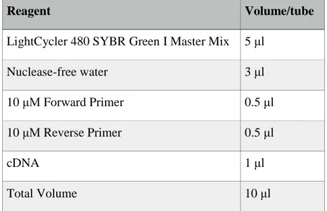
2.2.7.2 cDNA Synthesis ... 34
2.2.7.3 qPCR ... 34
2.2.8 ROC Curve Generation ... 36
2.2.9 Statistical Analyses ... 36
... 38
Results ... 38
3.1 Characterization of PC-3 and Urinary Exosomes ... 38
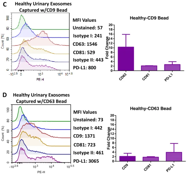
3.1.1 Surface Protein Characterization ... 38
3.1.2 Size Distribution of Exosomes ... 41
3.2 Determination of miRNA Levels in PC-3 Exosomes ... 45
3.3 Determination of miRNA Levels in Prostate Cancer Patients’ Exosomes ... 48
3.3.1 Cohort Profile in This Study ... 48
3.3.2 miRNA Levels in Patients’ Urinary Exosomes ... 49
3.3.3 Diagnostic Ability of Candidate miRNAs ... 54
3.3.4 miRNA Levels in Urinary Exosomes of Patients with Different PSA Levels ... 56
3.3.5 Diagnostic Ability of Candidate miRNAs for Patients with PSA Level Lower Than 10ng/ml……… ………65
xi
... 67
Discussion ... 67
APPENDIX ... 76
Appendix A.1- Cell Culture Solutions ... 76
Appendix A.2- Flow Cytometry ... 76
References ... 78
xii
List of Figures
Figure 1.1 Biogenesis of exosomes ... 6
Figure 1.2 Exosome biogenesis. ... 7
Figure 1.3 Canonical pathway of microRNA biogenesis ... 11
Figure 1.4 Mechanisms of tumor suppressor and oncogenic miRNAs ... 12
Figure 1.5 Roles of miRNAs in prostate cancer development. ... 14
Figure 2.1 Representative figure of chambers of hemocytometer. Cells within the chambers designated with red lines are counted in order to determine the cell number. ... 29
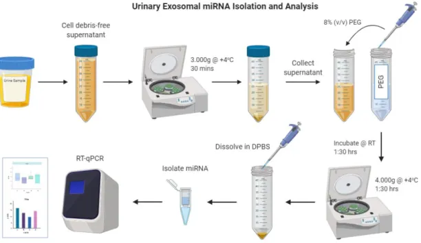
Figure 2.2 General scheme summarizing isolation and analysis of the urinary exosomes. ... 36
Figure 3.1 Presence of exosome-specific markers and PD-L1 in PC-3 exosomes and healthy urinary exosomes. ... 40
Figure 3.2 Size distributions of PC-3 exosomes, healthy and patient urinary exosomes. ... 42
Figure 3.3 . Representative Transmission Electron Microscopy (TEM) images of PC-3, healthy urinary and patient urinary exosomes. ... 43
Figure 3.4 Expressions relative to U6 snRNA of tumor suppressor microRNAs in healthy control urinary exosomes and PC-3 exosomes. ... 46
Figure 3.5 Expressions relative to U6 snRNA of oncogenic microRNAs in healthy control urinary exosomes and PC-3 exosomes. ... 47
Figure 3.6 Age-PSA and PSA-Gleason Score Correlations of the patients. ... 49
Figure 3.7 Expressions relative to U6 snRNA of tumor suppressor miRNAs in healthy control urinary exosomes and patient urinary exosomes... 51
Figure 3.8 Expressions relative to U6 snRNA of oncogenic miRNAs in healthy control urinary exosomes and patient urinary exosomes. ... 52
Figure 3.9 Fold changes of relative expression levels of candidate miRNAs for PC-3 and patients’ urinary exosomes. ... 53
Figure 3.10 Diagnostic ability of candidate miRNAs. ... 55
Figure 3.11 Expressions relative to U6 snRNA of tumor suppressor miRNAs in healthy control urinary exosomes and exosomes of patients with PSA levels lower than 10 ng/ml. ... 57
Figure 3.12 Expressions relative to U6 snRNA of oncogenic miRNAs in healthy control urinary exosomes and exosomes of patients with PSA levels lower than 10 ng/ml. ... 58
Figure 3.13 Expressions relative to U6 snRNA of tumor suppressor miRNAs in healthy control urinary exosomes and exosomes of patients with PSA levels between 10-15 ng/ml. ... 59
Figure 3.14 Expressions relative to U6 snRNA of oncogenic miRNAs in healthy control urinary exosomes and exosomes of patients with PSA levels between 10-15 ng/ml. ... 60
xiii
Figure 3.15 Expressions relative to U6 snRNA of tumor suppressor miRNAs in healthy control urinary exosomes and exosomes of patients with PSA levels higher than 15 ng/ml. ... 61 Figure 3.16 Expressions relative to U6 snRNA of oncogenic miRNAs in healthy control urinary exosomes and exosomes of patients with PSA levels higher than 15 ng/ml ... 62 Figure 3.17 Fold changes of relative expression levels of candidate miRNAs for prostate cancer patients with different PSA levels. ... 64 Figure 3.18 Diagnostic ability of candidate miRNAs in patients with PSA<10 ng/ml. ... 66
xiv
List of Tables
Table 2.1 Purified human-specific antibodies; their catalog numbers and clones. ... 25
Table 2.2 Fluorochrome-conjugated human-specific antibodies; their catalog numbers and clones. ... 25
Table 2.3 RT-PCR primer list ... 26
Table 2.4 qPCR primer list ... 27
Table 2.5 Reagents and volumes of them used in qPCR. ... 35
Table 2.6 qPCR conditions used in the experiments. ... 35
Table 3.1 Exosome diameters (in width and length scales) of PC-3 exosomes, healthy and patient urinary exosomes according to TEM images. ... 44
xv
Abbreviations
AGO2 Argonaute 2
AKT Protein Kinase B
ALIX ALG-2 Interacting Protein X
ANOVA Analysis of Variance
APAF1 Apoptotic Peptidase Activating Factor 1
AR Androgen Receptor
ASC Adipose-Derived Stromal Cells
AUC Area Under Curve
BCA Bicinchoninic Acid
BCL-XL B-cell Lymphoma-Extra Large
BCL-2 B-Cell Lymphoma 2
BSA Bovine Serum Albumin
CD Cluster of Differentiation
CDK Cyclin-Dependent Kinase
CT Cycle Threshold
CTL Cytotoxic T-Lymphocyte
CXCR4 Chemokine Receptor Type 4
DC Dendritic Cell
DGCR8 DiGeorge Syndrome Critical Region 8
DLS Dynamic Light Scattering
DNA Deoxyribonucleic Acid
xvi
DPBS Dulbecco’s Phosphate Buffered Saline
ERG ETS-Related Gene
ERSPC European Randomized Study of Screening for Prostate Cancer
ESCC Esophageal Squamous Cell Carcinoma
ESCRT Endosomal Sorting Complexes Required for
Transport
EV Extracellular Vesicle
FACS Fluorescence-activated Cell Sorting
FBS Fetal Bovine Serum
FDA US Food and Drug Administration
FOXD1 Forkhead Box D1
GS Gleason Score GTP Guanosine Triphosphate HCV Hepatitis C Virus HEPES 4-(2-Hydroxyethyl)-1-Piperazineethanesulfonic Acid IL Interleukin
ILV Intraluminal Vesicle
LNCaP Lymph Node Carcinoma of the Prostate
MEM Minimum Essential Medium
MET Hepatocyte Growth Factor Receptor (HGFR)
MFI Mean Fluorescence Intensity
MIF Macrophage Migration Inhibitory Factor
xvii
mRNA Messenger RNA
miRNA microRNA
MTDH Metadherin
mTOR Mammalian Target of Rapamycin
MV Microvesicle
MVB Multivesicular Bodies
NEAA Non-Essential Amino Acid
NF-κB Nuclear Factor Kappa B
NIH National Institute of Health
OncomiR Oncogenic microRNA
OSCC Oral Squamous Cell Carcinoma
PBS Phosphate Buffered Saline
PC-3 Prostate Cancer Cell Line
PCA-3 Prostate cancer gene 3
RT-qPCR Quantitative Reverse Transcription PCR
PCa Prostate Cancer
PDCD4 Programmed Cell Death Protein 4
PD-L1 Programmed Death-Ligand 1
PE Phycoerythrin
PEG Polyethylene Glycol
Pre-miRNA Precursor microRNA
Pri-miRNA Primary microRNA
PSA Prostate-Specific Antigen
xviii
PTEN Phosphatase and tensin homolog
RICTOR Rapamycin-Insensitive Companion of Mammalian Target of Rapamycin
RISC RNA-Induced Silencing Complex
RNA Ribonucleic Acid
RNase Ribonuclease
ROC Receiver Operating Characteristic RPMI Roswell Park Memorial Institute
RT Room Temperature
SENP1 Sentrin Specific Protease
SIRT1 Sirtuin 1
SIX1 Sineoculis Homeobox Homolog 1
snRNA Small Nuclear RNA
SOX5 SRY-Box Transcription Factor 5
SPSS Statistical Package for the Social Sciences
SYBR Synergy Brands
TEM Transmission Elctron Microscopy
TMPRSS2 Transmembrane Protease, Serine 2
TP53 Tumor Protein P53
TRBP RNA Binding Protein
TSPAN8 Tetraspanin 8
UA Uranyl Acetate
1
Chapter 1
Introduction
1.1 Cancer
Cancer is caused by abnormal cell growth and division and it is the second leading cause of death in 2018 [1]. There are many reasons of cancer and some of them are gene mutations, age, cellular stress (e.g. oxidative stress), diet and environmental factors such as radiation [2]. Although there are many checkpoints for regulation of cell division during cell cycle, when there are mutations in genes which regulate actions of these checkpoints, cell division continues in an uncontrolled manner and leads to tumor formation. One of the most important checkpoint is G1 checkpoint which is controlled by p53 gene so mutation in this gene cause tumorigenesis [3].
Several cancer subtypes have very high incidence and mortality rates. There were 18 million new cancer cases and 9.6 million deaths in 2018 worldwide [4]. In addition to its high incidence rate, cancer has high costs for the patients as well as governments. About $87.8 billion was spent for cancer-related health services in U.S. Moreover, patients paid roughly $4 billion for their cancer treatments [5]. These facts make cancer researches important and vital. Thus, American Cancer Society has funded $420.2 million for cancer researches in 2020 [6].
1.1.1 Prostate Cancer
Prostate cancer is one of the major cancer types that cause mortality and it is the second most incident cancer type in men worldwide. According to World Health Organization (WHO), prostate cancer is the second most diagnosed cancer type in men with 1.3 million diagnoses in 2018 and the fourth most diagnosed cancer type in total sex-independent cases [7]. Prostate cancer is classified based on factors such as aggressiveness, grade of the cancer and age of the patient. Mainly, prostate cancer classes can be listed as aggressive and nonaggressive prostate cancer (PCa), high and low-grade PCa, early onset
2
and indolent PCa (occurring after age of 55) [8]. The class and grade of the prostate cancer has a big role in prognosis of the disease and mortality because patients who are diagnosed in early stages have higher survival rates.
Development of prostate cancer is multifactorial which means that it is dependent on more than one condition; both genetic and environmental factors have essential roles as well as diet and age. According to American Cancer Society, 6 in 10 prostate cancer patients are 65 or older. Additionally, average diagnosis age is 66 and cases under age of 40 are rare [9]. Correlation of prostate cancer with age can be described with change in testosterone:oestrogen ratio with age. In men, serum testosterone level declines as the age gets older but oestrogen level is constant. It causes a change in testosterone:oestrogen ratio which is supposed to be one of the main reasons of prostate cancer. Testosterone is an androgen which binds to androgen receptors (AR). As testosterone level decreases, AR level increases to stabilize the AR-dependent signaling which causes DNA damage and development of prostate cancer consequently [10]. In addition to this, environmental factors such as radiation and cellular stress cause cells to divide in an uncontrolled manner and may cause prostate cancer.
1.1.1.1 Diagnosis of Prostate Cancer
Diagnosis of prostate cancer, especially early detection of it, is important for good prognosis of the disease. In addition, not only late diagnosis causes mortality but also overdiagnosis, which means diagnosis of a person as prostate cancer when he is healthy, and overtreatment; treatment of an individual as a result of screening whereas he would not have any symptom without treatment have huge impact [11]. Serum Prostate Specific Antigen (PSA) level and biopsy are currently used for diagnosis of prostate cancer. First step in diagnosis is checking the PSA level if it is out of the normal/expected range. Secondly, biopsy is performed if PSA level is out of range and grade of the cancer is identified. However, for some patients despite the fact that they have high PSA levels, the biopsy results show that there is no tumor in prostate or benign hyperplasia is detected so the person with high PSA level comes out to be healthy. This is because PSA level can change depending on many factors in addition to cancer so it can be misleading [12].
3
1.1.1.1.1 Prostate-Specific Antigen (PSA)
Prostate-specific antigen (PSA) is a protein, kallikrein‐like serine protease, produced by both normal and malignant prostate glands. This protein is produced by epithelial cells of prostate so it can be said that PSA is an organ-specific marker. On the other hand, production of PSA is dependent on many factors in addition to prostate cancer, so it is not a cancer-specific marker [13]. Besides, normal PSA level for healthy controls is not clear since it can fluctuate over time. Prostatitis, which is inflammation of prostate gland and urinary tract infection are diseases that increase PSA levels. Even biopsies are found to increase PSA level. These situations can lead to PSA level increase for reasons other than cancer and overdiagnosis of cases. On the other hand, benign prostate hyperplasia drugs such as finasteride and dutasteride cause decrease in PSA level and may result in misdiagnosis of prostate cancer. PSA level below 4 ng/ml is accepted as in normal range and above 4 ng/ml is considered as suspicious but biopsies are needed for the final decision [14].
There are many researches supporting that there are significantly many patients which are either misdiagnosed or overdiagnosed for prostate cancer and they have reduced life quality followed by high mortality. In one study, overdiagnosis of prostate cancer is investigated and it is found that 67% of total diagnosis cases is overdiagnosed [15]. In another study, when European Randomized Study of Screening for Prostate Cancer (ERSPC) results were evaluated, 4.1% of patients were found to be overtreated and the researchers conclude that PSA testing in Europe was associated with decrease in the life time of patients so it was not recommended for diagnosis [16].
1.1.1.1.2 Biopsy and Gleason Score
The second step in the diagnosis of prostate cancer after PSA test is identification of the prostate cancer grade by biopsy. Gleason Score is the score that pathologists give to the tumor parts obtained from 2 different sites. They decide on this according to how much tumor looks like a healthy tissue, so how much it is differentiated [17]. Gleason Score (GS) for each tumor part ranges from 1 to 5 and they are added for finding the GS of 2
4
sites. Gleason Score 1 is very similar to healthy tissues so that well differentiated whereas 5 is very poorly differentiated. Gleason scores can be 6 (3+3), 7 (3+4) or (4+3), 8 (4+4), 9 (4+5 or 5+4) and 10 (5+5) for 2 tumor sites. It is the most commonly used grading system for prostate cancer and score under 6 means the tissue is so similar to healthy tissue whereas score higher than 7 means that the cells are poorly differentiated and they look very different from healthy cells. After Gleason Scores for 2 tumor sites are summed up, they are graded according to the Gleason Grading System. The grading system for prostate cancer is as the following; Grade 1 if Gleason Score is 6, Grade 2 if Gleason Score is 7 (3+4), Grade 3 if Gleason Score is 7 (4+3), Grade 4 if Gleason Score is 8 (4+4) and Grade 5 if Gleason Score is 9 or 10. Gleason Grading is important for both diagnosis and prognosis of prostate cancer [18].
1.1.1.2 Therapy of Prostate Cancer
There are main therapy options in prostate cancer; radical prostatectomy, radical radiotherapy, conservative management, immunotherapy, hormonal therapy and chemotherapy (docetaxel). Radical prostatectomy means removing the tumor from the body by surgery. On the other hand, radical radiotherapy is the method that high-energy X-rays are used and it is a non-surgical therapy. On the other hand, conservative management is watching the disease progression and healing the symptoms rather than treating it [19].
Immunotherapy has been one of the best choices for therapy of prostate cancer in some patients and its efficiency depends on the cancer stage and history of the patient. Prostate has many tumor-associated antigens which can be targeted for vaccines. This is the reason that prostate cancer is a good cancer type for developing therapeutic cancer vaccines. In addition, development of prostate cancer is relatively slow so there is time for development of anti-tumor immune response in the body which is an advantage for cleaning the tumor. However, prostate cancer cells can prevent immune response by making the antigen presentation inefficient by downregulating human leukocyte antigen class I [20].
Sipuleucel-T (Dendreon Corp.) is the therapeutic vaccine approved by the US Food and Drug Administration (FDA). Phase III researches show that it is most efficient on early
5
prostate cancer because there was an increase in overall survival of patients with early prostate cancer. The mechanism of this therapeutic vaccine is not understood completely and it is known that tumor sizes or PSA levels are not affected but overall survival increases. For high-risk prostate cancer patients, combinational therapies such as chemotherapy and hormonal therapy (enzulatamid) combinations are applied frequently [21].
1.2. Extracellular Vesicles
Extracellular vesicles are vehicles which facilitate the communication between cells and regulate many biological mechanisms. Since they carry nucleic acids, lipids and proteins, they have been important for investigation of intercellular communication in both prokaryotes and eukaryotes. While some of these physiological pathways are understood, some of them are still unclear. Extracellular vesicles can be isolated from many types of biological fluids such as urine, plasma, saliva, breast milk and amniotic fluid. According to their sizes, EVs are divided into two main groups; microvesicles and exosomes [22]. Sizes of microvesicles can range from 100 nm to 1000 nm whereas exosomes are the smallest extracellular vesicle (EVs) with size of 30 nm-150 nm. Exosomes can be secreted from a cell as a part of normal conditions or as a result of a stimuli. For this reason, they are known to have roles in development and progression of many diseases including cancer [23]. In normal conditions, cargos of EVs, is very susceptible to degradation in the extracellular space if they were not encapsulated in EVs. For example, RNAs are one of the most sensitive molecules due to presence of RNase. However, EVs prevent degradation of RNAs and this characteristic of EVs makes them effective targets for therapeutic and diagnostic applications which will be outlined in Sections 1.3.2 and 1.3.3 [24].
1.2.1 Biogenesis of Exosomes
Exosomes are originated in endosomes and they firstly arise as intraluminal vesicles (ILVs) in multivesicular bodies (MVB); so that late endosomes. Destination of ILVs depends on fusion of MVB with lysosomal compartment. If a MVB fuses with lysosomal
6
compartment, its contents will be degraded whereas other MVBs will fuse with plasma membrane and result in exosome secretion. Endosomal V0V1-ATPase and some GTPases such as Rab GTPases are shown to have role in fusion with plasma membrane [25]. Contents of endosomes are known to be internalized by endocytosis into the MVBs and either degraded by lysosomes or secreted inside exosomes by exocytosis. (see Figure 1.1)
Figure 1.1 Biogenesis of exosomes [26].
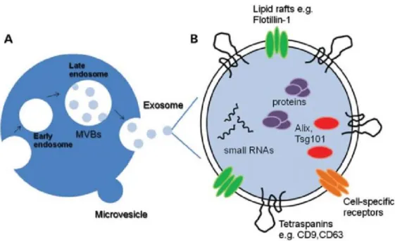
In exosome biogenesis, proteins in ESCRT (endosomal sorting complexes required for transport), lipid rafts enriched in ceramide, tetraspanin-enriched microdomains and ALIX, syntenin and syndecans also have essential roles. ALIX, syntenin and syndecans form a complex, link with ESCRT system and facilitate the endocytosis of nucleic acids and proteins into the exosomes [27]. CD9, CD63 and CD81 are tetraspanins found on endosomal membrane. The first biological event occurring on endosomal membrane is enrichment of the membrane with these tetraspanins in formation of ILVs (see Figure 1.2). They are also the surface markers of exosomes which can be used in characterization of exosomes. In comparison to exosomes, microvesicles (MVs) do not follow the endosomal pathway and they directly bud off from the plasma membrane [28].
7
Figure 1.2 Exosome biogenesis. (A) Comparison of microvesicle and exosome biogenesis. (B) Representation of common exosomal markers [29].
1.2.2 Exosomes in Health and Disease
Exosomes circulate in the body fluids and change activity and phenotype of recipient cells. They have important roles in diseases including autoimmune diseases, inflammation, cardiovascular diseases and metabolic disorders [30-32]. Exosomes also involve in viral infections. For instance, non-enveloped virions were detected in exosomes of Hepatitis C Virus (HCV)-infected cells and these virions were shown to be entering target cells [33]. Obesity was also shown to be related with exosomes and exosome number was found to be increasing in obesity [34]. Cancer is another disease that exosomes regulate. In cancer, tumorigenesis leads to increased exosome secretion [35]. Moreover, exosomes involve in tumor growth tumor microenvironment remodeling, cell migration and immune evasion [36-39].
8
1.2.3 Diagnostic Applications of Exosomes in Cancer
When exosomes were first recognized, they were defined as ‘trash bags’. However, their roles in intracellular communication has been investigated and it is shown that contents of exosomes can be good targets for diagnosis of diseases including cancer because they carry nucleic acids, proteins and lipids which give important information about the cells that they are released from. Biological fluids are the places that exosomes accumulate so by isolation and investigation of exosomes, diagnostic markers can be discovered as well as markers of disease progression [40].
Pancreatic cancer is one of the cancer types that exosomes are used for diagnosis and determination of metastasis. Macrophage migration inhibitory factor (MIF) was suggested to be upregulated in exosomes of pancreatic cancer patients. Also, surface proteins CD44v, CD44, MET, Tspan8 and CD133 which are the markers of pancreatic cancer initiating cells were detected in pancreatic cell originated exosomes [41]. Another cancer type with high mortal rate; lung cancer has also potential of being diagnosed by exosomal contents. Tumor cell-derived exosomes promote angiogenesis, invasion and proliferation in lung cancer. According to the studies made with healthy controls and lung cancer patients, candidate exosomal microRNAs (miRNAs) for early diagnosis of lung cancer were detected [42]. Additionally, exosomes were also shown to have role in apoptosis of tumor cells in breast cancer [43].
Since exosomes can protect miRNAs against RNase degradation and they regulate the expression of genes, exosomal miRNAs are one of the diagnostic markers of cancer. These miRNAs regulate the expression of genes playing role in pathways such as angiogenesis and apoptosis. miR-34a, miR-182 and miR-375 are some of the candidate miRNAs for diagnosis of prostate cancer [44]. In addition to miRNAs, some mRNA biomarkers from urine were also detected in prostate cancer such as PCA-3 and TMPRSS2:ERG [45]. However, there is no exosomal diagnosis kit used for prostate cancer so that overdiagnosis of prostate cancer cases is still ongoing as detailed in Section 1.1.1.
9
1.2.4 Therapeutic Applications of Exosomes in Cancer
Exosomes are efficient therapeutic agents for many diseases including pulmonary hypertension, diabetes and cancer [46]. Effects of exosomal therapy has been researched for various diseases and about 90 exosomal therapy studies are in clinical trial phases in 2020 according to NIH Clinical Trials database [47]. Exosomal therapy in cancer is one of the most studied field due to high incidence and mortality rates of cancer. In mouse models, Zitvogel et al showed that exosomes derived from tumor antigen peptide-stimulated DCs primed tumor-specific cytotoxic T-lymphocytes (CTL) and resulted in delay of tumor growth and tumor elimination [48]. This research was one of the leading studies about exosomal therapy and many similar ones have been conducted for different cancer subtypes.
Delivery of cargos to the target cells by loading of this cargo into exosomes has been one of the most preferred ways of exosomal therapy. Let-7a miRNA was loaded into exosomes of embryonic kidney cells and reduction in size of breast cancer cells was observed [49]. Additionally, Phosphatase and tensin homolog (PTEN) mRNA was loaded into mouse embryonic fibroblasts and glioma cell proliferation decreased [50]. Another example of exosomal therapies in cancer subtypes was reduction of the activity of Bcl-xL by adipose-derived stromal cells (ASCs) derived exosomal miR-145 and induction of prostate cancer cell apoptosis [51]. In addition to RNA delivery via exosomes, chemical drugs were also used as chemotherapeutic agents. For prostate cancer therapy, exosomes were loaded with Paclitaxel and decrease in prostate cancer cell growth was shown [52].
1.3 microRNAs
microRNAs are small, single-stranded, non-coding RNAs which are 22-25 nucleotides long [53]. They target mRNAs and either cleave or deadenylate the mRNAs or repress the translation [54]. By these ways, they facilitate gene silencing and they are one of the main regulators of biological processes. They have role in every cellular activity and are very important in maintaining cell homeostasis, differentiation and development [55]. They also play a vital role in development of many diseases including cancers,
10
autoimmune diseases such as rheumatoid arthritis, cardiovascular diseases, neurological disorders and liver diseases [56].
1.3.1 Biogenesis of microRNAs
There are 2 types of miRNAs; intragenic and intergenic miRNAs. Half of the currently discovered miRNAs are intergenic which means that they are found and processed from either introns or exons of the gene; but mostly from the introns. The other half of the miRNAs are intergenic and they are transcribed from a different gene regulated by a different promoter [57]. The most common pathway for biogenesis of miRNAs is the canonical pathway. In the canonical pathway, primary microRNAs (pri-miRNAs) transcribed by RNA Polymerase II are capped and polyadenylated. Mature miRNA sequences are found in the stem of a hairpin on the pri-miRNA [58]. Then, they are processed to single hairpins; pre-miRNAs, by the microprocessor complex. Microprocessor complex consists of two components; DiGeorge Syndrome Critical Region 8 (DGCR8) and Drosha which is a ribonuclease III enzyme. Pre-miRNAs are transported to the cytoplasm by Exportin 5/Ran GTP complex and the complex cleaved by RNase III endonuclease, Dicer and RNA Binding Protein, TRBP [59]. The length of the miRNA is the mature miRNA length after this step. Argonaute (Ago2) proteins load functional strand of the miRNA into the RNA-induced silencing complex (RISC) and lead it to the target mRNA (see Figure 1.3) [60]. Other biogenesis pathways are Drosha-independent and Dicer-Drosha-independent pathways but they are not as common as the canonical pathway.
11
Figure 1.3 Canonical pathway of microRNA biogenesis [61].
1.3.2 microRNAs and Cancer
MicroRNAs are shown to be good biomarkers and therapeutic agents for cancer [62]. Besides, dysregulation of some miRNAs are found to be the key player in progression of several cancer types. Cellular processes such as proliferation, growth, cell death, activation of invasion and metastasis are regulated by microRNAs [63]. miRNAs can function as oncogenes or tumor-suppressors. If a miRNA functions as an oncomiR, it targets a tumor suppressor gene and decreases its expression for inducing cell proliferation and repressing apoptosis. On the other hand, if it acts as a tumor suppressor, it targets an oncogene and reduces its expression [64]. The first study that investigated role of microRNAs in human cancers is from Calin et al. They revealed that miR-15 and miR-16 were downregulated in chronic lymphocytic leukemia [65]. After this study in 2002, many researches about miRNAs as cancer hallmarks have been conducted.
12
Studying microRNAs in cancer researches for diagnostic or therapeutic reasons has high potential and many advantages. First of all, microRNAs can be isolated from many biological fluids such as blood and urine, saliva which makes the noninvasive biomarker testing methods possible [66]. Secondly, miRNAs are very stable due to their small size so they make the technically challenging samples such as formalin fixed samples easier [67]. Additionally, microRNAs are found free in the circulation as well as encapsulated in extracellular vesicles. EV miRNAs give more information about tumor such as the tumor microenvironment. Moreover, microRNAs serve as biomarkers even for the grade of cancers. Finally, miRNAs are tissue-specific so that every tumor has different microRNA profile depending on the tissue type which make the profile specific for each tissue and more reliable for comparison with healthy profile [68].
13
1.3.2.1 microRNAs as Cancer Biomarkers
Tumor-suppressor and oncogenic properties of microRNAs make them good biomarker candidates. Differential expression of miRNAs results in changes of gene expression. There are hundreds of different targets for miRNAs and miRNA expression differs in each tissue; which means that different cancer types have different miRNA profiles [70]. Especially due to the advances in high-throughput studies in last years, miRNA biomarker panel researches for almost every cancer type has increased tremendously [71].
There are many promising researches and evidences about discovery of miRNA biomarkers for several cancers by examination of blood specimens. Heneghan and colleagues conducted a study for determining miRNA biomarkers of breast cancer. They reported that 195 expression was increased in cancer cohort [72]. Moreover, miR-215, miR-299-5p, and miR-411 expression levels was found to be downregulated by van Schooneveld et al [73]. Additionally, Konishi et al revealed that miR-21 concentration was significantly higher and miR-375 expression was significantly lower in esophageal squamous cell carcinoma when compared to healthy controls [74]. Besides, miR-486 was found to be a potential biomarker for non-small cell lung cancer and its expression was significantly increased in patients [75]. For gastric cancer, miR-204 level was significantly lower in the cancer cohort when compared to healthy donors [76]. Further, it has been documented that miR-145 levels were lower than the healthy controls as well as miR-204-5p levels [77, 78].
Although miRNA biomarker discovery from blood is more common, urinary biomarker researches gathered speed because it is a non-invasive method and has high potential for being used as biomarker specimen. Erbes et al stated that 21-5p, 125b-5p, miR-155-5p, and miR-451-5p were differentially expressed in urine of breast cancer patients [79]. Indeed, in bladder cancer patients, it was observed that miR-10b and miR-34b were upregulated compared to controls [80]. Furthermore, Andreu et al revealed that miR-375 level was significantly lower in high-grade bladder cancer patients. On the other hand, miR-146a was significantly upregulated in low-grade bladder cancer patients [81]. For prostate cancer, a study pointed up diagnostic power of miR-222-3p*miR-24-3p/miR-30c-5p biomarkers in cell free urine for prostate cancer diagnosis [82].
14
1.3.2.2 microRNAs in Prostate Cancer Diagnosis
Prostate cancer has been one of the most studied cancer subtype for biomarker discovery due to its high prevalence and mortality rate. When low reliability of PSA testing is also taken into consideration, increase in researches about this field is inevitable [83]. Huge progress has been made in miRNA biomarker in recent years and promising evidences showed that microRNAs are strong biomarker candidates for prostate cancer diagnosis just like other cancer subtypes [84]. By loss-of-function and gain-of-function experiments in human cancer cells, mouse xenografts, transgenic mouse models and knockout mouse models, it was shown that miRNAs have essential roles in tumor formation and prognosis of cancer [85]. Besides, advances in technology led to improvement in studies with high number of prostate cancer patient and healthy volunteers so number of researches which are statistically more reliable increased. Several miRNA profiling studies have been conducted for prostate cancer as well as high throughput profiling analysis in last 15 years [86-89]. Figure 1.5 shows the mechanism of actions of several miRNAs in prostate cancer.
15
1.3.2.2.1 Tumor Suppressor microRNAs
Tumor suppressor microRNAs have oncogene targets and they reduce cancer cell proliferation, inhibit metastasis and increase survival. In cancers, downregulation of tumor-suppressor miRNAs is expected [91]. When taken into account that locations of more than 50% of miRNA genes are in cancer-associated genomic regions or in fragile sites, tumor-suppressor microRNA studies have gained speed for diagnosis and treatment of cancer [64]. In prostate cancer, miR-145 was detected as a tumor-suppressor miRNA which targets SENP1 (Sentrin Specific Protease) gene and causes growth arrest in PC-3 cell line [92]. Besides, Guan et al revealed that miR-218 targets RICTOR which is the component of mTOR pathway and prevents angiogenesis in prostate cancer [93]. miR-152 is another tumor suppressor miRNA which binds to 3’-UTR of DNA (cytosine-5)-methyltransferase 1 (DNMT1) and decreases its expression level which might be responsible for aggressiveness of prostate cancer [94]. In our study, expression of 5 oncomiRs; miR-30a-5p, miR-107, miR-139-5p, miR-145-5p and miR-204-5p were investigated and their functions are explained in more detail in the following sections.
1.3.2.2.1.1 hsa-miR-30a-5p
One of the candidate tumor suppressor miRNAs in prostate cancer is hsa-miR-30a-5p which is found on Chromosome 6 [95]. In general, this miRNA is downregulated in many cancer subtypes which means it functions as a tumor suppressor miRNA [96]. One of its targets is SIX1, a gene which has role in development and inhibits cell apoptosis in organ development [97]. Other examples of miR-30a-5p targets are MYBL2, FOXD1, and SOX4 which also induce cell proliferation [98].
Downregulation of miR-30a in prostate cancer patients was shown by Xu et al [99]. This finding was supported by the research conducted by Zhao et al and Zhu et al [97]. When exosomal hsa-miR-30a-5p expression was examined, it was found to be responsible for development of some cancer subtypes such as ovarian cancer and oral squamous carcinoma [100, 101]. However, role of exosomal miR-30a, especially urinary exosomal miR-30a dysregulation, in prostate cancer is yet to be understood.
16
1.3.2.2.1.2 hsa-miR-107
hsa-miR-107 is found on Chromosome 10 and it has only one mature sequence [102]. miR-107 plays a vital role in many cancers including colorectal cancer, gastric cancer, pancreatic cancer and hepatocellular carcinoma. miR-107 was found to be having a dual role in cancer; so it can be a tumor suppressor in some cancer types whereas it was upregulated and functioning as an oncomiR in other cancers. For instance, although its overexpression was shown to induce migration and invasion in hepatocellular carcinoma, it had a tumor suppressor effect on colorectal, pancreatic cancer and non-small cell lung cancers [103-106].
There is a contradiction about dysregulation of hsa-miR-107 in prostate cancer. There are researches which claim it can act as an oncomiR whereas some studies demonstrate tumor suppressor role of miR-107 in prostate cancer. Zhang et al showed that miR-107 targeted Cyclin E1 and represses cell proliferation in prostate cancer so it acted as a tumor suppressor [107]. However, Lodes et al stated that miR-107 was upregulated in prostate cancer patients compared to healthy controls [108]. The diagnostic ability of miR-107 in prostate cancer is not well studied and needs to be further investigated.
1.3.2.2.1.3 hsa-miR-139-5p
hsa-miR-139-5p is a 23 nucleotides long miRNA found on Chromosome 11 [109]. Its tumor suppressor effects have been studied in many researches of different cancer subtypes including breast cancer, parathyroid carcinoma and esophageal squamous cell carcinoma (ESCC). These studies revealed that hsa-miR-139-5p was downregulated in cancer [110-112].
In prostate cancer, miR-139 was also found to be behaving as a tumor suppressor. In the study which aims identification of proliferation inhibitory mechanism of hsa-miR-139-5p in LNCaP prostate cancer cell line, it was shown that cyclin D1 level was decreased by miR-139 activity so miR-139 inhibits cell proliferation by targeting Notch1 and interferes with the cell cycle [113]. Similarly, in another study, Nom et al indicated that growth inhibition effect of hsa-miR-139-5p in prostate cancer was due to downregulation of activated AKT and cyclin D1 and upregulation of the p21 which is a CDK inhibitor [114]. Progression of prostate cancer was found to be regulated by miR-139 too. In
17
prostate cancer, miR-139 was found to inhibit SOX5 expression and TWIST was downregulated. By this way, miR-139 repressed Epithelial-Mesenchymal Transition [115]. In the literature, there is no research about the role of exosomal hsa-miR-139-5p in prostate cancer although there are many researches which support the tumor suppressor effects of it on prostate cancer cells.
1.3.2.2.1.4 hsa-miR-145-5p
hsa-miR-145 is a miRNA found on the Chromosome 5 [116]. Almost all studies aiming determination of the role of hsa-miR-145-5p in tumorigenesis and progression of several different cancers ended up with the same conclusion; miR-145-5p was downregulated in cancer and it was a future-promising biomarker candidate. In breast cancer cells, miR-145 was downregulated and acted as a tumor suppressor [117]. Likewise, miR-miR-145 level decrease in ovarian cancer was reported by Hang et al and they claimed downregulation of miR-145-5p in cancer cells and also their derived exosomes leads to ovarian cancer development [118].
Effects of miR-145-5p in prostate cancer is similar to other cancer types. In LNCaP prostate cancer cells, overexpression of miR-145-5p resulted in inhibition of proliferation and invasion and also facilitated early apoptosis of the cells [119]. In addition, Pan et al found that miR-145-5p targeted Metadherin (MTDH) in prostate cancer cells. This gene was shown to be upregulated in several tumor cells and it was responsible from proliferation, and migration of tumor cells [120]. In another study, overexpression of miR-145–5p caused SOX2 repression and inhibition proliferation of prostate cancer cells [121]. There are a few researches about exosomal miRNA in different cancer subtypes. However, there is no research about correlation of urinary exosomal miR-145-5p and prostate cancer until today.
18
1.3.2.2.1.5 hsa-miR-204-5p
miR-204-5p is the last tumor suppressor miRNA candidate in this study and it is found on the Chromosome 9 [122]. In the literature, there are evidences about tumor suppressor effects of miR-204-5p in different cancer types. Overexpression of miR-204-5p caused repression of proliferation, migration and viability in human and murine breast cancer cells [123]. Similarly, the expression of miR-204-5p was lower in oral squamous cell carcinoma (OSCC) cells and it was claimed that miR-204-5p targeted chemokine receptor type 4 CXCR4 [124]. Moreover, miR-204-5p was shown to be a good biomarker candidate for non-small cell lung cancer [125].
Role of hsa-miR-204-5p has been investigated and its tumor suppressor activities and its mechanisms have been reported. In prostate cancer cells, apoptosis was induced by miR-204-5p. In the same study, researchers clarified the mechanism of apoptosis inhibition by concluding that miR-204-5p was targeting BCL2 in these cells [77]. Besides, miR-204 targeted SIRT1/p53 pathway in doxorubicin-treated prostate cancer cells and enhanced mitochondrial apoptosis by this way [126]. Supporting this finding, Wu et al claimed that 204 expression level decreased in chemoresistant prostate cancer tissues [127]. miR-204-5p plays an important role in metastasis in prostate cancer as well as tumorigenesis. miR-204-5p was shown to be targeting NF-κB signaling in prostate cancer and repressing bone metastasis [128]. Diagnostic ability of urinary exosomal miR-204-5p in prostate cancer is yet to be identified.
1.3.2.2.2 Oncogenic microRNAs
Oncogenic microRNAs, also known as oncomiRs are key players in tumor formation, carcinogenesis and metastasis [129]. They generally target mRNA of tumor-suppressor genes and lead to RNA degradation or translational repression via binding to the 3′ untranslated region of these mRNAs [130]. miR-221/miR-222 are oncogenic microRNAs which have been examined by several studies and it was concluded that they facilitate cell cycle progression at G-S phase in prostate cancer [131-133]. Another common oncomiR cluster is MiR-106b/miR-25 which was found to be targeting caspase 7 mRNA [134, 135]. In addition, miR-18a and miR-32 were found to be candidate biomarkers for aggressive prostate cancer and they might be a target for drug development [136, 137].
19
Not only patient researches but also preliminary cell lines studies have vital role in biomarker discovery. One good example of these studies is by Seashols-Williams et al which includes high-throughput sequencing and miRNA panel analysis. According to the authors, miR-9 was an oncomiR validated by miRNA panel, RT-qPCR as well and high-throughput sequencing analysis. This study was conducted on tumorigenic and metastatic subline M12 as well as other prostate cancer cell lines DU-145, PC-3 and human P69 prostate cell line as a control [138]. In our study, expression levels of 3 oncomiRs; miR-21-5p, miR-375-5p and miR-574-3p were investigated and functions of these miRNAs are explained in the following sections.
1.3.2.2.2.1 hsa-miR-21-5p
miR-21 is one of the most frequently overexpressed microRNA in prostate cancer patients compared to healthy controls. This miRNA is located on the Chromosome 17 and has a length of 22 nucleotides [139]. Li et al stated that tissue samples obtained from prostate cancer patients had higher miR-21 expression than healthy controls and it was significantly associated with stage of the cancer, Gleason score and biochemical recurrence [140]. Moreover, another study made with PC-3 cell line revealed the mechanism of oncogenic property of miR-21. miR-21 targets and inhibits the expression of tumor suppressor gene PTEN and promotes cell division and cancer progression [141]. Metastasis is also related with miR-21. miR-21 attenuates expression of tumor suppressor PDCD4 (programmed cell death) which is an inhibitor of prometastatic factor. [142] In addition to researches made on blood samples for miR-21, urine is also a good specimen for diagnosis of prostate cancer. In prostate cancer patients’ urine samples, miR-21 was found to be upregulated in prostate cancer patients in comparison to benign prostate hyperplasia [143].
Exosomal miR-21 was also found to be overexpressed in prostate cancer. Agaoglu et al showed that miR-21 level was higher in serum exosomes of prostate cancer patients with metastasis compared to patients with localized tumor [144]. A study revealed that miR-21 suppresses APAF1 (apoptotic peptidase activating factor 1) and PDCD4 expression levels in order to to inhibit apoptosis [145]. Despite the fact that there are some researches about exosomal miR-21 dysregulation in prostate cancer, especially urinary miR-21
20
deregulation and its mechanism should be investigated in more detail in order to translate this biomarker into clinic.
1.3.2.2.2.2 hsa-miR-375-5p
Another oncomiR investigated in this study, hsa-miR-375-5p is found on Chromosome 2. Hsa-miR-375 has 2 mature strands; hsa-miR-375-5p and hsa-miR-375-3p [146]. Although hsa-miR-375-3p is more abundant than hsa-miR-375-5p, miR-375-5p was examined in this study. There are evidences that less abundant strand of miRNAs can also be functional and may have roles in diseases [147-149].
hsa-miR-375 was found to be dysregulated in many cancer types including prostate cancer [150-152]. miR-375 has dual role in cancer so it was shown to be acting as tumor suppressor as well as oncogenic miRNA in different cancer types such as liver cancer, breast cancer and colon cancer [153-155]. In prostate cancer, many researches supported the hypothesis that miR-375 was acting as an oncomiR and upregulating genes which promote cell proliferation. [156-158] Exosomal miR-375 was also studied in prostate cancer. Lie et al found that there was an increase in miR-375 in LNCaP cell-derived exosomes [159]. According to another study conducted by Zhan et al, exosomal miR-375 level in serum samples was also associated with metastasis in prostate cancer [160]. Diagnostic potential of urinary exosomal miR-375-5p is yet to be studied.
1.3.2.2.2.3 hsa-miR-574-3p
hsa-miR-574-3p which has length of 22 nucleotides is found on Chromosome 4 [161]. This microRNA was shown to have tumor-suppressor or oncogenic effects in different cancer subtypes such as osteosarcoma, ovarian cancer and breast cancer [162-164]. In prostate cancer, it functions as an oncogene according to both high-throughput and RT-qPCR results. In both serum and urinary cells of prostate cancer patients, miR-574-3p level was found to be significantly higher than healthy controls [165]. In serum samples of prostate cancer patients, fifteen miRNAs including miR-574-3p were found to be upregulated at the end of the microarray analysis. This result was complementary with
21
prostate cancer cell line 22Rv1 data [108]. miR-574-3p has important tumor suppressor targets including TP53 targets (TP53TG3, TP53TG3B, TP53TG3D) [166].
When extracellular vesicles of prostate cancer patients were investigated, miR-574-3p level was shown to be a good biomarker candidate in prostate cancer patients as a result of investigation of it in serum microvesicles [167]. miRNAs found in extracellular vesicles, especially in urinary exosomes, are not well studied in prostate cancer and need to be further examined.
22
1.4. Aims and Outline of the Study
Exosomes, which are the smallest members of extracellular vesicles with 30-150 nm dimeter size, carry microRNAs and dysregulation of these miRNAs play role in cancer development. Tumorigenesis, tumor microenvironment remodeling and metastasis are examples for effects of exosomal miRNAs. Studies with exosomal miRNAs isolated from several biological fluids such as blood and urine prove their efficiency in both diagnosis and therapy of different cancer types including prostate cancer. Prostate specific antigen (PSA) is the current biomarker used for diagnosis of prostate cancer. However, PSA level was shown to be altered due to many reasons in addition to prostate cancer so it can be misleading in several cases. Thus, a new biomarker research for prostate cancer research would be invaluable and it has become a necessity.
In this study, we focused on the diagnostic potential of urinary exosomal miRNAs in prostate cancer. The first aim of the study was investigation of candidate exosomal miRNA levels in PC-3 cell line which is a prostate cancer cell line and comparison of them with healthy urinary exosomal miRNA levels. The second aim of the study was to reveal differences of candidate miRNAs relative expressions in prostate cancer patient and healthy urinary exosomes. miRNA level changes in patients would make these miRNAs potential exosomal biomarkers for prostate cancer. Another aim of the study was to identify differential expression of candidate miRNAs in urinary exosomes of prostate cancer patients with different PSA levels and comparison of them between each other. Finally, we aimed to test the diagnostic ability of candidate urinary exosomal miRNAs and compare the efficiency of them when they were used as single and as combination for discrimination of patients and healthy controls.
For the purposes stated above, first of all, exosomes were isolated from supernatants of PC-3-cell lines by ultracentrifugation. Besides, morning urine specimens were collected from 14 healthy male individuals and urinary exosomes were isolated by precipitation with PEG. Exosome characterization was performed via flow cytometry, qNano, DLS and TEM. Then, miRNA was isolated from exosomes of PC-3 cell line and healthy controls. Finally, relative expressions of them were analyzed by RT-qPCR.
For the second purpose of the study, morning urine specimens were collected from 17 prostate cancer patients with their consent. Exosome isolation was performed with the same procedure of healthy urinary exosome isolation with PEG. Following miRNA
23
isolation and RT-qPCR, relative expressions of candidate miRNAs were calculated according to the efficiency-corrected CT method and expression levels of urinary exosomal miRNAs in prostate cancer patients and healthy controls were compared. Finally, in order to determine the diagnostic ability and diagnostic potential of miRNAs, ROC curve was generated for each miRNA and their different combinations and AUC values were analyzed as predictors as diagnostic potential of miRNAs.
24
Materials and Methods
2.1 Materials
2.1.1 Cell Culture Solutions
For preparation of cell culture media, RPMI1640 with phenol red (#21875-034), Sodium Pyruvate (100mM, #11360-070), MEM Non-Essential Amino Acids (100X, #11140-050), Penicillin-Streptomycin (10,000U/mL, #15140-122) were used from Gibco, Thermo Fisher Scientific (Waltham, MA, USA). HEPES Buffer (1M, #03-025-1B), FBS (#04-127-1A, Lot 1808243) was purchased from Biological Industries (Beit-Haemek, Israel). RPMI media was prepared by adding appropriate amount of FBS so that 10% of the total volume is FBS and Sodium Pyruvate, MEM Non-Essential Amino Acids (NEAA) , Penicillin-Streptomycin, HEPES were added to the media by diluting 1/100 each of them. 10X Trypsin without phenol red (2.5%, #15090-046) and DPBS (#14190-250) were also from Gibco, Thermo Fisher Scientific (Waltham, MA, USA).
2.1.2 Antibodies
In order to capture exosomes for exosome characterization purposes, purified exosome-specific antibodies were used. Purified human-exosome-specific antibodies used for exosome characterization were listed in Table 2.1
25
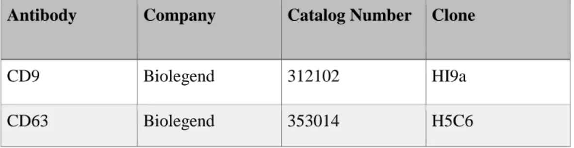
Table 2.1 Purified human-specific antibodies; their catalog numbers and clones.
Antibody Company Catalog Number Clone
CD9 Biolegend 312102 HI9a
CD63 Biolegend 353014 H5C6
For flow cytometry analysis, fluorochrome conjugated antibodies were used. (Table 2.2)
Table 2.2 Fluorochrome-conjugated human-specific antibodies; their catalog numbers and clones.
RT-PCR and qPCR primers were synthesized by Oligomer Biotechnology (Ankara, Turkey). Primer sequences for RT-PCR, qPCR and their optimal conditions are listed in Table 2.3 and Table 2.4 respectively.
Antibody Company Catalog
Number
Clone
CD9-PE Biolegend 312106 HI9a
CD63-PE Biolegend 353004 H5C6 CD81-PE Biolegend 349506 5A6 CD274 (PD-L1)-PE Biolegend 329706 29E.2A3
Mouse IgG1, κ -PE
(Isotype Control) Biolegend
400112 MOPC-21
Mouse IgG2b, κ -PE
(Isotype Control) Biolegend
![Figure 1.1 Biogenesis of exosomes [26].](https://thumb-eu.123doks.com/thumbv2/9libnet/5880399.121367/25.892.253.684.312.628/figure-biogenesis-exosomes.webp)
![Figure 1.3 Canonical pathway of microRNA biogenesis [61].](https://thumb-eu.123doks.com/thumbv2/9libnet/5880399.121367/30.892.240.679.126.567/figure-canonical-pathway-microrna-biogenesis.webp)
![Figure 1.4 Mechanisms of tumor suppressor and oncogenic miRNAs [69].](https://thumb-eu.123doks.com/thumbv2/9libnet/5880399.121367/31.892.190.735.444.1024/figure-mechanisms-tumor-suppressor-oncogenic-mirnas.webp)
![Figure 1.5 Roles of miRNAs in prostate cancer development [90].](https://thumb-eu.123doks.com/thumbv2/9libnet/5880399.121367/33.892.148.784.607.1020/figure-roles-mirnas-prostate-cancer-development.webp)