REALISTIC RENDERING OF A
MULTI-LAYERED HUMAN BODY MODEL
a thesis
submitted to the department of computer engineering
and the institute of engineering and science
of bilkent university
in partial fulfillment of the requirements
for the degree of
master of science
By
Mehmet S¸ahin YES¸˙IL
August, 2003
I certify that I have read this thesis and that in my opinion it is fully adequate, in scope and in quality, as a thesis for the degree of Master of Science.
Assist. Prof. Dr. U˘gur G¨ud¨ukbay (Supervisor)
I certify that I have read this thesis and that in my opinion it is fully adequate, in scope and in quality, as a thesis for the degree of Master of Science.
Prof. Dr. B¨ulent ¨Ozg¨u¸c
I certify that I have read this thesis and that in my opinion it is fully adequate, in scope and in quality, as a thesis for the degree of Master of Science.
Prof. Dr. ¨Ozg¨ur Ulusoy
Approved for the Institute of Engineering and Science:
Prof. Dr. Mehmet Baray
ABSTRACT
REALISTIC RENDERING OF A
MULTI-LAYERED HUMAN BODY MODEL
Mehmet S¸ahin YES¸˙IL
M.S. in Computer Engineering
Supervisor: Assist. Prof. Dr. U˘gur G¨ud¨ukbay
August, 2003
In this thesis study, a framework is proposed and implemented for the real-istic rendering of a multi-layered human body model while it is moving. The proposed human body model is composed of three layers: a skeleton layer, a muscle layer, and a skin layer. The skeleton layer, represented by a set of joints and bones, controls the animation of the human body model using inverse kine-matics. Muscles are represented by action lines, which are defined by a set of control points. The action line expresses the force produced by a muscle on the bones and on the skin mesh. The skin layer is modeled in a 3D modeler and deformed during animation by binding the skin layer to both the skeleton layer and the muscle layer. The skin is deformed by a two-step algorithm according to the current state of the skeleton and muscle layers. In the first step, the skin is deformed by a variant of the skinning algorithm, which deforms the skin based on the motion of the skeleton. In the second step, the skin is deformed by the underlying muscular layer. Visual results produced by the implementation is also presented. Performance experiments show that it is possible to obtain real-time
Keywords: Human body modeling and animation, multi-layered modeling,
¨
OZET
C
¸ OK KATMANLI B˙IR ˙INSAN MODEL˙IN˙IN GERC
¸ EKC
¸ ˙I
G ¨
OR ¨
UNT ¨
ULENMES˙I
Mehmet S¸ahin YES¸˙IL
Bilgisayar M¨uhendisli˘gi B¨ol¨um¨u, Y¨uksek Lisans
Tez Y¨oneticisi: Yrd. Do¸c. Dr. U˘gur G¨ud¨ukbay
A˘gustos, 2003
Bu tez ¸calı¸smasında, insan v¨ucudunun hareket ederken ger¸cek¸ci olarak g¨or¨unt¨ulenmesi (boyanması) i¸cin ¸cok katmanlı bir insan modeli ¨onerilmi¸s ve ger¸cekle¸stirilmi¸stir. ¨Onerilen insan modeli, iskelet katmanı, kas katmanı ve deri katmanı olmak ¨uzere ¨u¸c katmandan olu¸smaktadır. Bir grup eklem ve kemikle temsil edilen iskelet katmanı, insan modelinin ters kinematik y¨ontemler kul-lanılarak hareket ettirilmesini saˇglamaktadır. Kaslar, bir grup kontrol noktasıyla tanımlanan etki do˘grularıyla modellenmi¸stir. Bu etki do˘gruları, kas tarafından kemik ve deri y¨uzeyi ¨uzerine uygulanan kuvvetleri temsil etmektedir. Deri kat-manı ¨u¸c boyutlu bir modelleyici kullanılarak modellenmi¸stir ve bu katkat-manın iskelet katmanı ile kas katmanına ba˘glanması suretiyle canlandırma s¨uresince derinin deforme edilmesi sa˘glanmı¸stır. Deri y¨uzeyi, iskelet ve kas katmanının mevcut durumlarına ba˘glı olarak iki a¸samalı bir algoritma kullanılarak deforme edilmi¸stir. Birinci a¸samada, deriyi iskeletin hareketine g¨ore deforme eden bir deri kaplama algoritması kullanılmı¸stır. ˙Ikinci a¸samada ise, deri y¨uzeyi, alttaki kas
(deri katmanında 33,000 ¨u¸cgen i¸ceren) bir model i¸cin ger¸cek zamanlı hızlarda ¸calı¸stıˇgını g¨ostermi¸stir.
Anahtar kelimeler: ˙Insan v¨ucudunun modellenmesi ve canlandırılması, ¸cok
kat-manlı modelleme, eklemli v¨ucut, kinematik, ters kinematik, etki do˘grusu, deri kaplama, deformasyon.
ACKNOWLEDGMENTS
I gratefully thank my supervisor Assist. Prof. Dr. U˘gur G¨ud¨ukbay for his supervision, guidance, and suggestions throughout the development of this thesis.
I am also gratefully thankful to Prof. Dr. B¨ulent ¨Ozg¨u¸c for his support, guidance and great help throughout my master’s study. I would also like to give special thanks to my thesis committee member Prof. Dr. ¨Ozg¨ur Ulusoy for his valuable comments.
Besides, I would like to thank to Aydemir Memi¸so˘glu for implementing the testbed for my research and for his appreciative support and Mustafa Akba¸s for his comments and support on improving this thesis.
And very special thanks to my friend, ¨Ozg¨ur T¨ufek¸ci, for his patience and support.
Contents
1 INTRODUCTION 1
1.1 Human Body Modeling in General . . . 2
1.2 Our Approach . . . 3
1.3 Organization . . . 5
2 RELATED WORK 6 2.1 Stick Figure Models . . . 7
2.2 Surface Models . . . 7
2.2.1 Rigid Body Animation . . . 8
2.2.2 Stitching . . . 8
2.2.3 Skinning . . . 10
2.2.4 Deformation Based on Keyshapes . . . 12
2.3 Volume Models . . . 13
2.3.1 Implicit Surfaces . . . 13
2.4 Physically-Based Models . . . 15 2.5 Multi-Layered Models . . . 16 2.5.1 Skeleton . . . 16 2.5.2 Muscle . . . 17 2.5.3 Skin . . . 18 2.6 Summary . . . 19
3 MODELING AND ANIMATION OF THE SKELETON 21 3.1 Modeling of the Skeleton Layer . . . 21
3.1.1 Articulated Structure Based on the H-Anim Specification . 22 3.1.2 The Articulated Figure Model . . . 24
3.2 Animation of the Skeleton Layer . . . 25
3.2.1 Motion Control . . . 25
3.2.2 Motion of the Skeleton . . . 28
4 MODELING AND ANIMATION OF THE MUSCLES 30 4.1 Modeling of the Muscle Layer . . . 30
4.1.1 Action Lines . . . 32
4.2 Animation of the Muscle Layer . . . 33
5.1.1 Skin Attaching . . . 37
5.2 Animation of the Skin Layer . . . 42
5.2.1 Skeletal Deformation . . . 43 5.2.2 Muscular Deformation . . . 46 6 EXPERIMENTAL RESULTS 47 6.1 Visual Results . . . 47 6.2 Performance Analysis . . . 51 7 CONCLUSION 54 APPENDICES 56 A HUMAN MODEL IN XML FORMAT 56 A.1 Document Type Definition (DTD) of Human Model . . . 56
A.2 XML Formatted Data of Human Model . . . 57
B THE SYSTEM AT WORK 60 B.1 Overview . . . 60
B.2 The Main Menu . . . 62
B.3 The Motion Control and The Skinning Toolbox . . . 62
B.4 The Keyframe Editor . . . 64
List of Figures
2.1 Stitching components: a) bone structure and b) attached mesh
structure (reproduced from [24]) . . . 9
2.2 Skinned arm mesh bent to 90 degrees and 120 degrees (reproduced from [24]) . . . 11
2.3 The elbow collapses when the forearm is twisted (reproduced from [24]) . . . 12
2.4 The elbow collapses upon flexion (reproduced from [24]) . . . 12
2.5 The modeling of volume-preserving ellipsoid (reproduced from [41]) 18 3.1 The H-Anim Specification 1.1 hierarchy (from [47]) . . . 23
3.2 A portion of humanoid data in XML format. . . 25
3.3 The front and left view of the skeleton . . . 26
3.4 The articulated figure and the Cardinal spline curves . . . 28
4.1 Action line abstraction of a muscle . . . 32
4.2 The structure of an action line: control points and forces on these points . . . 33
4.3 The deformation of an action line and force field changes: a) rest position and initial force fields of an action line, b) action line shortens and forces increases due to muscle contraction, and c) ac-tion line lengthens and forces decreases due to muscle elongaac-tion . 35
5.1 The shaded point and solid view of the skin model . . . 37
5.2 First step of the attaching algorithm: the right upper arm part is bound with the right shoulder joint and the left thigh part is bound with the left hip joint. . . 39
5.3 Second step of the attaching algorithm: the front and left view of some critical joints and their influence areas. The darken rectan-gles denotes the vertices that are attached with the corresponding joint. . . 40
5.4 The holes at the backside of the model while walking due to unattached vertices . . . 41
5.5 Binding skin vertices with action lines. Point j is attached to its nearest three control points, v1, v2 and v3. . . 43
5.6 The skeletal animation: a) reference position and b) after rotation 44
5.7 The skinning algorithm . . . 45
5.8 The skinning method used in our implementation: a) vertex as-signment and b) after rotation . . . 46
6.1 The front view of the human walking behavior. . . 48
6.4 Showing muscular deformation on the skin mesh. . . 51
6.5 Different resolution valued skin meshes: a) high resolution (origi-nal model), b) middle resolution, and c) low resolution. . . 53
B.1 Top level user interface of the system. . . 61
B.2 The motion control and the skinning toolbox . . . 63
B.3 The keyframe editor . . . 65
List of Tables
Chapter 1
INTRODUCTION
For the Computer Graphics society, human body modeling and animation has long been an important and challenging area. The reason why human body modeling still remains one of the greatest challenges is two-fold. First, the human body is so complex that no current model comes even close to its true nature. The second reason is that we, as human beings, are all experts when it comes to human shapes. The sensitivity of our eyes to human figures is such that we can easily identify the unrealistic body shapes (or body motions).
Nowadays, virtual humans are more necessary for our lives. There are lots of fields in which 3D virtual characters are in action: video games, films, televi-sion, virtual reality, ergonomics, medicine, biomechanics, etc. We can classify all these applications in the following three categories: film production, real-time applications and simulations involving humans.
The first class, the film industry, has widely used the computer-generated images. Films like Jurassic Park, Titanic and Final Fantasy featured virtual humans in a very realistic manner. In addition, due to the decreasing cost of
computer-generated imagery, virtual characters are also used increasingly in com-mercials. For example, safety instructions on most long-distance flights are now demonstrated by digital actors.
The applications that require real-time interaction form the second category. Video games and virtual environments are fundamental examples. Most of the virtual reality applications need representation of the user and the interaction with the environment in real-time (for example virtual surgery). Military simu-lation is another example of real-time applications. That means virtual reality applications are mostly used for training people for specific tasks.
Lastly, virtual humans are necessary for most of the simulation applications. From computer aided ergonomics to automotive industry, virtual actors play important role. Medicine and sports also benefit from the same technology in order to get more accurate results.
1.1
Human Body Modeling in General
These necessities also shaped the computer graphics area, and the modeling and animation of human figure has been an important research problem in this area. In the seventies, Badler and Smoliar [3] and Herbison-Evans [18] firstly worked for representing 3D models by volumetric primitives. Since then many new methods and models were introduced by the researchers. The problems of these researches could be divided into two key parts: realistic modeling of the human figure, and the motion of this model. Human body modeling consists of the basic structure modeling; i.e., the definition of the joints and the segments, their positions and orientations and the hierarchy between these components. Then, the body volume, that is compound of muscles and fat, and the skin part of the model should be defined. The second part of the problem, simulating
human motion, is also a complex task. It is very difficult to take into account all the interactions with the environment involved in a simple movement.
In the early stages, we see that humans have been represented as simple articulated bodies made of segments and joints (stick figures). These articulated bodies were simulated using kinematics based methods. More recently, dynamic
methods have been used to improve the reality of the movement. However, since
the human body is a collection of complex rigid and non-rigid components that are very difficult to model, these dynamic and kinematics models did not meet the needs. Consequently, researchers began to use human anatomy to produce human models with more realistic behaviors.
1.2
Our Approach
This study proposes an anatomically based, layered structure of the human body. The structure is composed of three layers: a skeleton layer, a muscle layer and a skin layer. Our model is similar to the multi-layered anatomically based models of Wilhelms and Van Gelder [60] and Scheepers et al. [41].
The skeleton layer is composed of joints and bones and controls the motion of the body by manipulating the angles of the joints. The basis for the articu-lated structure is relied on the Humanoid Animation (H-Anim) standard, which is developed by Humanoid Animation Working Group of Web3D Consortium. In our model, by conforming the hierarchy and naming conventions of H-Anim standards, we used Extensible Markup Language (XML) data format to repre-sent the skeleton. In the system, the motion of the skeleton is controlled by inverse kinematics methods. Spline-driven animation techniques are used, at the low-level control, to generate position and velocity curves of the joints [30].
named Inverse Kinematics using Analytical Methods (IKAN), that provides the required functionality for controlling the motion of the body parts like arms and legs.
The muscle layer represents the muscular structure of the human body. We control the deformation of the muscle through the joint angles of the skeleton layer under the following constraint: the insertion/origin points of the muscle on the bones are attached during animation and form the action line. In our system, the muscle layer is represented only by action lines and the muscle shape is not considered. An action line denotes the imaginary line along which the force exerted onto the bone is produced. This line also includes the forces exerted onto the skin layer that create the skin deformation due to muscular motion during the animation. It is a polyline that includes some control points, which are responsible for determining the forces mentioned above. We represented some fusiform muscles in the upper and lower parts of the legs and arms.
The skin layer is created through a 3D modeler and contains 53 different parts. The skin layer is deformed by the positions of the joints in the skeleton layer and the forces applied by the muscle layer. To deform the skin layer real-istically during animation, we bind the vertices on the skin layer to the joints of the skeleton layer and to the action lines of the muscle layer simultaneously. This binding operation is performed in three steps. In the first step, for each object of the skin mesh, a particular joint of the skeleton is determined and the vertices of the object are attached to this joint. The second step deals with binding some vertices, especially the ones closer to the joints, with more than one joint. In the last step, some problematic vertices are handled manually. After attaching skin vertices to the skeleton, we also bind the required vertices with the muscles. At the end, the skin mesh become available for representing both skeletal and muscular deformations.
In this study, we aimed to choose the appropriate human model by studying human joints and motion capabilities. In addition to this, we tried to design an efficient method that leads us realistically and automatically deform the skin of the model based on the transformation of the inner layers. This method should also be computationally efficient.
1.3
Organization
The organization of the thesis is as follows: In Chapter 2, related work on human body modeling is given. Chapter 3 deals with the modeling and animation of the skeletal layer, which is the first layer of our multi-layered body model. Chapter 4 focuses on the modeling and animation of the second layer, muscles. In Chapter 5, the modeling and animation of the skin mesh is presented. Chapter 6 presents the visual results and the results of the performance experiments. Chapter 7 concludes and gives the future research areas. In Appendix A, the human skeleton model in XML format is presented. In Appendix B, the implementation details are given and the functionality of the different parts of the system are described.
Chapter 2
RELATED WORK
Representation and deformation of human body shape is a well studied subject in computer graphics. The models proposed up to now can be divided into four categories:
• stick figure models, • surface models, • volume models,
• and multi-layered models.
Due to technological limitations and the demand for rapid feedback, most of the time, curves, stick figures, and simple geometric primitives are used to represent human body at the cost of losing the realism of the animation. In order to increase realism, surface and volume models have been proposed. Finally, layered approaches that combine the skeleton layer with different layers such as muscle, skin or clothing layer have been proposed [9, 39, 44, 58]. These additional layers are used to improve the realism of the model.
2.1
Stick Figure Models
The first studies on human animation were made in 1970s [3]. Technological limitations of those days allow only stick figures with only a few joints and segments and geometric primitives. These models are built by using hierarchical set of rigid segments connected at joints. Such models are called articulated
figures and their complexity depends on the number of joints and segments used.
They provide easy control of movement at the expense of losing from realism of the animation. Studies on directed motions of articulated figures by Korein et al. [22] and stick figure model by Thalmanns [52] are good examples for this category.
2.2
Surface Models
Surface models were proposed as an improvement to the stick models. A new
layer, which represents human skin, was introduced in addition to the skeleton layer [4]. Therefore, this model is based on two layers: a skeleton, which is the backbone of the character animation and a skin, which is a geometric envelop of the skeleton layer. The deformations in the skin layer are governed by the motion of the skeleton layer. This model can be examined under three topics:
• points and lines (the simplest surface model), • polygons (used in Rendezvous a Montreal [54]),
• and curved surface patches (Bezier, Hermite, bi-cubic, B-spline and
2.2.1
Rigid Body Animation
The skin layer can be modeled as a collection of polygonal meshes laying on top of the skeleton. Motion of the skin layer is achieved by attaching each mesh vertex to a specific joint. By this way, the motion of the skin layer fits the motion of the articulated structure. However, muscle behaviors is not modeled in this approach and body parts may disconnect from each other during some motions. Contrary to these deficiencies, these models are still very common in Web applications [47].
A solution to the deformation problem of this model is to use a continuous deformation function with respect to the joint points. First, Komatsu [21] used this method in order to deform the control points of biquartic B´ezier patches. Then, the concept of Joint-dependent Local Deformation (JLD) was introduced by Thalmanns [53]. Both in Komatsu’s approach and in JLDs, the skin is de-formed algorithmically. First of all, the skin vertices are mapped to the skeleton segments in order to limit the influence of the joint connecting corresponding seg-ments. Next, a function of the joint angles is used to deform the vertices. These studies showed that using specialized algorithms may achieve more realistic skin deformations. However, there are two main limitations: first, mathematical func-tions are too limited to accurately describe the complexity of a real human body, and second, a graphic designer cannot easily control the deformations because they are specified via an algorithm.
2.2.2
Stitching
Stitching is defined as the operation on a continuous mesh attached to a bone
structure as seen in Figure 2.1. In rigid body animation, polygons are attached to bones. Transformation of those polygons is accomplished by changing the matrix representing the corresponding bone. In stitching, each vertex of a polygon can
Figure 2.1: Stitching components: a) bone structure and b) attached mesh struc-ture (reproduced from [24])
be attached to a different bone. Therefore, each vertex can be transformed by a different matrix representing the bone that the vertex is attached. This type of attachment enables us to create a single polygon that “stitch” multiple bones at the same time by attaching different vertices to different bones. A requirement for a polygon is that it should fill the gap formed as a result of a manipulation of the bones [12].
Rigid body animations and stitching differ in the data topology used for rep-resenting a character. In rigid body animations, a pointer to the bone structure that is to be animated is essential. The matrix representing the corresponding bone transforms the geometry. On the other hand, in stitching, one must store the data that lists the vertices and the corresponding bones. Once this data is available, each vertex is transformed by the orientation and the animation ma-trices of corresponding bones successively. Breaking up a continuous skin so that
the vertices exist in the local space of the bone that they are attached simplifies the implementation of stitching.
2.2.3
Skinning
Although stitching is a valid technique, it is not perfect and it has some prob-lems. Unnatural geometries appear during extreme joint rotations. For example, rotating a forearm by 120 degrees using stitching technique results in a shear ef-fect at the elbow. A solution proposed to solve this problem is to allow a vertex to be effected by more than one bone. This solution is called full skinning and compatible with the behavior of the human body, because, for example, the skin on one’s elbow is not affected by only a single bone but both the upper and the lower arm bones affect it.
In order to implement this type of a technique, each vertex in a skinned mesh must contain a list of bones affecting it. In addition to listing the bones affecting the vertex, the weight for the bone that specifies the amount of the effect must be included. The position of each vertex is calculated using Equation 2.1 [12].
new vertex =
N −1X i=0
weighti× matrixi× vertex (2.1)
where, in the case of linear skinning, the sum of all weights is 1.0.
The technique outlined above produces the skinned forearm given in Fig-ure 2.2. It should be noted that even in the extreme 120-degree case, the conti-nuity of the elbow is still maintained.
Skinning is a very simple technique that has a very low computational cost. For this reason, it is used in video games. Along with the recent graphics cards with matrix blending support, the computational cost is further reduced. Skin-ning is applicable not only to human skin deformation but also to cloth defor-mation. Current research focuses on the improvement of the skinning procedure
Figure 2.2: Skinned arm mesh bent to 90 degrees and 120 degrees (reproduced from [24])
by increasing the the speed of computations. Sun et al. [49] use the concept of normal-volume, i.e., they restrict the number of computations by mapping a high-resolution mesh onto a lower-resolution control mesh. Singh and Kokkevis choose surface-based FFDs to deform skins [45]. A very useful property of the surface-oriented control structures is that they bear a strong resemblance to the geometry they deform. In addition, they can be constructed from the deformable geometry automatically. An advantage of the method is that a high-resolution object can be deformed by skinning the low-resolution surface control structure.
Assigning weights is a semi-automatic process that requires huge amount of human intervention, which is a big limitation for the skinning technique. In addition, a combination of weights for highly mobile portions of the skeleton may be very appropriate for one position of the skeleton but the same weights may not be acceptable for another position. Therefore, there is no single com-bination of weights that provides an acceptable performance for all parts of the
Figure 2.3: The elbow collapses when the forearm is twisted (reproduced from [24])
Figure 2.4: The elbow collapses upon flexion (reproduced from [24])
body. Two examples are shown in Figure 2.3 and 2.4. Contrary to these defi-ciencies, the skinning method is still one of the most acceptable techniques for skin deformation due to its simplicity.
2.2.4
Deformation Based on Keyshapes
Using predefined keyshapes have recently been proposed as a new technique for skin deformation [24, 46]. Keyshapes are some triangular meshes in some skeletal positions. They are obtained via a digitization procedure [50]. The idea behind this technique is that new shapes are created by interpolation or extrapolation.
Deformation-by-keyshapes technique differs from 3D morphing algorithms in that it is limited to smooth interpolation problems.
Deficiencies of the skinning techniques are not present in this approach. More-over, it performs better than multi-layer deformation models [46]. There is no limitation on the number of keyshapes, the designer can add as many keyshapes as he wants. This makes the technique quite flexible.
2.3
Volume Models
Controlling the surface deformation across joints is the major problem with the surface models. In volume models, simple volumetric primitives like ellipsoids, spheres and cylinders are used to construct the shape of the body [61]. A good example to volume models is metaballs. They can do better than surface models. However, it is really a hard job to control a large number of primitives during the animation. In the very early studies, volumetric models were built from geometric primitives such as ellipsoids and spheres to approximate the shape of the body. These models were proposed when the computer graphics were in its early stages. Therefore, they were constrained by the limited computer hardware. Along with the advances in computer hardware technology, implicit surfaces present an interesting generalization of these early models. Today, volumetric models often allow to handle collisions.
2.3.1
Implicit Surfaces
An implicit surface is also known as an iso-surface. It is defined with the help of a function that assigns a scalar value to each 3D point in space. Then, an iso-surface is extracted from the level set of points that are mapped to a same scalar
the scalar field. Each skeleton produces a potential field whose distribution is determined by the field function. A common choice for the field function is to use a high-order, generally more than four, polynomial of distance to the skeleton. This approach is known as the metaball formulation.
Due to their natural smoothness, implicit surfaces are often used in the repre-sentation of organic forms. One of the first examples of this method is a “blobby man” created by Blinn [5]. It is generated from an implicit surface that is con-structed using an exponentially decreasing field function. In one of the later studies, Yoshomito showed that a complete realistic-looking virtual human body can be created with metaballs at a lower storage cost [61]. A more complicated implicit formulation introduced by Bloomenthal [6, 7].
Implicit surfaces have many properties that give rise to successful body mod-eling. The most useful property of such surfaces is their continuity, which is the main requirement for obtaining realistic shapes. There are two advantages worth to mention: first, due to the compact formulation of the field functions, lit-tle memory is required and second, they are simple to edit since they are defined by point or polygon skeletons. However, undesired blending may be observed during animations. Hybrid techniques that are mixtures of surface deformation models and implicit surfaces are proposed as a solution to this problem [27, 55].
2.3.2
Collision Models
Handling collisions between different models or different parts of a same model and generating deformed surfaces in parallel is well achieved by some volumetric models. Elastic properties are included into the formulation of distance-based implicit surfaces by Cani-Gascuel [16]. In this work, a correspondence between the radial deformation and the reaction force is established. A non-linear finite element model of a human leg derived from the Visible Human Database [36] was
recently proposed by Hirota et al. [19]. It also achieves a high level of realism in the deformation.
2.4
Physically-Based Models
Simulation of the physical properties of object models is a new and attractive area of computer graphics. Shape deformation and motion of such models are simulated by obeying the physical rules that govern the real motion or defor-mation [2]. Therefore, anidefor-mations become more realistic. Physical quantities such as forces, velocities, and collisions can also be included in this model. Barr, Terzopoulos, Platt, Fleischer [51], and Haumann [17] have applied substance properties of the object to model flexible elastic behavior. The discrete molecu-lar components of the object can be considered as point masses interconnected by springs with stiffness and damping attributes. Values of spring properties are calculated from the physical properties of the object. Flexible materials and elastic surfaces can be successfully modeled using mass-spring models. Nedel simulated muscles using a mass-spring system composed of angular springs [34].
Physically based models are very good at animating “inanimate” or “not
con-sciously moving” objects. Some fields of biomechanics tries to model the phys-ical properties of body tissues. The major difficulties arising in modeling the dynamics of soft body parts are the structural inhomogeneity and the inelastic, time-dependent behaviors of tissues. Different types of tissues behaves differ-ent. Thus, complex models such as those presented by Fung [14] are required for accurate physical simulations of these mixed structures.
2.5
Multi-Layered Models
Muscle layer was first used by Chadwick et al. [9]. They coated their character with additional muscle layer. After that study, layered approach to character modeling became popular. Lasseter emphasized that computers provide the ad-vantage of working an animation in layers [26]. Complex motions are easily cre-ated using the advantage of additively building an animation in different layers. The animator just specifies different kinds of constraint relationships between different layers. Then, he/she can control the global motion from a higher level.
Layered approach has been accepted both in construction and animation of computer generated characters. There are two major groups of models. The first group relies on a combination of ordinary computer graphics techniques like skinning and implicit surfaces, and tends to produce a single layer from several anatomical layers. The other group is inspired by the actual biology of the human body and tries to represent and deform every major anatomical layer, and to model their dynamic interplay.
2.5.1
Skeleton
The skeleton layer is an articulated structure that provides the foundation for controlling the motion [55]. Sometimes, the articulated structure is covered by material bones approximated by simple geometric primitives [19, 37, 40]. Studies on the skeleton layer is focused on the accurate characterization of the range of motion of specific joints. Different models of joint limits have been suggested in the literature: spherical polygons as boundaries for the spherical joints like the shoulder are used by Korein [23] whereas joint sinus cones for the shoulder and scapula joints were used by Maurel and Thalmann [29].
2.5.2
Muscle
The foundations for the muscles and deformations used in a muscle layer are based on free form deformations (FFD) [42]. Muscles construct a relationship between the control points of the FFDs and the joint angles. The possibility that the FFD box does not approximate the muscle shape very well and the FFD control points have no physical meaning can be listed as the limitations of the method.
Moccozet modeled the behavior of the hand muscles using Dirichlet free form deformation (DFFD), which is a generalization of FFD that provides a more local control of the deformation and removes the strong limitation imposed on the shape of the control box [33]. It seems that the most appropriate method for modeling organic forms is implicit surfaces. Therefore, they are widely used to model the muscle layer. In [56], implicit primitives like spheres and super-quadrics are used to approximate muscles. On the other hand, in [55], the gross behavior of bones, muscles, and fat are approximated by grouped ellipsoidal metaballs with a simplified quadratic field function. This technique does not produce realistic results for highly mobile parts of the human body, in which each ellipsoidal primitive is simultaneously influenced by several joints.
Since an ellipsoid approximates quite well the appearance of a fusiform mus-cle, muscle models tend to use the ellipsoid as the basic building block when the deformation is purely geometric. Moreover, the analytic formulation of an ellipsoid provides a scaling ability. When the primitive is scaled along one of its axes, the volume of the primitive can easily be preserved by adequately adjust-ing the lengths of the two remainadjust-ing axes (see Figure 2.5). Followadjust-ing a similar way, the ratio of the height to width can be kept constant. This is the rea-son why a volume-preserving ellipsoid for representing a fusiform muscle is used
Figure 2.5: The modeling of volume-preserving ellipsoid (reproduced from [41])
A polyline called “action line” is introduced by Nedel and Thalmann [34]. The action line is used for abstracting muscles. It represents the force produced by the muscle on the bones, and a surface mesh deformed by an equivalent mass-spring network. An noteworthy feature of this mass-spring system is the introduction of angular springs. These angular springs are very useful to smooth out the surface and to control the volume variation of the muscle.
B-spline solids can also be used for modeling muscles, as described by Ng-Thow-Hing [35]. The advantage of using B-spline solids for modeling muscles is that B-spline solids can capture the multiple muscle shapes (fusiform, triangular, etc.) and can also render various size of attachments.
2.5.3
Skin
Polygonal [9, 19, 60], parametric [15, 41, 55], subdivision [13, 27], and implicit [7, 8] surfaces have been used for modeling the skin.
Polygonal surfaces are preferred due to the fact that they are processed by the graphics unit. Therefore, when speed or interactivity are needed, polygon meshes are most suitable. However, some surface discontinuities, which are required to be smoothed out, may arise when polygonal surfaces are used.
Parametric surfaces yield very smooth shapes. This makes them very at-tractive for modeling the skin. Shen and Thalmann [55] derive a lower degree polynomial field function for the inner layers.
As mentioned previously, implicit surfaces are very appropriate for represent-ing organic forms. The main limitation of their usage is that it is difficult or even impossible to apply texture maps. Therefore, they are very seldom used for directly extracting a skin and used frequently for invisible anatomical layers.
Subdivision surfaces can be considered as the most proper representation for the skin layer. They have several advantages such as: i) smoothness can be guaranteed by recursively subdividing the surface, ii) a polygonal version suitable for rendering is automatically derived without further computations, and iii) interpolating schemes can be used [13].
There are three ways of deforming the skin in multi-layered models [1]:
• First, surface deformation models are applied to the skin. Then, the skin
is projected back onto the inner anatomical layers.
• A mechanical model is used to deform the skin while keeping the skin a
certain distance away from the material beneath.
• The skin is defined as the surface of a volume finite element (mass-spring
model of the body).
2.6
Summary
Thalmanns and Badler were the first who generated realistic human models. Marilyn and Humphrey models created by Thalmanns were implemented with
used ellipsoids to model bones, muscles and fatty tissue in animal modeling [58]. The polygonal skin surface is attached to the inner layers, such as muscles and bones. The skin is deformed and transformed automatically when the body moves. Chadwick and his coworkers developed a structured layered model based on anatomy [9]. In his model, each layer is assigned a specific purpose; motion control is the responsibility of the skeleton layer, model shape is controlled by the skin layer which is deformed using free-form deformations (FFD). Singh et al. [43], Thalmann et al. [55] and Turner [57] were also developed multi-layer models. Scheepers’s model was an improvement of multi-layered models. The improvement was the introduction of muscular deformation. In this model, the skeleton layer controls the motion of the human as before; the difference is that in addition to changes in the skeleton layer, changes in the muscle layer also affects the deformation of the skin layer [31].
Chapter 3
MODELING AND
ANIMATION OF THE
SKELETON
3.1
Modeling of the Skeleton Layer
The skeleton, which is defined by a set of joints and bones, is the major layer that determines the general shape of a body. A tree structure is used to represent the relationships between the joints. The root node of the tree corresponds to the center of the body whereas leaf nodes correspond to the limbs, for example, hands or feet.
In an adult body, there exist 206 bones. In addition to supporting the body weight and enabling movement, the skeleton protects the internal organs and store vital nutrients. There are two main parts of the skeleton: i) the central bones of skull, ribs, and vertebral column, and ii) bones of the arms and legs, along with the scapula, clavicle, and pelvis.
A joint or articulation is defined as the location where two body parts meet. Human joints are categorized into three groups (immovable, slightly movable, or freely movable) according to their range of motion. It is also possible to classify the joints according to their structure. Immovable joints are placed in the skull. A typical place where slightly movable joints is found is the vertebral column. The surfaces of bones in a freely movable joint are completely separated and bound by ligaments (strong fibrous bands).
3.1.1
Articulated Structure Based on the H-Anim
Spec-ification
A hierarchical articulated structure entirely controls the motion of a character. In this study, the H-Anim standard is accepted as the basis for the articulated structure.
The H-Anim specification [47] is the product of a joint study to standardize the representation and animation of virtual humans (see Figure 3.1). The speci-fication is based on the Virtual Reality Modeling Language (VRML). However, if the VRML file containing the human body definition can be parsed and correctly interpreted, the standard can be used by any software for human body model-ing and animation. The ability to reuse any animation produced for H-Anim standard is one of the attractive benefits of using an H-Anim compliant skeleton.
The joints in an H-Anim file is listed in a hierarchical manner. Besides, a list of segments gathering all the information concerning the representation of body parts are arranged hierarchically, and a list of specific landmarks on the body are available. In order to support the needs of a broad range of applications, four different levels of animation are suggested. Finally, new joints (only as leaves) can be added to the hierarchical structure.
3.1.2
The Articulated Figure Model
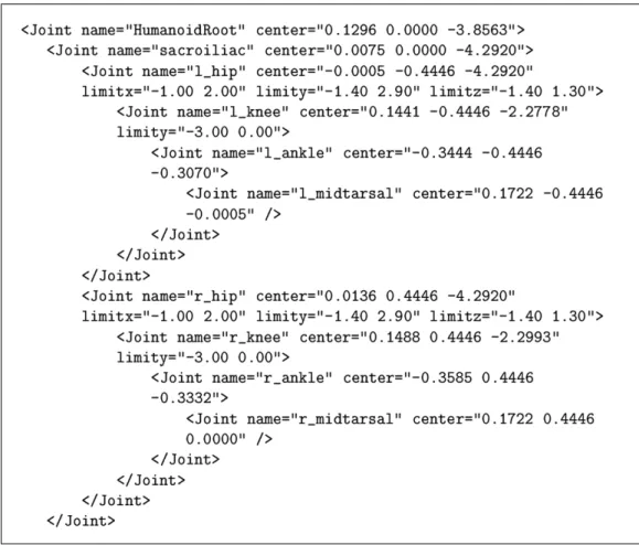
In our studies, the human body model that is constructed by Memi¸soˇglu [30] is used. It is based on the H-Anim 1.1 Specification. The model is constructed by conforming the hierarchy of joints and segments of human body and their naming conventions given in the H-Anim 1.1 Specification. XML data format is used to represent the skeleton. XML provides assistance for describing and defining the skeleton data due to its structured, and self-descriptive format. A portion of the human model data in XML format is given in Figure 3.2 and the complete human model data in XML format can be found in Appendix A.2.
The main elements in H-Anim 1.1 Specification, namely Humanoid, Joint, and Segment, are used to arrange a strict hierarchy to construct the skeleton. Each element has the following attributes. center attribute is used for posi-tioning the associated element. name attribute specifies the name of associated element. limitx, limity, and limitz attributes are used for setting the upper and lower limits for the joint angles, respectively.
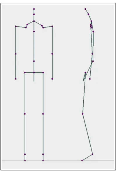
The complete hierarchy is too complex for most of the applications. There-fore, we do not need completely define all the joints in our application. However, it should be noted that the data format stated above can be used for represent-ing the full skeleton specified in the H-Anim 1.1 Specification. A front and left
Figure 3.2: A portion of humanoid data in XML format.
view of the skeleton corresponding to the XML based model in Appendix A.2 are shown in Figure 3.3.
3.2
Animation of the Skeleton Layer
3.2.1
Motion Control
The system designed by [30] aims to establish a method for the low-level motion control using inverse kinematic methods. Positions, joint angles, forces, torques and other motion parameters are specified manually. Spline-driven animation
Figure 3.3: The front and left view of the skeleton
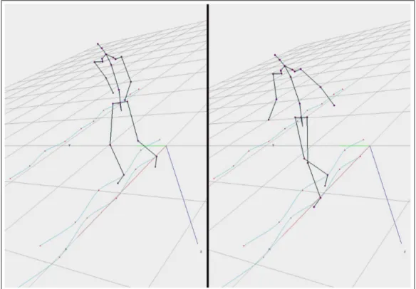
pelvis, ankle and wrist motions are specified by Cardinal splines. Cardinal splines are a class of cubic splines that are very powerful and have easily controllable input control points fitting mechanism with a tension parameter. Cubic splines are mostly preferred due to less computation cost and low memory requirement. More detailed information about splines and the description of a high-level walk-ing animation mechanism that makes use of these low-level techniques can be found in [30].
In addition to position curves generated for pelvis, ankle and wrist, there exists a velocity curve specified independently for each body part. Thus, char-acteristics of the motion can be changed just by modifying the velocity curve. This method is called as double interpolant method [48].
However, arclength problem arises when moving an object along a given posi-tion spline due to the parametric nature of cubic splines. The arclength problem appears when we have a path specified by a spline and we are looking for a set of points along the spline such that the distance travelled along the curve between consecutive frames is constant. The arclength parameterization of spline curve is a remedy to this problem.
Spline curves provide useful functionality for motion control. They can be constructed with a few set of control points and can be manipulated easily. Figure 3.4 indicates an example to these motion control issues. Following this approach, the system provides a low-level animation system to the user in which one can obtain any kind of motion by specifying a set of spline curves for position, distance and joint angles over time.
Furthermore, a high-level motion mechanism for walking that uses these low-level techniques is provided. In the system, high low-level control of walking is achieved by allowing the user to specify a few number of locomotion parameters. Specifying the straight travelling path on flat ground without obstacles, and the speed of locomotion, the system generates the walking automatically by computing the 3D path information and the low-level kinematics parameters of the body elements. Furthermore, some extra parameters such as the size of the walking step, the time elapsed during double-support, rotation, tilt, and lateral displacement of pelvis can be adjusted by the user to have a different walking motion. The characteristics of the walking motion of the system is explained in detail in [30].
Figure 3.4: The articulated figure and the Cardinal spline curves
3.2.2
Motion of the Skeleton
In this study, kinematics methods are used for the motion of the skeleton. Kine-matics methods involve the study of motion specification independent of under-lying forces that produced the motion. Forward and inverse kinematics are two different approaches for the animation of a figure. In forward kinematics, the animator explicitly defines the position, rotation angles, and etc. whereas in inverse kinematic methods, the goal positions are specified and the position and rotation of joints are computed accordingly. Moving a hand to grab an object or placing the foot to a desired position requires the usage of inverse kinematics methods. Thus, our system uses inverse kinematics for motion control.
Memi¸soˇglu [30] uses an inverse kinematics package named IKAN software, which was developed at the University of Pennsylvania, to implement his design
[10]. IKAN is a complete set of inverse kinematics algorithms for an anthropo-morphic arm or leg. IKAN provides functionality to control the arm and leg of my human model. For the arm, with the goal of putting the wrist in the desired location, IKAN computed the joint angles for the shoulder and elbow. In the case of leg, rotation angles for hip and knee is calculated. The details of how IKAN is incorporated with our implementation is explained in [30].
Chapter 4
MODELING AND
ANIMATION OF THE
MUSCLES
4.1
Modeling of the Muscle Layer
While the skeleton creates the general structure of the body, muscles determine the general shape of the surface mesh. As a biological fact, human body muscles account for nearly half of the total mass of the body and fill the gap between the skeleton and the skin [38].
Human body movements require the muscles to perform different tasks, that is why our body includes three types of muscles; cardiac, smooth and skeletal muscles [28]. Cardiac muscles, which are used only in the heart, perform the pumping of the blood throughout the body. The second class of muscles, smooth
muscles, is a part of the internal organs and found in the stomach, bladder, and
blood vessels. Both of these two muscles are called involuntary muscles, because they cannot be consciously controlled. On the other hand, Skeletal muscles put
into practice the voluntary movements. They are attached to bones by tendons and provide us the capability to perform various actions by simply contracting and pulling the bones they are attached to towards each other. In this study, only skeletal muscles are modeled because we aimed to represent only the external appearance of the human body.
Skeletal muscles are located on top of the bones and other muscles, and they are structured side by side and in layers. There are approximately 600 skeletal muscles in the human body and they make up 40% to 45% of the total body weight. A typical skeletal muscle is an elastic, contractile material that originates at fixed origin locations on one or more bones and inserts on fixed insertion locations on one or more other bones [38]. The relative positions of these origins and insertions determine the diameter and shape of the muscle. In a real human, muscle contraction causes joint motion but in our articulated figure, muscles deform due to the joint motion, in order to result realistic skin deformations during animation.
There are two types of contraction: isometric (same length) and isotonic (same tonicity) contraction. In isotonic contraction, the muscle belly changes shape that causes the total length of the muscle shortens. As a result, the bones to which the muscle is attached are pulled towards each other. On the other hand, due to isometric contraction, the changes in the shape of the muscle belly because of the tension in the muscle does not change the length of the muscle, so no skeletal motion is produced [1]. Although most body movements requires both isometric and isotonic contraction, in our application only isotonic contraction is considered. Because, isometric contractions do not have much influence on the appearance of the body model during the animation.
In most muscle models, a muscle is represented using two levels: the action line and the muscle shape. However, we represented the muscle layer only with
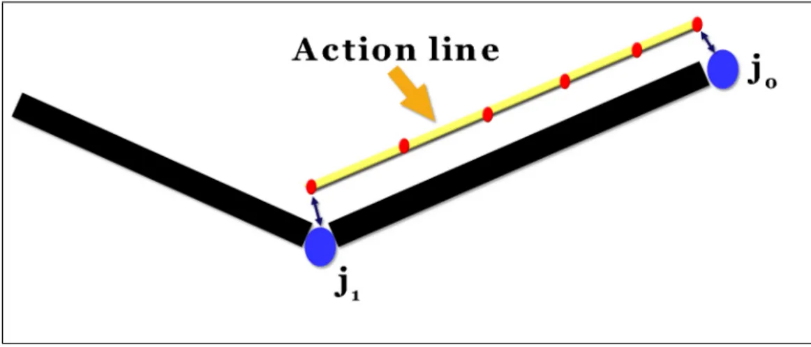
Figure 4.1: Action line abstraction of a muscle
We modeled muscles only in the upper arm, upper leg and lower leg parts of the human body. The key idea of our approach is that the deformations of the skin mesh are driven by only the underlying action lines and no muscle shape is needed. This muscle structure will be explained in the next section.
4.1.1
Action Lines
An action line denotes the imaginary line along which the force applied onto the bone is produced. Nevertheless, the definition of this line is not so clear [1]. Many specialists assume the action line as a straight line, but the most com-mon definition receives the action line as a series of line segments (or polyline in computer graphics terminology) [11]. These segments and their number are determined through the anatomy of the muscle. As a matter of fact, action line is the representative of the muscle force at a cross-section.
In this study, muscles are represented by an action line that simulates the muscle forces and is basically defined by an origin and an insertion point. How-ever, as it is stated above, we have decided to represent the actions of the muscles by using polylines, as described in [11]. For this purpose, we used some control points, which are responsible for guiding the line and including the forces exerted
Figure 4.2: The structure of an action line: control points and forces on these points
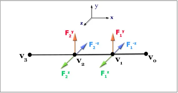
onto the skin mesh. These force fields are inversely proportional to the length of the corresponding action line segment. An example of this kind of action line is shown in Figure 4.2.
4.2
Animation of the Muscle Layer
Animation of the muscles is a more difficult task than the modeling. It is a very complicated process to derive the position and deformation of a muscle in any possible frame. As in the most graphics applications, we used the method that the deformations of the muscles are inferred from the the motion of the skeleton, which is contrary to the real life.
In our approach, the deformations of the skin layer are driven by the un-derlying bones and action lines. This makes the three-dimensional nature of the deformation problem to be reduced to one dimension. As it is stated, each action line is modeled by a polyline and these polylines include some number of control
muscle are attached to the skeleton joints so that their motion is dictated by the skeleton. The positions of all the remaining control points are obtained through a linear interpolation formulation for each animation frame.
First of all, since the positions of the control points of the action line provide information as to how the surface mesh will expand or shrink over time, we need to determine the local frame of each action line control point.
After the local frames are constructed, action line becomes ready to animate in correspondence with the underlying skeleton. Since the insertion and ori-gin points of the action line are fixed on the skeleton, when the skeleton layer moves, this movement is reflected on the action line as decrease or increase in length. Parallel to this change in action line length, the lengths of each action line segment also change. Since the force fields on each control point are in-versely proportional with the segment length, this variation in length also causes a change in the force fields as demonstrated in Figure 4.3. The next step in animation is the deformation in the skin mesh due to the changes on the forces that are exerted on skin vertices by the action line control points. This deforma-tion is automatically propagated on the skin layer via the anchors between skin vertices and action line. Detailed information about these anchors are presented in Subsection 5.1.1. If the segment length shortens, the force fields increase and make the skin mesh to bump. Similarly, the elongation in segment length results in decrease in force fields and relaxation of skin mesh.
Figure 4.3: The deformation of an action line and force field changes: a) rest position and initial force fields of an action line, b) action line shortens and forces increases due to muscle contraction, and c) action line lengthens and forces decreases due to muscle elongation
Chapter 5
MODELING AND
ANIMATION OF THE SKIN
5.1
Modeling of the Skin Layer
The skin is a continuous external sheet that covers the body. The skin accounts for about 16% of the body weight and has surface area from 1.5 to 2.0 m2 in
adults [25]. Its thickness varies depending on the location.
In computer graphics, there are basically three ways to model a skin layer. Designing from scratch or modifying an existing mesh in a 3D modeler is the first method. Another way is to laser scan a real person that produces a dense mesh that truly represents a human figure. The last method for modeling the skin layer is extracting it from underlying components if exist.
Our skin model is modeled in 3D modeler, Poser [32]. We used P3 Nude Man model, which is one of the standard characters of the Poser software. The shaded points and the solid view of the model are seen in Figure 5.1. The model contains 17,953 vertices and 33,234 faces. The whole body is composed of 53
Figure 5.1: The shaded point and solid view of the skin model
parts like hip, abdomen, head, right leg, left hand, etc. This structure provides us the required functionality for binding vertices to the inner layers.
5.1.1
Skin Attaching
After surface creation, in a second stage called attaching, each vertex in the skin is associated with the closest underlying body components (muscle and bone). Basically, the attachment of a particular skin vertex is the nearest point
changes in the underlying component are propagated through these anchors to the corresponding skin vertices. The attaching procedure will be examined in more detail in the sequel.
In our system, skin vertices are first bound with the joints of the skeleton layer in a multi-step algorithm. Basically, to attach the skin to the joints, skin vertices are transformed into the joint coordinate system. As it is stated in Section 5.1, the skin model is decomposed into parts. Parts are groups of vertices of the skin mesh correspond to some part of the body. In the first step of the attaching algorithm, for each part of the skin, we determined a particular joint and attached the vertices of the object to this joint, i.e., right arm object is bound with right shoulder joint and left thigh object is anchored to left hip joint as seen in Figure 5.2. The mathematical details of this and proceeding attachment procedures are explained in Section 5.2. This binding procedure is a virtual process and dictates the position of the body parts when the underlying joint orientation changes.
However, the first step is not sufficient for realistic deformation of the skin layer. In order to perform full skinning method, it is required for some vertices to be bound with more than one joint. Especially, the vertices near the joints are need to be attached to two adjacent joints. The second step of the attaching algorithm is focused on binding the required vertices with two joints. For this purpose, we determine a distance value as a threshold for the joints; left ankle, right ankle, left knee and right knee (see Figure 5.3). For the vertices of each part that are closer to these joints, if the distance between the vertex and the joint is smaller than the threshold value then this vertex is bound with the joint with a weight although the vertex is belonging to another object. The weight of the joints are determined with inversely proportional to the distance between the vertex and the joint.
Figure 5.2: First step of the attaching algorithm: the right upper arm part is bound with the right shoulder joint and the left thigh part is bound with the left hip joint.
Figure 5.3: Second step of the attaching algorithm: the front and left view of some critical joints and their influence areas. The darken rectangles denotes the vertices that are attached with the corresponding joint.
Figure 5.4: The holes at the backside of the model while walking due to unattached vertices
The second step of the algorithm also has some problems. We aimed to bind the near vertices to the joints but we could not determine the appropriate distance value for the binding operation. If we use a smaller threshold value, some of the vertices that should be attached are not attached with the joints. For example, some vertices of the back part of the hip need to be attached to the left hip or right hip joints in order to generate a realistic walking motion. However, since the distance of these vertices to the corresponding joints are bigger than the threshold value, undesired renderings were generated during animation (see Figure 5.4). The unattached vertices caused holes at the backside of the model, also some in the shoulders. As a solution we tried to increase the threshold value. Unfortunately, this attempt caused unnatural results for the other parts of the body during movement.
Therefore, contrary to the first two steps, which are fully automatic, we implemented a manual step to overcome the deficiencies. This last step includes selecting some of the unattached vertices manually by using mouse and then
the capability for picking skin vertices on the screen by clicking on them with mouse. When all the necessary vertices are selected, they are attached to the related joints.
Anchoring points to action lines of muscles can be achieved by a similar process, which is described next. An action line can be considered as a skeleton; control points denote joints and line segments correspond to bones. This allows us to reuse algorithms developed for mapping skin vertices to skeleton joints with some extensions. In the skin-skeleton mapping algorithm, each point of an object are bound with a particular joint. In skin-action line mapping algorithm, each vertices of an object are again attached to a particular action line, but since an action line is composed of a set of control points and each control point has a different force field on the skin mesh (see Figure 4.2), we have to bind each vertex to a number of control points of the action line. As it is seen from Figure 5.5, we decided to select the nearest three action line control points of a vertex. Then, the vertex is influenced by the sum of the effects of these three points. The details of the effects of control points on skin vertices are given in Subsection 5.2.2.
5.2
Animation of the Skin Layer
Due to the deformation of the underlying skeletal and muscular layers, we deform the skin mesh according to the following steps: vertex positioning on the surface due to skeletal movement and displacement along the normal to the surface so as to simulate the action of the muscular system. The first step simulates the adherence of the skin to the inner layers. In the second stage, the skin is shaped by the underlying muscles.
Figure 5.5: Binding skin vertices with action lines. Point j is attached to its nearest three control points, v1, v2 and v3.
5.2.1
Skeletal Deformation
Before applying muscle-induced deformations to the skin layer, we displace each skin vertex based on the skeletal position. In this step we aimed two goals: first generating a smooth skin appearance and second, simulating the adherence of the skin layer to the skeleton layer. We now introduce the skinning based surface deformation model, which is based on [20]
Skeleton and skin layers are designed in some reference position. Here, skeletal animation aimed to move only the skeleton and propagate this movement to the skin. For this purpose, assume that the parent of joint j is denoted by
p(j), the root joint has index zero, and R(j) is the 4 × 4 matrix describing the
transformation from joint p(j) to joint j. Then, the transformation from the world coordinate system to the local frame of joint j is as follows:
Figure 5.6: The skeletal animation: a) reference position and b) after rotation
where R(j) denotes the coordinate transformation from p(j) to j.
Besides, we use transformation matrix T (j) that expresses the rotation of the bone starting at joint j to move a character. So, the final transformation of the joint with the movement applied to it and all its parents is expressed by:
F(j) = F(p(j))R(j)T(j) (5.2)
Now the actual posture of the vertex v can be determined when it is trans-formed to vertex v0 by Equation 5.3, assuming that v is attached to joint j.
v0 = F(j)A(j)−1v (5.3)
However, the basic skeletal animation method does not generate a realistic shape near the joints due to sudden changes in attached bones as seen in Fig-ure 5.6. Since we aimed to model smooth skin deformation using a segmented skeleton, we used the skinning method as we explained briefly in Subsection 5.1.1. To be more precise, allowing attachment of one vertex to more than one bone and averaging the results produces smooth skin appearance near the joints. In this scheme, if vertex v is attached to joints j1, ..., jn then the formula for the
Figure 5.7: The skinning algorithm v0 = n X i=0 w(i)F(ji)A(ji)−1v (5.4)
where w(1), ..., w(n) are weights of the attachment to particular joint where total weight is equal to 1 and n is 2 for our case. Skin deformation with this technique is illustrated in Figure 5.7. In 5.7, the vertices that are near the joints were assigned to both bones with equal weights 0.5 and 0.5. The white vertices represent the results of averaging.
In our system, we implemented a more complicated skinning algorithm. The vertices that are closer to a joint more than a threshold value are attached to two joints and deformed by the movements of both bones. The process is demon-strated in Figure 5.8.
The interpretation of Figure 5.8 is that, first, we select the vertices that are in some neighborhood of a particular joint. These vertices can be either in one of the adjacent parts of the joint. If we can not select all the necessary vertices au-tomatically by means of distance measurement, we select the remaining vertices manually by picking up vertices and binding them to the corresponding joint
Figure 5.8: The skinning method used in our implementation: a) vertex assign-ment and b) after rotation
vertices. As a result, these selected vertices are influenced by both of the nearby joints and the parent of these joints.
5.2.2
Muscular Deformation
The muscular deformation is based on the control points of the action lines. Due to the skeletal movements, the length of the action line and the lengths of each segment of the action line change. There exists an inverse proportionality between the length of the segment and the force fields of the control points. The forces that are exerted on the skin mesh by the action line may increase or decrease, due to length changes. These changes are propagated through the skin layer via the anchors between the skin vertices and action line control points. As a result of this propagation, the skin mesh is deformed in order to create a realistic appearance during animation. The visual results of the muscular deformation are presented in Section 6.1.
Chapter 6
EXPERIMENTAL RESULTS
6.1
Visual Results
The front and back views of the walking behavior produced by our system is illustrated in Figures 6.1 and 6.2.
The visual results in Figures 6.1 and 6.2 show that our system is capable of generating realistic rendering of the human walking behavior. There is no holes or wrinkles on the skin mesh, even near the highly moveable joints like shoulders, hips and knees.
The postures of the arm of the human model generated in a multi-dimensional motion are presented in Figure 6.3. The deformation of the skin mesh can be seen more clear in these figures.
Figure 6.4: Showing muscular deformation on the skin mesh.
In order to demonstrate muscular and skeletal deformations, we show a series of stills taken from animations rendered in our system. In the animation seen in the Figure 6.4, the right arm of the character is raised forward and then flexed in order to demonstrate muscular deformation on the skin mesh.
6.2
Performance Analysis
# of # of # of Vertices # of Vertices Frame Vertices Triangles Bound with Bound with Rate
One Joint Two Joint (fps) High 17,953 33,234 15,305 2,648 19 Resolution Middle 8,923 15,203 7,623 1,270 48 Resolution Low 5,133 7,625 4,189 944 92 Resolution
Table 6.1: Performance evaluation results
human model is composed of a skin mesh that contains 17,953 vertices and 33,234 faces, a muscle layer, and a skeleton structure with 25 joints and 23 bones. Since bones are simulated with lines, they do not have an significant effect on total performance. The most tedious and time consuming operation is the binding process of the skin layer with the underlying layers. However, this does not effect the frame rate since it is done as a preprocessing step before the animations. We tested the system with different skin meshes ranging from low to high resolutions (see Figure 6.5). The average frame rates given in terms of frames per second (fps) for different meshes are given in Table 6.1.
Figure 6.5: Different resolution valued skin meshes: a) high resolution (original model), b) middle resolution, and c) low resolution.
Chapter 7
CONCLUSION
In this study, we proposed an animation system for a human body model. The system is based on an anatomically based approach that builds layers of the hu-man body: the skeleton layer, the muscle layer and the skin layer. The skeleton layer consist of an articulated figure that is modeled based on the H-Anim 1.1 Specification. By using the skeleton layer, we control the motion of the human body, and with the muscle layer, we can deform the skin layer according to the motion. We introduced a simple muscle model that approximates the human anatomy. The deformation of the skin is mostly driven by the muscle action lines. Our skin layer is modeled in an 3D modeler. The vertices of the skin mesh is attached with the inner layers in such a way that, the attachment of a particular skin vertex is the nearest point on its underlying component. This provides the functionality that if the shape of the underlying component changes, these changes are propagated through these anchors to the skin layer. Perfor-mance evaluations showed that the multi-layered model produces realistic skin deformation in real-time.
We suggest a few directions for future research. We did not deal with the problem of collision detection and response between different body parts, which is
called body awareness. Besides, other muscle forms and more number of muscles could be simulated.
Appendix A
HUMAN MODEL IN XML
FORMAT
In this appendix, the document type definition (DTD) of the human model used in our implementation and XML formatted data of the human model are given.
A.1
Document Type Definition (DTD) of
Hu-man Model
<!ELEMENT Humanoid (author, name, center, Joint*)> <!ELEMENT author (#PCDATA)>
<!ELEMENT name (#PCDATA)> <!ELEMENT center (#PCDATA)>
<!ATTLIST center value CDATA #REQUIRED> <!ELEMENT Joint (Joint*, Segment*, Site*)> <!ATTLIST Joint name CDATA #REQUIRED> <!ATTLIST Joint center CDATA #REQUIRED> <!ATTLIST Joint limitx CDATA #IMPLIED>
![Figure 2.1: Stitching components: a) bone structure and b) attached mesh struc- struc-ture (reproduced from [24])](https://thumb-eu.123doks.com/thumbv2/9libnet/5986566.125613/25.892.321.640.149.534/figure-stitching-components-structure-attached-struc-struc-reproduced.webp)
![Figure 2.2: Skinned arm mesh bent to 90 degrees and 120 degrees (reproduced from [24])](https://thumb-eu.123doks.com/thumbv2/9libnet/5986566.125613/27.892.187.773.153.514/figure-skinned-arm-mesh-bent-degrees-degrees-reproduced.webp)
![Figure 2.3: The elbow collapses when the forearm is twisted (reproduced from [24])](https://thumb-eu.123doks.com/thumbv2/9libnet/5986566.125613/28.892.184.773.157.329/figure-elbow-collapses-forearm-twisted-reproduced.webp)
![Figure 2.5: The modeling of volume-preserving ellipsoid (reproduced from [41])](https://thumb-eu.123doks.com/thumbv2/9libnet/5986566.125613/34.892.188.773.155.366/figure-modeling-volume-preserving-ellipsoid-reproduced.webp)
![Figure 3.1: The H-Anim Specification 1.1 hierarchy (from [47])](https://thumb-eu.123doks.com/thumbv2/9libnet/5986566.125613/39.892.189.774.212.1035/figure-h-anim-specification-hierarchy.webp)
