IMMUNOMODULATORY POTENTIAL OF HUMAN UMBILICAL CORD
TISSUE-DERIVED MESENCHYMAL STROMAL CELL (UCX
®) EXOSOMES
IN COMBINATION WITH IMMUNOSUPPRESSIVE “A151”
OLIGODEOXYNUCLEOTIDE
A THESIS SUBMITTED TO
THE GRADUATE SCHOOL OF ENGINEERING AND SCIENCE OF BILKENT UNIVERSITY
IN PARTIAL FULLFILMENT OF THE REQUIREMENTS FOR THE DEGREE OF
MASTER OF SCIENCE IN
MOLECULAR BIOLOGY AND GENETICS
By Özlem Bulut
i
IMMUNOMODULATORY POTENTIAL OF HUMAN UMBILICAL CORD TISSUE-DERIVED MESENCHYMAL STROMAL CELL (UCX®) EXOSOMES IN COMBINATION
WITH IMMUNOSUPPRESSIVE “A151” OLIGODEOXYNUCLEOTIDE
By Özlem Bulut
July 2019
We certify that we have read this thesis and that in our opinion it is fully adequate, in scope
and in quality, as a thesis for the degree of Master of Science.
________________________
İhsan Gürsel (Advisor)
_______________________
Kamil Can Akçalı
________________________
Onur Çizmecioğlu
Approved for the Graduate School of Engineering and Science
________________________
Ezhan Karaşan
ii
ABSTRACT
IMMUNOMODULATORY POTENTIAL OF HUMAN UMBILICAL CORD
TISSUE-DERIVED MESENCHYMAL STROMAL CELL (UCX
®) EXOSOMES IN
COMBINATION WITH IMMUNOSUPPRESSIVE “A151” OLIGODEOXYNUCLEOTIDE
Özlem BulutM.Sc. in Molecular Biology and Genetics Advisor: Prof. İhsan Gürsel
July 2019
Mesenchymal stromal or stem cells (MSCs) modulate immune responses apart from their regenerative capacities. Accumulating evidence suggests that MSCs exert their paracrine effects through extracellular vesicles known as exosomes. In this study, we utilized a particular human umbilical cord tissue-derived
MSC type termed as UCX®. UCX® is superior to the gold-standard bone marrow-derived MSCs in
terms of immunosuppressive properties. We aimed to characterize and employ UCX® exosomes as
cell-free immunosuppressive therapeutic agents. Another aim was to compare the functionalities of exosomes either isolated from 2-dimensional (2D) cultures or isolated from 3-dimensional (3D) spheroid cultures. 3D culture provides better cell-to-cell and cell-to-matrix interactions thereby mimics the in vivo environment better. A synthetic oligodeoxynucleotide called A151 ODN, which consists of 4 repeats of the mammalian telomeric TTAGGG motif, has broad immunosuppressive effects. Delivery of A151 ODN within liposomes or exosomes protects it from degradation by nucleases and improve the
desired outcome. We also aimed to combine the immunomodulatory potentials of UCX®exosomes and
A151 ODN through direct loading of A151 ODN into exosomes with ~95% efficiency via a dehydration-rehydration-based lyophilization method. First, we determined the binding and internalization kinetics of exosomes with different immune cells. 3D-exosomes interacted with the target cells much faster and
more efficiently. Next, we investigated how UCX® exosomes influence Toll-like receptor (TLR)
signaling in mouse splenocytes and bone marrow-derived macrophages (BMDMs). 3D-exosomes compared to 2D-exosomes were more potent to suppress IFNγ, IL6, IL12, and to a lesser extent TNFα production mediated by TLR1/2, TLR4, TLR7/8 and TLR9 but not by TLR3 triggering. A151 ODN-loading to either 2D- or 3D-exosomes improved the inhibition of all the above mentioned pro-inflammatory cytokines. Especially 3D-exosomes downregulated co-stimulatory molecules CD80 and CD86 along with MHC-II on BMDMs following TLR stimulation. Macrophage polarization
experiments revealed that UCX® exosomes reprogram BMDMs to produce high amounts of nitric oxide
and arginase-1 which are the key immunomodulatory factors induced by myeloid-derived suppressor cells (MDSCs) to inhibit T- and NK-cell activity. Besides shifting macrophages to an MDSC-like suppressive phenotype, exosomes also supported expansion of MDSC populations in vivo upon
intraperitoneal injection. Next, we tested the therapeutic efficiency of UCX® exosomes with or without
iii
mice. Exosomes could not alleviate zymosan-induced peritonitis which is an acute and severe inflammation. However, 3D-exosomes and A151 ODN-loaded versions of both 2D- and 3D-exosomes remarkably prevented DSS-induced colitis progression. A151 ODN itself was also therapeutic, albeit to a lesser degree. Standalone 3D-exosomes and A151 ODN-loaded exosomes prevented weight loss and colon shortening. All treatments except for 2D-exosomes could restore DSS-induced loss of T-cell numbers and cytokine-producing capacities in mesenteric lymph nodes and spleen. All treatments except for A151 ODN prevented DSS-induced macrophage accumulation in the lymph nodes. 3D-exosomes and A151 loaded versions of both exosomes normalized serum IL6 levels while only A151 ODN-loaded 3D-exosomes could impact the cytokine production capacities of macrophages. Finally, we
tested the effects of UCX® exosomes with or without A151 ODN on wound healing. In vitro, 2D- and
3D-exosomes differentially upregulated the productions of wound healing-related cytokines and growth factors such as IL1α, TGFβ and VEGFα from fibroblast and keratinocyte cell lines. In vivo, in an excisional wound healing model, free or A151 ODN-loaded exosomes did not accelerate wound closure. However, they caused systemic immunosuppression at the late stages of wound healing. Systemic outcomes include reduced inflammatory capacity of macrophages and higher granulocytic MDSC numbers in spleen. A151 ODN-loaded 3D-exosomes also reduced T-cell numbers in spleen and pro-inflammatory cytokine levels in circulation. Taken together, this study revealed that 2D- but more importantly 3D-culturing of umbilical cord MSCs result in functionally different exosomes, 3D culture-derived exosomes display higher immunosuppressive potential, A151 ODN-loading into these
exosomes improves immunosuppressive capacity and A151 ODN-loaded UCX® exosomes could be a
valuable therapeutic agent for inflammatory and autoimmune disorders.
Keywords: Mesenchymal stromal cells, mesenchymal stem cells, 3D culture, exosomes, A151 ODN, TLR, MDSCs, inflammation, immunosuppressive, immunomodulatory
iv
ÖZET
İNSAN GÖBEK KORDONU DOKUSUNDAN ELDE EDİLEN MEZENKİMAL KÖK
HÜCRE (UCX
®) EKSOZOMLARININ İMMÜNBASKILAYICI
OLİGODEOKSİNÜKLEOTİD A151 İLE BİRLİKTE İMMÜN DÜZENLEYİCİ ETKİLERİ
Özlem BulutMoleküler Biyoloji ve Genetik, Yüksek Lisans Tez Danışmanı: Prof. İhsan Gürsel
Temmuz 2019
Mezenkimal stromal hücreler veya mezenkimal kök hücreler (MKHler) doku yenileyici özelliklerinin yanında immün yanıtı düzenleyici özelliklere de sahiptir. Araştırmalar MKHlerin etkilerinin çoğunun parakrin bir şekilde ve eksozom adı verilen hücre dışı kesecikler ile sağlandığını göstermektedir. Bu
çalışmada, patentli ve özel bir MKH türü olan, göbek kordonu dokusundan elde edilen ve UCX® adı
verilen MKHler kullanılmıştır. UCX® hücreleri immün baskılayıcılık açısından altın standart olan kemik
iliği MKHlerinden üstündür. Çalışmanın amacı UCX® eksozomlarını karakterize etmek ve immün
düzenleyici terapi yöntemi olarak test etmektir. Bir başka amaç da 2 boyutlu (2D) hücre kültürü ile 3 boyutlu (3D) hücre kültüründen elde edilen eksozomların fonksiyonel olarak karşılaştırılmasıdır. 3D kültür hücreler arasında ve hücreler ile hücre dışı matris arasında daha fazla etkileşim sağladığı için canlı organizmadaki duruma daha yakındır. A151 ODN adı verilen ve memeli telomerlerindeki TTAGGG motifinin 4 tekrarından oluşan sentetik oligodeoksinükleotid geniş kapsamlı immün baskılayıcı etkilere sahiptir. A151 ODN’in lipozom veya eksozomlara yüklenmesi nükleazlar tarafından
parçalanmasını önleyerek etkinliğini artırmaktadır. Bu çalışmada, A151 ODN’i UCX® eksozomlarına
yükleyerek iki ajanın immün düzenleyici etkilerini birleştirmek ve sinerjik etki elde etmek amaçlanmıştır. Grubumuzca geliştirilen yükleme tekniğiyle A151 ODN eksozomlara yaklaşık %95
verimle yüklenebilmiştir. Öncelikle, farklı immün hücrelerin UCX® eksozomlarına bağlanma ve
eksozomları içine alma kapasiteleri incelenmiş ve 3D eksozomların hedef hücrelerle daha çabuk ve daha fazla etkileşime girdiği görülmüştür. Eksozomların Toll-benzeri reseptör (TLR) yolaklarına etkisi fare dalak hücrelerinde ve kemik iliğinden farklılaştırılmış makrofajlarda (BMDMler) incelendiğinde, eksozomların TLR1/2, TLR4, TLR7/8 ve TLR9 yolaklarının aktivasyonu sonucu gerçekleşen IFNγ, IL6, IL12, ve daha az ölçüde TNFα üretimini bastırabildiği ve 3D eksozomların genel olarak 2D eksozomlardan daha etkin olduğu görülmüştür. Eksozomlara A151 ODN yüklenmesi enflamatuar sitokinlerin baskılanmasını artırmıştır. Ayrıca eksozomlar, özellikle 3D eksozomlar, BMDMlerde TLR yolaklarının aktive edilmesi üzerine artan yardımcı uyarıcı CD80 ve CD86 molekülleri ile antijen sunucu MHC-II molekülünün miktarlarını azaltmıştır. Makrofaj polarizasyonu deneyleri ise eksozomların BMDMleri yüksek miktarda nitrik oksit ve arjinaz üretecek şekilde programladığını göstermiştir. Bu iki faktör, myeloid kökenli baskılayıcı hücrelerin (MDSCler) T ve NK hücrelerini baskılamakta kullandıkları ana faktörlerdir. Makrofajları MDSC benzeri baskılayıcı bir fenotipe
v
çekmelerinin yanı sıra, eksozomlar farelerde karın içi enjeksiyon yoluyla uygulandıklarında MDSC
popülasyonlarını artırmışlardır. UCX® eksozomlarının A151 ODN varlığında ve yokluğunda immün
düzenleyici etkileri farelerde zymosan ile uyarılmış peritonit modeli ve dekstran sodyum sülfat ile uyarılmış kolit modelinde test edilmiştir. Akut ve şiddetli bir enflamasyon olan peritonit modelinde eksozomlar enflamasyonu baskılayamasa da, kolit modelinde özellikle tek başına 3D-eksozomlar ve iki eksozom tipinin de A151 ODN yüklü halleri hastalığın ilerlemesini durdurabilmişlerdir. Boş veya A151 ODN yüklü 3D-eksozomlar kilo kaybını ve kolon boyutlarındaki kısalmayı engellemiş, lenf nodları ve dalaktaki DSS kaynaklı T-hücre kaybını ve T-hücrelerinden sitokin salımındaki disregülasyonu onarabilmiştir. A151 ODN yüklenmiş veya yüklenmemiş bütün eksozomlar ise lenf nodlarına DSS kaynaklı makrofaj birikmesini önlemiştir. 3D eksozomlar ve iki eksozom tipinin de A151 ODN yüklü halleri serumda DSS ile artan IL6 miktarını normalize edebilmiş ama sadece A151 ODN yüklü 3D-eksozomlar makrofajların tekrar uyarım sonucu sitokin üretim kapasitesinde etkili olmuştur. Son olarak, eksozomların A151 ODN varlığında ve yokluğunda yara iyileşmesine etkileri araştırılmıştır. 2D ve 3D eksozomlar fibroblast ve keratinosit hücre hatlarında yara iyileşmesi ile alakalı IL1α, TGFβ ve VEGFα gibi faktörlerin üretimini farklı derecelerde artırmıştır. Farede yara iyileşmesi modelinde A151 ODN varlığında ya da yokluğunda eksozomlar yara iyileşmesini hızlandırmamış olsa da iyileşmenin son safhalarında sistemik bir immün baskılanmaya sebep olmuşlardır. Bu sistemik etkilerin içerisinde makrofajların enflamatuar kapasitlerinde azalış ve granülositik MDSC popülasyonunda artış vardır. A151 ODN yüklü 3D eksozomlar ayrıca T hücre sayılarını ve dolaşımdaki enflamatuar sitokin miktarlarını da düşürmüştür. Topluca ele alındığında bu çalışma 2D ve 3D hücre kültürlerinden elde edilen eksozomların fonksiyonel açıdan farklı olduğunu, 3D kültürden izole edilen eksozomların daha immün baskılayıcı olduğunu, A151 ODN yüklenmesinin eksozomların immün baskılayıcı kapasitlerini
artırdığını ve A151 ODN yüklü UCX® eksozomlarının enflamatuar ve otoimmün hastalıklarda değerli
bir tedavi yaklaşımı olabileceğini göstermektedir.
Anahtar Sözcükler: Mezenkimal stromal hücre, mezenkimal kök hücre, 3 boyutlu hücre kültürü, eksozom, A151 ODN, TLR, MDSC, enflamasyon, immün baskılayıcı, immün düzenleyici
vi
vii
Acknowledgements
First and foremost, I would like to express my gratitude to have Prof. İhsan Gürsel as my supervisor. He is a father figure to me beyond being a scientific mentor. I am forever indebted to him for his support, encouragement and guidance both inside and outside the lab. It was an honor to work with him and to learn from him at the beginning of my career. I would also like to thank our collaborators Asst. Prof.
Joana Miranda and Sérgio Camões from University of Lisbon for providing the UCX® cells and for all
their efforts on this joint project. I want to thank the jury members of my thesis defense, Prof. Kamil Can Akçalı and Asst. Prof. Onur Çizmecioğlu for their interest and valuable suggestions.
I am grateful for Dr. Gözde Güçlüler, Dr. Banu Bayyurt Kocabaş and Dr. Tamer Kahraman’s knowledge and guidance in the early periods of my graduate studies. I also want to thank THORLAB members Göksu Gökberk Kaya, Havva Özgen Kılgöz, Muzaffer Yıldırım, Seda Sabah Özcan and especially İrem Evcili and Naz Bozbeyoğlu for their friendship and help. Without them, the long working hours in the lab would be unbearable. I would also like to thank İhsan Cihan Ayanoğlu for his help and guidance with atomic force microscopy, and Ferda Topal Çelikkan for the histological analyses. I want to express my gratitude for our veterinary physician Gamze Aykut and Ulaş Saçıntı for their help with animal experiments. I am thankful to the lab technicians Abdullah Ünnü and Okan Erşahan, lab managers Pelin Makas and Seda Şengül Birkan for their invaluable help and support about everything, and to Yavuz Ceylan for always cheering me up and watching out for me.
Through good and bad, my biggest pillars of support are my best friends Dilara Boğa and Said Tiryaki. Success or happiness is only real if they are by my side to share it. I am thankful to Çağdaş Yalçınkaya for teaching me patience and persistence. I will always cherish the strength he has helped me build. I am very lucky to have found my irreplaceable lab partner Gizem Kılıç. No one can motivate me better and more easily than she can. Without her uplifting me, I would not be able to be as productive and hopeful towards work and life. It gives me great joy to know that we will continue working alongside each other for our PhD degrees and I am hoping for many more and better years together. Another special thank-you is for Buğra Oben Karasaka who is my medicine in times of stress, who has been with me and done so much for me these past years as a good friend and as my partner. I deeply appreciate and treasure his luminous presence and love.
My biggest share of gratitude is for my dear family. I will be forever in debt for my mother Ayşe Bulut and my father Kemal Bulut’s unconditional love and their belief in me and my goals. I am also very lucky to have my big brother Önder Bulut. He is the person who encouraged me to be a scientist at a very young age and whose advice I rely on the most. Without my family’s encouragement and sacrifices, none of my accomplishments would be possible.
viii
Table of Contents
ABSTRACT ... ii
ÖZET ... iv
Acknowledgements ... vii
Table of Contents... viii
List of Figures ... xii
List of Tables ... xv
Abbreviations ... xvi
Chapter 1 ... 1
Introduction ... 1
1.1 The Immune System ... 1
1.1.1 Innate Immunity ... 1
1.1.1.1 Pattern Recognition Receptors (PRRs)... 2
1.1.1.1.1 Toll-like Receptors (TLRs) ... 2
1.1.1.2 Anti-Inflammatory Myeloid Cells ... 4
1.1.1.2.1 Alternatively Activated Macrophages (M2) ... 4
1.1.1.2.2 Myeloid Derived Suppressor Cells (MDSCs) ... 5
1.2 Mesenchymal Stromal Cells (MSCs) ... 5
1.2.1 MSC Types and Niches in Human Body... 6
1.2.2 Human Umbilical Cord Tissue-Derived MSCs (UCX®) ... 6
1.2.2.1 3D Culturing of UCX® Cells ... 7
1.2.3 Immunomodulatory Actions of MSCs ... 8
1.2.3.1 Interactions of MSCs with Innate Immunity ... 8
1.2.3.2 Interactions of MSCs with Adaptive Immunity ... 9
1.2.4 Clinical Uses of MSCs ... 10
1.3 Exosomes... 11
1.3.1 Exosome Biogenesis ... 12
1.3.2 Role of Exosomes in Health and Disease ... 13
1.3.3 Therapeutic Applications of Exosomes ... 14
1.3.4 Use of Exosomes as Nanocarriers ... 16
1.3.5 Therapeutic Potential of MSC-derived Exosomes ... 16
1.4 Immunosuppressive Oligodeoxynucleotides (Sup ODNs) ... 18
1.4.1 A151 ODN ... 18
1.4.1.1 Mechanisms of Action ... 18
1.4.1.2 Therapeutic Applications ... 19
ix
1.5.1 Ulcerative Colitis ... 20
1.5.2 Peritonitis ... 21
1.5.3 Wound Healing ... 21
1.5.3.1 Inflammation in Wound Healing ... 21
1.5.3.2 MSCs in Wound Healing ... 22
1.6 Aims and Outline of the Study ... 23
Chapter 2 ... 25
Materials and Methods ... 25
2.1 Materials ... 25
2.1.1 Cell Culture Media and Solutions ... 25
2.1.2 TLR Ligands ... 25
2.1.3 Flow Cytometry Antibodies ... 26
2.1.4 ELISA Antibodies and Reagents ... 27
2.1.5 PCR Primers ... 28
2.2 Methods ... 29
2.2.1 Cell Culture ... 29
2.2.1.1 2D UCX® Cell Culture ... 29
2.2.1.2 3D UCX® Cell Culture ... 29
2.2.1.3 NIH3T3 Fibroblast Cell Line Culture ... 30
2.2.1.4 HaCaT Keratinocyte Cell Line Culture ... 30
2.2.1.5 Bone Marrow Derived Macrophage (BMDM) Generation ... 30
2.2.1.6 Single Cell Isolation from Mouse Spleen ... 30
2.2.1.7 Isolation of Mouse Peritoneal Exudate Cells (PECs) ... 31
2.2.1.8 Isolation of Mouse Mesenteric Lymph Nodes ... 31
2.2.1.9 Single Cell Isolation from Mouse Skin ... 31
2.2.1.10 Isolation of Peripheral Blood Mononuclear Cells (PBMCs) from Human Blood ... 31
2.2.1.11 Cell Counting ... 32
2.2.2 Isolation of Exosomes from UCX® Conditioned Media ... 33
2.2.3 Determination of Exosomal Protein Content ... 33
2.2.4 Characterization of Exosomes ... 34
2.2.4.1 Size Distribution Analysis by Dynamic Light Scattering ... 34
2.2.4.2 Atomic Force Microscopy (AFM) ... 34
2.2.4.3 Analysis of Surface Markers by Flow Cytometry ... 34
2.2.5 Determination of Exosome Binding and Uptake with Mouse Splenocytes, BMDMs and Human PBMCs ... 34
2.2.6 Loading of A151 ODN into Exosomes ... 35
2.2.7 Determination of A151 ODN Loading Efficiency into Exosomes ... 36
x
2.2.8.1 Determination of the Presence of Immunomodulatory Surface Proteins on Exosomes
36
2.2.8.2 Determination of UCX® Exosomes’ Effect on BMDM Polarization ... 36
2.2.8.3 Determination of UCX® Exosomes’ Effect on Cytokine Production from Fibroblasts and Keratinocytes ... 37
2.2.8.4 In Vitro Stimulations with TLR Ligands and UCX® Exosomes ... 37
2.2.8.5 Determination of Cytokine Levels by ELISA ... 37
2.2.8.6 Determination of Gene Expression ... 38
2.2.8.6.1 RNA Isolation and cDNA Synthesis ... 38
2.2.8.6.2 RT-qPCR ... 38
2.2.8.7 Analysis of Cell Surface Markers by Flow Cytometry ... 39
2.2.8.8 Measurement of Nitric Oxide Levels by Griess Assay ... 39
2.2.9 In Vivo Experiments ... 40
2.2.9.1 Determination of Cell Phenotypes Following Intraperitoneal Injection of UCX® Exosomes... 40
2.2.9.2 Dextran Sulfate Sodium (DSS)-Induced Colitis Model ... 40
2.2.9.3 Zymosan-Induced Peritonitis Model ... 40
2.2.9.4 Excisional Wound Healing Model ... 41
2.2.10 Statistical Analysis ... 41
Chapter 3 ... 42
Results ... 42
3.1 Characterization of UCX® Exosomes... 42
3.1.1 Size Distribution ... 42
3.1.2 Surface Protein Characterization ... 44
3.2 Exosome Binding and Uptake Kinetics ... 47
3.2.1 Exosome Binding and Uptake by Immune Cell Populations in Human PBMCs ... 47
3.2.2 Exosome Binding and Uptake by Immune Cell Populations in Mouse Splenocytes .... 49
3.2.3 Exosome Binding and Uptake by Mouse BMDMs ... 51
3.3 Determination of A151 ODN Loading Efficiency into Exosomes ... 52
3.4 Immunomodulatory Effects of UCX® Exosomes on TLR Signaling ... 53
3.4.1 Dose-Dependency Studies ... 53
3.4.2 Effects of Free or A151 ODN-Loaded UCX® Exosomes on Mouse Splenocytes ... 57
3.4.3 Effects of Free or A151 ODN-Loaded UCX® Exosomes on Mouse BMDMs ... 62
3.5 Effects of Free or A151 ODN-Loaded UCX® Exosomes on Fibroblasts and Keratinocytes 73 3.6 Effects of UCX® Exosomes on Macrophage Polarization ... 77
3.7 Changes in Cell Phenotypes of Mouse PECs Following Intraperitoneal Injection of Free or A151 ODN-Loaded UCX® Exosomes ... 81
3.8 In Vivo Immunomodulatory Potential of Free or A151 ODN-Loaded UCX® Exosomes in DSS-Induced Colitis Model ... 83
xi
3.9 In Vivo Immunomodulatory Potential of Free or A151 ODN-Loaded UCX® Exosomes in
Excisional Wound Healing Model ... 96
Chapter 4 ... 102 Discussion ... 102 Appendices ... 113 Appendix A ... 113 Additional Figures ... 113 Appendix B ... 121
Buffers for ELISA and Flow Cytometry ... 121
Bibliography ... 122
xii
List of Figures
Figure 1.1. Signaling pathways of TLRs 1-9………4
Figure 1.2. Regulation of innate immune cells by MSCs……….9
Figure 1.3. Regulation of adaptive immune cells by MSCs……….10
Figure 1.4. Composition and biogenesis of exosomes………13
Figure 1.5. Mechanisms of A151 ODN-mediated immunosuppression…………...19
Figure 1.6. Mechanisms of wound healing promotion by MSCs……….23
Figure 2.1. Representative figure of the counting chamber of hemocytometer………32
Figure 2.2. Representative gating strategy for cell counting………33
Figure 2.3. Exosome loading with controlled rehydration-dehydration method……….36
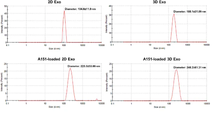
Figure 3.1. Size distribution of UCX exosomes before and after A151 ODN loading...….43
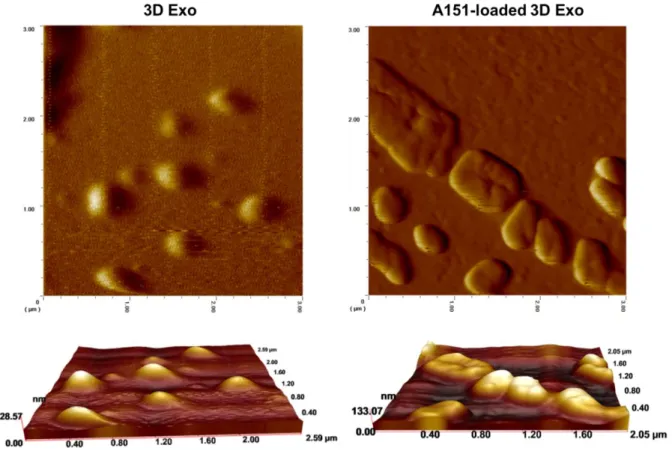
Figure 3.2. AFM images of 3D UCX exosomes before and after A151 ODN loading…...44
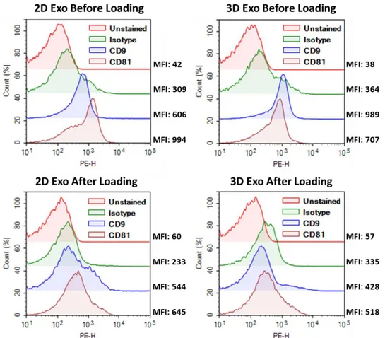
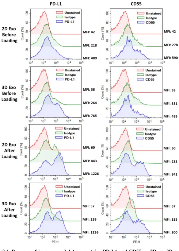
Figure 3.3. Presence of conventional exosome-specific surface markers on UCX exosomes before or after the lyophilization based loading procedure……….………45
Figure 3.4. Presence of immunomodulatory proteins PD-L1 and CD55 on 2D- or 3D-exosomes before or after the lyophilization based loading procedure...………...46
Figure 3.5. Time- and dose-dependent binding and uptake of 2D- and 3D-exosomes by different cell populations in human PBMCs…...48
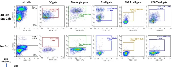
Figure 3.6. Representative flow cytometry plots related to Figure 3.5 showing exosome positivity in different cell types...49
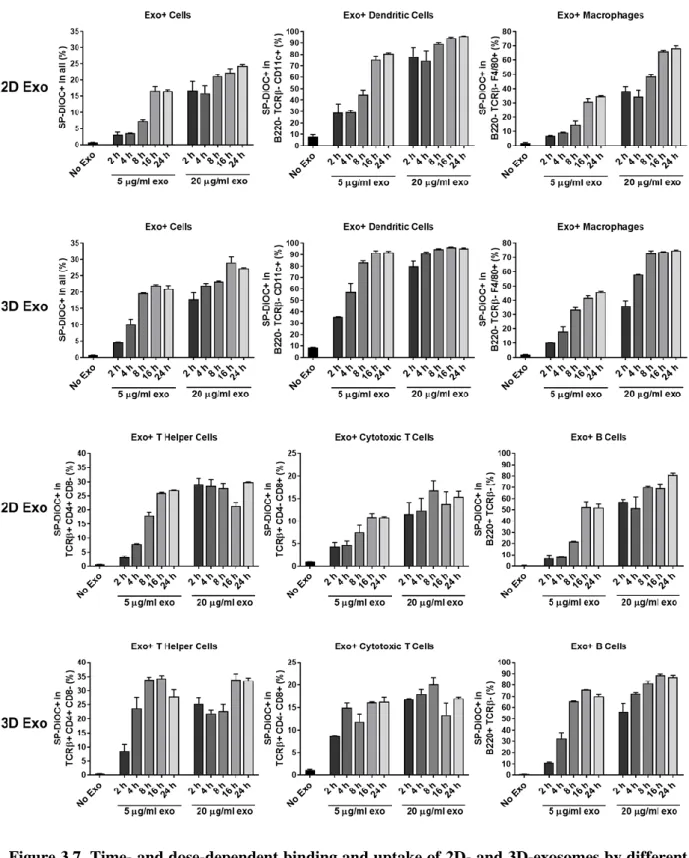
Figure 3.7. Time- and dose-dependent binding and uptake of 2D- and 3D-exosomes by different immune cell populations in mouse splenocytes...50
Figure 3.8. Representative flow cytometry plots related to Figure 3.7 showing exosome positivity in different cell types...51
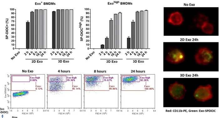
Figure 3.9. Time-dependent binding and uptake of 2D- and 3D-exosomes by mouse BMDMs……...52
Figure 3.10. Representative flow cytometry plots displaying the loading efficiency of A151 ODN into 2D- and 3D-exosomes...53
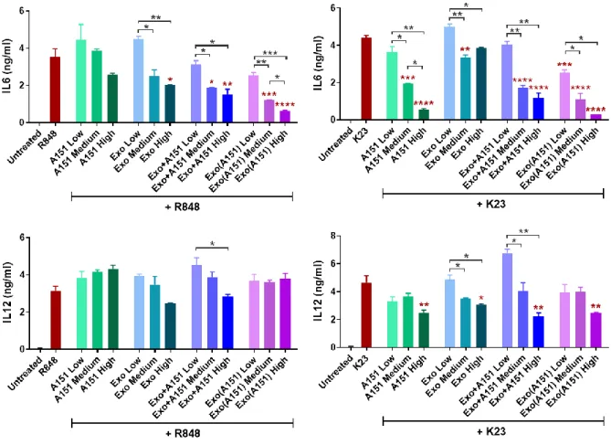
Figure 3.11. Dose-dependent effects of A151 ODN, 3D-exosomes and their combinations on IL6 and IL12 secretions from mouse splenocytes upon stimulation with TLR7/8 ligand R848 and TLR9 ligand K23...55
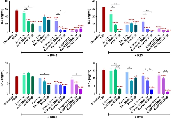
Figure 3.12. Dose-dependent effects of A151 ODN, 3D-exosomes and their combinations on IL6 and IL12 secretions from mouse BMDMs upon stimulation with TLR7/8 ligand R848 and TLR9 ligand K23...56
Figure 3.13. Effects of A151 ODN, 2D- or 3D-exosomes and their combinations on IFNγ secretion from splenocytes upon stimulation with various TLR ligands...58
Figure 3.14. Effects of A151 ODN, 2D- or 3D-exosomes and their combinations on IL6 secretion from splenocytes upon stimulation with various TLR ligands...59
Figure 3.15. Effects of A151 ODN, 2D- or 3D-exosomes and their combinations on IL12 secretion from splenocytes upon stimulation with various TLR ligands...60
Figure 3.16. Effects of A151 ODN, 2D- or 3D-exosomes and their combinations on TNFα secretion from splenocytes upon stimulation with various TLR ligands...61
Figure 3.17. Effects of A151 ODN, 2D- or 3D-exosomes and their combinations on IL6 secretion from BMDMs upon stimulation with various TLR ligands...63
Figure 3.18. Effects of A151 ODN, 2D- or 3D-exosomes and their combinations on IL12 secretion from BMDMs upon stimulation with various TLR ligands...64
xiii
Figure 3.19. Effects of A151 ODN, 2D- or 3D-exosomes and their combinations on TNFα secretion from BMDMs upon stimulation with various TLR ligands...65 Figure 3.20. Effects of A151 ODN, 2D- or 3D-exosomes and their free or loaded combinations on CD80, CD86 and MHC-II cell surface expressions in BMDMs...67 Figure 3.21. Effects of A151 ODN, 2D- or 3D-exosomes and their free or loaded combinations on CD80, CD86 and MHC-II cell surface expressions in BMDMs upon stimulation with PAM3CSK4...68 Figure 3.22. Effects of A151 ODN, 2D- or 3D-exosomes and their free or loaded combinations on CD80, CD86 and MHC-II cell surface expressions in BMDMs upon stimulation with poly(I:C)...69 Figure 3.23. Effects of A151 ODN, 2D- or 3D-exosomes and their free or loaded combinations on CD80, CD86 and MHC-II cell surface expressions in BMDMs upon stimulation with LPS...70 Figure 3.24. Effects of A151 ODN, 2D- or 3D-exosomes and their free or loaded combinations on CD80, CD86 and MHC-II cell surface expressions in BMDMs upon stimulation with R848...71 Figure 3.25. Effects of A151 ODN, 2D- or 3D-exosomes and their free or loaded combinations on CD80, CD86 and MHC-II cell surface expressions in BMDMs upon stimulation with K23 CpG ODN...72 Figure 3.26. Effects of A151 ODN, 2D- or 3D-exosomes and their combinations on nitric oxide and cytokine production from NIH3T3 fibroblast cell line...74 Figure 3.27. Effects of A151 ODN, 2D- or 3D-exosomes and their combinations on wound healing-related gene expressions in NIH3T3 fibroblast cell line...75 Figure 3.28. Effects of A151 ODN, 2D- or 3D-exosomes and their combinations on nitric oxide and cytokine production from HaCaT keratinocyte cell line...76 Figure 3.29. Effects of A151 ODN, 2D- or 3D-exosomes and their combinations on wound healing-related gene expressions in HaCaT keratinocyte cell line...77 Figure 3.30. Effects of A151 ODN, 2D- or 3D-exosomes and their combinations on M1/M2 polarization in terms of nitric oxide and cytokine secretion...79 Figure 3.31. Effects of A151 ODN, 2D- or 3D-exosomes and their combinations on M1/M2 polarization in terms of mRNA signatures...80 Figure 3.32. Effects of A151 ODN, 2D- or 3D-exosomes and their combinations on mouse peritoneal cell populations...82 Figure 3.33. Representative flow cytometry plots displaying the effects of A151 ODN, 2D- or 3D-exosomes and their combinations on MDSC populations in PECs...83 Figure 3.34. Therapeutic effects of A151 ODN, 2D- or 3D-exosomes and their A151 ODN-loaded forms on mice with DSS-induced colitis...84 Figure 3.35. Histological scoring and representative histological images displaying the therapeutic effects of A151 ODN, 2D- or 3D-exosomes and their A151 ODN-loaded forms on mice with DSS-induced colitis...86 Figure 3.36. T-lymphocyte populations in the mesenteric lymph nodes of mice with DSS-induced colitis treated with A151 ODN, free UCX exosomes or their A151 ODN-loaded forms...87 Figure 3.37. Dendritic cell and macrophage populations in the mesenteric lymph nodes of mice with DSS-induced colitis treated with A151 ODN, free UCX exosomes or their A151 ODN-loaded forms...88 Figure 3.38. B and T-lymphocyte populations in the spleens of mice with DSS-induced colitis treated with A151 ODN, free UCX exosomes or their A151 ODN-loaded forms...89 Figure 3.39. Macrophage, dendritic cell and MDSC populations in the spleens of mice with DSS-induced colitis treated with A151 ODN, free UCX exosomes or their A151 ODN-loaded forms...91 Figure 3.40. Serum IL6 and TNFα levels of mice with DSS-induced colitis treated with A151 ODN, free UCX exosomes or their A151 ODN-loaded forms...92 Figure 3.41. IFNγ production capacities of splenocytes isolated from mice with DSS-induced colitis treated with A151 ODN, free UCX exosomes or their A151 ODN-loaded forms...93
xiv
Figure 3.42. IL6 production capacities of splenocytes isolated from mice with DSS-induced colitis treated with A151 ODN, free UCX exosomes or their A151 ODN-loaded forms...94 Figure 3.43. TNFα production capacities of splenocytes isolated from mice with DSS-induced colitis treated with A151 ODN, free UCX exosomes or their A151 ODN-loaded forms...95 Figure 3.44. IL10 production capacities of splenocytes isolated from mice with DSS-induced colitis treated with A151 ODN, free UCX exosomes or their A151 ODN-loaded forms...96 Figure 3.45. Effects of exosome-depleted 2D- or conditioned media, A151 ODN, 2D- or 3D-exosomes and their A151 ODN-loaded forms on mice with excisional wounds...97 Figure 3.46. Serum IL6, TNFα and IL10 levels of mice with excisional wounds treated with exosome-depleted 2D- or 3D-conditioned media, A151 ODN, 2D- or 3D-exosomes or their A151 ODN-loaded forms...98 Figure 3.47. Changes in macrophage, MDSC and T cell populations at day 14 in spleens of mice with excisional wounds treated with exosome-depleted 2D- or 3D-conditioned media, A151 ODN, 2D- or 3D-exosomes or their A151 ODN-loaded forms...100 Figure 4.1. UCX exosomes promote an MDSC-like macrophage phenotype but not M1 or M2 differentiation of macrophages in vitro...109 Figure 4.2. A151 ODN-loaded UCX exosomes restore DSS-induced pathologies in mice...111 Figure A1. Effects of 2D- and 3D-exosomes and their free mixtures with A151 ODN on IL6 and IL12 secretions from mouse BMDMs upon stimulation with TLR7/8 ligand R848...113 Figure A2. Agarose gel electrophoresis results showing the TLRs present on 2D- or 3D-cultured UCX cells...114 Figure A3. Effects of 2D- and 3D-exosomes on mouse peritoneal cell populations in mice with zymosan-induced acute peritonitis...114 Figure A4. Wound images of mice with excisional wounds treated with exosome-depleted 2D- or 3D-conditioned media, A151 ODN, 2D- or 3D-exosomes or their A151 ODN-loaded forms and sacrificed at day 7...115 Figure A5. Wound images of mice with excisional wounds treated with exosome-depleted 2D- or 3D-conditioned media, A151 ODN, 2D- or 3D-exosomes or their A151 ODN-loaded forms and sacrificed at day 14...116 Figure A6. Leukocyte, neutrophil, macrophage and MDSC populations at day 7 in the wound tissues of mice with excisional wounds treated with exosome-depleted 2D- or 3D-conditioned media, A151 ODN, 2D- or 3D-exosomes or their A151 ODN-loaded forms...117 Figure A7. Leukocyte, neutrophil, macrophage and MDSC populations at day 14 in the wound tissues of mice with excisional wounds treated with exosome-depleted 2D-or 3D-conditioned media, A151 ODN, 2D- or 3D-exosomes or their A151 ODN-loaded forms...118 Figure A8. Macrophage, MDSC and T-cell populations at day 7 in spleens of mice with excisional wounds treated with exosome-depleted 2D- or 3D-conditioned media, A151 ODN, 2D- or 3D-exosomes or their A151 ODN-loaded forms...119 Figure A9. Venn diagram showing the unique and shared protein numbers among exosomes, exosome-depleted media and whole conditioned media obtained from 2D and 3D UCX cultures...120
xv
List of Tables
Table 1.1. Completed, ongoing and planned clinical trials utilizing exosomes or microvesicles as
therapeutic agents...………...15
Table 2.1. List of TLR ligands used throughout the study..………...25
Table 2.2. List of ODNs used throughout the study...……….26
Table 2.3. Human and mouse-specific flow cytometry antibodies used throughout the study...26
Table 2.4. Human and mouse-specific ELISA antibodies and reagents used throughout the study...27
Table 2.5. Human and mouse-specific PCR primers and conditions used throughout the study...28
Table 2.6. Components and differential volumes of the qPCR reaction...38
xvi
Abbreviations
αGC α-galactosylceramide 2D Two dimensional 3D Three dimensional ACK Ammonium-chloride-potassiumAICDA Activation induced cytidine deaminase
AP-1 Activator protein 1
ARG1 Arginase 1
ATMP Advanced therapy medicinal product
BM Bone marrow
BMDM Bone marrow-derived macrophage
BP Base pair
BSA Bovine serum albumin
CD Cluster of differentiation
cDNA Complementary DNA
cGAS Cyclic GAMP synthase
CHIL3 Chitinase-like 3
CIA Collagen-induced arthritis
CM Conditioned media
COX2 Prostaglandin-endoperoxide synthase 2 / Cyclooxygenase 2
DAMP Danger-associated molecular pattern
DC Dendritic cell
DMEM Dulbecco’s modified eagle medium
DNA Deoxyribonucleic acid
DPBS Dulbecco’s phosphate buffered saline
DSS Dextran sodium sulfate
DTH Delayed-type hypersensitivity
ECM Extracellular matrix
xvii
ESC Embryonic stem cell
ESCRT Endosomal sorting complexes required for transport
EV Extracellular vesicle
FasL Fas ligand
FBS Fetal bovine serum
FGF Fibroblast growth factor
FSC Forward scatter
G-MDSC Granulocytic myeloid-derived suppressor cell
GM-CSF Granulocyte-macrophage colony stimulating factor
GPx Glutathione peroxidase
GvHD Graft-versus-host disease
HEPES 4-(2-hydroxyethyl)-1-piperazineethanesulfonic acid
HGF Hepatocyte growth factor
HMGB1 High mobility group protein B1
HSC Hematopoietic stem cell
HSP Heat-shock protein
I/R Ischemia/reperfusion
ICAM Intercellular adhesion molecule
IFI16 IFNγ-inducible protein 16
IFN Interferon
IL Interleukin
ILC Innate lymphoid cell
ILV Intraluminal vesicle
iNKT Invariant natural killer T
iNOS Inducible nitric oxide synthase
IP Intraperitoneal
IRF Interferon regulatory factor
IV Intravenous
KGF Keratinocyte growth factor
LBPA Lysobisphosphatidic acid
xviii
LPS Lipopolysaccharide
LRR Leucine-rich repeats
M-CSF Macrophage colony stimulating factor
M-MDSC Monocytic myeloid-derived suppressor cell
MBV Multivesicular body
MCP1 Monocyte chemoattractant protein 1
MDSC Myeloid-derived suppressor cell
MEM Minimum essential medium
MHC Major histocompatibility complex
miRNA microRNA
MMP Matrix metalloproteinase
MSC Mesenchymal stromal/stem cell
MyD88 Myeloid differentiation factor 88
NF-κB Nuclear factor kappa-light-chain-enhancer of activated T-cells
NOD Nucleotide-binding oligomerization domain
NLR NOD-like receptor
NK Natural killer
NO Nitric oxide
NOS Nitric oxide synthase
ODN Oligodeoxynucleotide
OVA Ovalbumin
PAM3CSK4 Pam3CysSerLys4
PAMP Pathogen-associated molecular pattern
PBMC Peripheral blood mononuclear cell
PBS Phosphate buffered saline
PD-L1 Programmed death-ligand 1
PDGF Platelet-derived growth factor
PEC Peritoneal exudate cell
PGE2 Prostaglandin E2
PI3K Phosphoinositide 3-kinase
xix
PMA Phorbol 12-myristate 13-acetate
PNPP p-nitrophenyl phosphate
Poly(I:C) Polyinosinic:polycytidylic acid
PRR Pattern recognition receptor
R848 Resiquimod
RA Rheumatoid arthritis
RCF Relative centrifugal force
RLR RIG-I-like receptor
RNA Ribonucleic acid
ROS Reactive oxygen species
RPM Revolutions per minute
RPMI Roswell Park Memorial Institute
RT Room temperature
RT-qPCR Real time quantitative polymerase chain reaction
SA-ALP Streptavidin - alkaline phosphatase
SCF Stem cell factor
SD Standard deviation
SLE Systemic lupus erythematosus
SOCS Suppressor of cytokine signaling
SOD Superoxide dismutase
SP-DiOC 3,3'-dioctadecyl-5,5'-di(4-sulfophenyl)oxacarbocyanine sodium salt
SSC Side scatter
STAT Signal transducer and activator of transcription
STING Stimulator of interferon genes
Sup ODN Immunosuppressive oligodeoxynucleotide
TBS Tris buffered saline
TBS-T Tris buffered saline - Tween 20
TCR T-cell receptor
TGFβ Transforming growth factor beta
TIR Toll-IL-1 receptor
xx
TNBS 2,4,6-trinitro benzene sulfonic acid
TNFα Tumor necrosis factor alpha
Treg Regulatory T-cell
TRIF TIR domain-containing adaptor protein
TSG Tumor susceptibility gene
UC Umbilical cord
UCX® Human umbilical cord tissue-derived mesenchymal stromal cells
1
Chapter 1
Introduction
1.1
The Immune System
The immune system is made up of myriads of molecules and effector cells that work to detect and eliminate infectious agents or toxins and thus protect the body from the damage they cause. Immune cells, also termed leukocytes, originate from pluripotent hematopoietic stem cells (HSCs) in the bone marrow and most of them develop and mature there. Mature immune cells can circulate in blood and the lymphatic system or they can be tissue-resident cells. HSCs give rise to two main leukocyte lineages: myeloid and lymphoid. B- and T-lymphocytes, natural killer cells and innate lymphoid cells (ILCs) belong to the lymphoid lineage whereas platelets, erythrocytes, neutrophils, eosinophils, basophils, mast cells, monocytes and macrophages originate from the myeloid lineage [1]. Dendritic cells (DCs) are generally considered to be myeloid cells but they may also originate from lymphoid progenitors [2]. In terms of antigen specificity, rapidity and the length of response, immune system is divided into innate and adaptive arms. Innate immunity recognizes common microbial patterns via various pattern recognition receptors (PRRs) and mounts a response that is not antigen-specific while cells of adaptive immunity have specific antigen receptors and they are capable of developing immunological memory for the antigens that they recognize. Innate responses occur more acutely upon immediate recognition of a pathogen or danger-associated molecular pattern but adaptive responses require days for proliferation and activation of antigen-specific cells upon antigen recognition and for memory cell formation [3]. Although the adaptive response occurs later, the protection it confers can be lifelong through memory B- and T-cells.
1.1.1 Innate Immunity
Many invading pathogens are detected and eliminated by the innate immune system within hours or even minutes before they could cause any damage. Earlier innate immune defense includes physical mucosal and epithelial barriers to prevent the entry of pathogens. Epithelial cells and phagocytes at these first lines of defense secrete antimicrobial peptides and proteins causing the disruption of cell walls or membranes of bacteria and fungi [4]. Complement system comprising of many plasma proteins is another component of the innate immunity. The complement cascade marks pathogens for lysis or phagocytosis [5]. Since many pathogens have evolved to evade these defense mechanisms, physical and chemical barriers may fail. In that case, the pathogen-associated molecular patterns (PAMPs) present on pathogens or danger-associated molecular patterns (DAMPs) resulting from pathogen- or injury-caused tissue damage are recognized by germline-encoded pattern recognition receptors (PRRs) found
2
on innate immune cells [6]. Once activated through PAMP or DAMP recognition, phagocytes may destroy pathogens directly. They also produce numerous chemokines, cytokines and upregulate co-stimulatory molecules in order to help initiate an adaptive response if innate immunity fails to clear the infection.
1.1.1.1 Pattern Recognition Receptors (PRRs)
PRRs that recognize PAMPs and DAMPs are mainly expressed on phagocytes which are dendritic cells, macrophages and neutrophils. In terms of downstream function, PRRs can be classified in two groups as phagocytic PRRs and signaling PRRs. Phagocytic receptors include scavenger receptors that can bind to numerous bacterial ligands, complement receptors recognizing complement-coated pathogens, C-type lectins such as Dectin-1 which recognizes β-glucans of fungal cell walls and mannose receptor which recognizes mannosylated ligands found on bacteria, fungi and viruses [7, 8].
The most heavily studied signaling PRRs are Toll-like receptors (TLRs). They are described in detail in Section 1.1.1.1.1. NOD-like receptors (NLRs) are another class of PRRs. Cytoplasmic NOD1 and NOD2 can recognize bacterial cell wall components and lead to production of pro-inflammatory cytokines through NFκB pathway [9]. Some NLR proteins such as NLRP3, NLRP1, NLRP6 and NLRC4 react to various cellular damage-associated signals and induce pro-inflammatory cytokine production and cell death through a multiprotein complex called the inflammasome [10]. RIG-I-like receptors (RLRs), another PRR family, are able to sense viral RNA in cytoplasm and induce production of type I interferons [11]. Type I interferons are also induced when cyclic GAMP synthase (cGAS) recognizes cytosolic bacterial or viral DNA and activates the stimulator of interferon genes (STING) [12].
1.1.1.1.1 Toll-like Receptors (TLRs)
Toll receptor was initially discovered in Drosophila melanogaster as an inducer of host defense mechanisms [13]. Then, its homologs Toll-like receptors (TLRs) were identified in mammals. Humans express 10 TLRs while mice express 12. Each TLR recognizes distinct PAMPs. TLRs are found either on the cell surface or on the endosomal membrane. Extracellular regions of TLRs are composed of leucine-rich repeats (LRR) and the cytoplasmic tails contain a TIR domain which can interact with other TIR domain-containing signaling molecules such as MyD88 and TRIF. TLRs might act as homodimers or heterodimers such as TLR1:TLR2 and TLR2:TLR6. Signaling events downstream of TLR activation result in the activation of interferon regulatory factor (IRF), nuclear factor κ-light-chain-enhancer of activated T-cells (NF-κB) and activator protein 1 (AP-1) transcription factors leading to expression of type I interferons or pro-inflammatory cytokines such as tumor necrosis factor α (TNFα), 6 and IL-12 [14]. Signaling pathways of TLRs are displayed in detail in Figure 1.1.
Cell surface mammalian TLRs are TLR1, TLR2, TLR4, TLR5 and TLR6. Heterodimers of TLR1 or TLR6 with TLR2 recognize various PAMPs including lipoteichoic acid of Gram-positive bacteria,
3
diacyl or triacyl lipopeptides of Gram-negative bacteria, lipomannans of mycobacteria and β-glucans of fungal or bacterial cell walls [15]. They are found in macrophages, DCs, mast cells and granulocytes. TLR4, also found on macrophages, DCs, mast cells and eosinophils, binds to lipopolysaccharide (LPS) of Gram-negative bacteria but it also needs an accessory protein called MD-2 [16]. TLR5 which is the receptor for bacterial flagellin is present on macrophages, DCs and epithelial cells [17]. The endosomal TLRs TLR3, TLR7, TLR8 and TLR9 are all nucleic acid sensors. TLR3 which is mainly expressed on macrophages, DCs and epithelial cells is activated by viral double-stranded RNA [18]. TLR7 and TLR9 are expressed in T-cells, plasmacytoid DCs and eosinophils while TLR8 is mainly found in macrophages and neutrophils. TLR7 and TLR8 sense single-stranded RNA of viral origin and in cases such as tissue damage they also respond to self-derived single stranded RNA [19]. TLR9’s ligand is the unmethylated CpG motifs present in bacterial DNA [20]. Synthetic ligands are available for studying TLRs such as PAM3CSK4 for TLR1:TLR2, poly(I:C) for TLR3, Resiquimod (R848) for TLR7/8 and CpG oligodeoxynucleotides (ODNs) for TLR9 all of which are used in this study. Apart from the hematopoietic cells mentioned above, certain types of TLRs, primarily TLR3 and TLR4, can be found on other cell types including mesenchymal stem cells, fibroblasts and keratinocytes all of which are addressed in this study [21, 22]. TLR functions on these cells are critical for immune modulation, tissue remodeling and wound healing which is explained in more detail in Sections 1.2.3 and 1.5.3.
4
Figure 1.1. Signaling pathways of TLRs 1-9 [23].
1.1.1.2 Anti-Inflammatory Myeloid Cells
1.1.1.2.1 Alternatively Activated Macrophages (M2)
Cells of monocyte/macrophage lineage have great plasticity and therefore heterogeneity in the populations. Differential states of macrophage activation have been broadly divided into two and termed as classically activated macrophages (M1) and alternatively activated macrophages (M2) [24].
M1 activation is induced by type 1 T helper (Th1) cells producing the pro-inflammatory cytokine IFNγ in response to intracellular infection. Microbial patterns such as LPS and cytokines like TNFα also contribute to M1 induction. M1 cells produce microbicidal nitric oxide (NO) and pro-inflammatory cytokines such as IL-6 and TNFα thereby contributing to type 1 inflammation and tumor resistance [25]. Alternative M2 activation is promoted by Th2 cytokines IL-4 and IL-13. M2-macrophages promote type 2 immunity, parasitic clearance, tissue repair and remodeling, and tumor progression in the form of tumor-associated macrophages [26]. They produce anti-inflammatory cytokines such as IL-10, transforming growth factor β (TGFβ), IL-1 receptor antagonist (IL-1Ra), and upregulate scavenger receptors and mannose receptor CD206 which aid endocytosis and clearance [27, 28]. M2-macrophages are critically different from M1-macrophages in terms of arginine metabolism. Arginase-1 (Arg1) which metabolizes l-arginine is upregulated in M2 macrophages. Arginine metabolites ornithine and proline
5
help tissue remodeling and repair by increasing tissue contractility and promoting collagen synthesis [29]. Moreover, arginase-1 inhibits T-cell responses by depleting l-arginine required for T-cell receptor (TCR) expression and function [30].
Even after macrophages are polarized into M1 or M2 state, it is not a terminal differentiation. Due to this high degree of plasticity depending on environmental signals, ways of manipulating the M1/M2 balance provide attractive therapeutic opportunities.
1.1.1.2.2 Myeloid Derived Suppressor Cells (MDSCs)
MDSCs are an intriguing heterogenous population of immature myeloid cells with immunosuppressive functions that are expanded in cases of infection, inflammation and cancer in order to negatively regulate the immune response [31]. MDSCs emerge when usual differentiation of immature myeloid cells to dendritic cells, macrophages or granulocytes is blocked due to a pathological condition. However, under appropriate cytokine milieu, MDSCs can differentiate into mature myeloid cell populations. In mice, two MDSC subpopulations are defined: granulocytic MDSCs (G-MDSCs) that are polymorphonuclear
and CD11b+ Ly6G+ Ly6Clow, and monocytic MDSCs (M-MDSCs) CD11b+ Ly6G– Ly6Chigh with
monocyte-like morphology [32]. Signals that induce MDSC expansion include IL-6, macrophage colony stimulating factor (M-CSF), granulocyte-macrophage colony stimulating factor (GM-CSF), stem cell factor (SCF), vascular endothelial growth factor (VEGF) and prostaglandin E2 (PGE2) [33]. All these factors activate JAK/STAT signaling, particularly signal transducer and activator of transcription 3 (STAT3) which is crucial for MDSC expansion and function [34].
Main players of MDSC-mediated immunosuppression are arginase-1 and inducible nitric oxide synthase both of which metabolize l-arginine. Arginase-1 converts l-arginine to l‐ornithine and urea while iNOS generates citrulline and NO [35]. MDSCs inhibit T-cell function through l-arginine depletion causing CD3 ζ-chain downregulation and cell cycle arrest while also inhibiting T-cell proliferation and IFNγ production [36]. NO produced by iNOS blocks T-cell proliferation via disrupting JAK3/STAT5 signaling [37]. Another mechanism of T-cell inhibition by MDSCs depend on the engagement of programmed death-ligand 1 (PD-L1) upregulated on the MDSC surface with its receptor PD-1 on T-lymphocytes [38]. MDSCs also induce regulatory T-cell (Treg) development by producing TGFβ and IL-10 [39]. The remarkable T-cell suppression capacity of MDSCs involving both contact-dependent and independent mechanisms make these cells appealing therapy targets.
1.2
Mesenchymal Stromal Cells (MSCs)
Mesenchymal stromal cells, also called mesenchymal stem cells (MSCs), are multipotent plastic-adherent cells capable of self-renewal and differentiation to mesenchymal lineages, mainly adipogenic, chondrogenic and osteogenic [40]. In addition to this tri-lineage differentiation potential, MSCs can also give rise to other cell types including hepatocytes [41] and neurons [42]. Different MSC populations with distinct phenotypes and differentiation capacities exist in the body in various niches (detailed in
6
Section 1.2.1). However, these cells share common features that are used to characterize a cell as an MSC. According to International Society for Cellular Therapy’s minimal criteria, in addition to having self-renewal and tri-lineage differentiation capacity, MSCs are characterized as expressing CD73, CD90 and CD105 surface markers while lacking the pan-leukocyte marker CD45, primitive hematopoietic and endothelial marker CD34, monocytic marker CD14, T-cell marker CD19 and class II major histocompatibility complex (MHC) molecule HLA-DR [43]. Due to their remarkable differentiation capacity, MSCs have long been an interest for regenerative medicine purposes. In the past two decades, their role in immune system has also attracted a great amount of attention. Regenerative and immunomodulatory capacities of MSCs are discussed in more detail in the following sections.
1.2.1 MSC Types and Niches in Human Body
MSCs were first discovered in the bone marrow in 1970 as colony forming units that are able to form fibroblastoid colonies [44]. Since then, they have been isolated from umbilical cord blood [45], umbilical cord tissue Wharton’s jelly [46], adipose tissue [47], dental pulp [48], synovial fluid [49], amniotic fluid [50], endometrium [51], menstrual blood [52] and placenta [53]. MSCs of varied sources appear to have differences in gene expression profiles, secretomes and differentiation capacities [54]. For instance, umbilical cord MSCs (UC-MSCs) has higher proliferation capacity and could be cultured for longer periods in vitro in contrast to bone marrow MSCs (BM-MSCs) [55, 56]. Moreover, UC-MSCs from Wharton’s jelly and adipose tissue MSCs are more potent immuno-suppressors compared to BM-MSCs [57, 58]. Comparative studies imply that the source of BM-MSCs should be carefully considered for the clinical applications being pursued.
A stem cell niche is a defined microenvironment supporting and regulating stem cell self-renewal and differentiation upon relevant stimuli. Other cell types present in the niche also help maintain stem cell behavior. The most studied stem cell niche in the body is the bone marrow hosting hematopoietic stem cells (HSCs). MSCs also contribute to the bone marrow niche but the particular MSC niche is proposed to be perivascular. MSCs are found to reside around the microvasculature of their various tissues of origin such as adipose tissue, dental pulp and placenta [59], however, they are also reported in articular cartilage which is an avascular tissue [60]. Therefore, the existence of distinct tissue-specific niches instead of a generic niche is likely and sensible considering that MSCs are not a homogenous population like HSCs but rather have distinct phenotypes, functions and migratory abilities depending on the tissue of origin.
1.2.2 Human Umbilical Cord Tissue-Derived MSCs (UCX
®)
UCX® cells that are used in this study are derived from the human umbilical cord tissue Wharton’s jelly
and isolated with an optimized and patented method [61]. The procedure results in a high yield of cells after a single adhesion and expansion phase in a much shorter period compared to the techniques that have been in use so far. MSC cultures are problematic in the sense that they lose their stemness and
7
therapeutic abilities in late passages. UCX® are advantageous in that way, since they can be cultured up
to 50 generations and still preserve their genomic stability, self-renewal and differentiation capacities, and immunomodulatory effects [62]. Therefore, it is a reproducible and robust method for stem cell
banking and use in clinical studies. The procedure was further refined for the production of UCX® as an
advanced therapy medicinal product (ATMP) safe for autologous and allogeneic therapy [63].
Several preclinical studies demonstrated the therapeutic potentials of UCX®. UCX® transplantation had
cardioprotective effects in a murine myocardial infarction model [64]. This was due to pro-angiogenic
and anti-apoptotic paracrine mechanisms of UCX®. UCX® was also shown to have motogenic and
proliferative effects on BM-MSCs, keratinocytes and fibroblasts, therefore displaying potency for tissue regeneration in vivo [65]. In addition to producing molecules such as hepatocyte growth factor (HGF),
TGFβ, VEGF and IL-6 that play important roles during different stages of wound healing, UCX®, in
contrast to BM-MSCs, secrete high amounts of keratinocyte growth factor (KGF), epidermal growth factor (EGF) and basic fibroblast growth factor (FGF2) which act in earlier phases of the wound healing
process. UCX® were also proved to be less immunogenic than BM-MSCs and more potent
immuno-suppressors. In an 𝜆-carrageenan-induced acute inflammation model, UCX® showed greater
immunosuppressive action compared to BM-MSCs [62]. The same study revealed enhanced expression of several immunomodulatory proteins such as CD274 (PD-L1), CD273 (PD-L2), leukemia inhibitory
factor and TGFβ2 by UCX®. The reports so far imply that UCX® might be a more potent alternative for
regenerative and immunomodulatory clinical applications compared to the current gold standard BM-MSCs.
1.2.2.1 3D Culturing of UCX
®Cells
A three-dimensional (3D) culture system for UCX® where the cells self-assemble into spheroids inside
spinner flasks was developed and characterized in an attempt to allow more cell-to-cell interactions, mimicking the complex in vivo environment better than the traditional two-dimensional (2D) adherent culture system. Some previous studies reported that 3D culturing of MSCs results in critical changes in gene expression profiles and enhances anti-inflammatory properties and therapeutic potential [66, 67] while retaining the multi-lineage differentiation capacity [68].
3D-cultured UCX® cells are found to secrete higher amounts of VEGFα, matrix metalloproteinases
(MMPs) 2 and 9, HGF, TGFβ1, FGF2, and granulocyte colony stimulating factor (G-CSF) which are critical for tissue remodeling, angiogenesis and immune regulation [69]. Furthermore, conditioned
media of UCX® 3D culture (CM3D) was able to induce keratinocyte and fibroblast migration better than
CM of UCX® 2D culture (CM2D). The same study demonstrates more complete tissue regeneration by
CM3D compared to CM2D in a rat excisional wound model. Another recent study revealed that CM3D contains more anti-inflammatory proteins such as LIF, IL-10 and alleviates a murine model of rheumatoid arthritis (RA), while CM2D contains higher levels of IL-6, IL-21 and monocyte
8
chemoattractant protein 1 (MCP1) which have pro-inflammatory effects in RA [70]. In summary, 3D-culturing strategy could be utilized to further potentiate the regenerative and immunomodulatory actions
of UCX® for clinical applications.
1.2.3 Immunomodulatory Actions of MSCs
MSCs are seen as promising therapeutic agents for inflammatory and autoimmune disorders, although they may exert pro-inflammatory effects in some cases depending on the factors in the microenvironment. During inflammation and tissue injury, PAMPs and DAMPs trigger the recruitment and response of immune cells via TLR signaling. In a similar way, MSCs are thought to be regulated by the TLRs they express, particularly TLR3 and TLR4. A classification similar to the M1/M2 paradigm of macrophages has been proposed for MSCs where the MSCs with pro-inflammatory responses are termed MSC1 and the anti-inflammatory MSCs are termed MSC2 [71]. TLR-4 primed MSCs (MSC1) secrete higher amounts of pro-inflammatory cytokines and chemokines such as IL-6, CXCL8, CXCL10 and RANTES, and support immune cell migration and activation. In contrast, TLR-3 primed MSCs (MSC2) potently produce immunosuppressive molecules including indoleamine 2,3-dioxygenase (IDO) and PGE2. IDO is involved in tryptophan catabolism and generates suppressive tryptophan catabolites that are cytotoxic for T-cells [72]. PGE2 inhibits IL-2 production and responsiveness in T-cells while also suppressing NK, macrophage, DC and granulocyte functions [73]. Interactions of MSCs with innate and adaptive immunity are detailed in sections 1.2.3.1 and 1.2.3.2.
1.2.3.1 Interactions of MSCs with Innate Immunity
In homeostatic conditions and during inflammation or injury, MSCs regulate numerous aspects of innate immunity. For instance, MSCs can partially inhibit complement cascade and protect themselves from complement-mediated lysis via cell surface expression of complement inhibitors CD46, CD55 and CD59 and secretion of the soluble mediator Factor H [74, 75]. MSCs recruit neutrophils to wound sites by secreting CXCL8, causing quick neutrophil accumulation and microbial clearance at the wound site [76]. On the other hand, they can also restrain neutrophil-mediated excess tissue damage by producing superoxide dismutase 3 (SOD3) which reduces superoxide anion presence, inhibits neutrophil extracellular trap formation and release of tissue-damaging proteases [77]. MSCs can promote the migration of monocytes and macrophages to wound sites by secreting chemokines such as CCL2 (MCP-1), CCL3 and CCL12 to enhance wound repair [78]. Additionally, MSCs inhibit cytokine production, IgE secretion and degranulation of mast cells, which are the key players of allergic inflammation, in a prostaglandin-endoperoxide synthase 2 (COX2), PGE2 and TGFβ1-dependent manner [79, 80]. IL-2 induced T-cell proliferation, their cytokine production and cytotoxicity are all inhibited by MSCs which cause downregulation of activating NK receptors such as NKG2D in an IDO and PGE2-dependent manner [81]. Upon contact with MSCs, T-cells gain CD73 expression which leads to anti-inflammatory adenosine production [82, 83]. Moreover, TLR4 expression and activation in MSCs were found to be correlated with T-cell suppression [84].
9
MSCs downregulate the expressions of CD80, CD86, HLA-DR and CD40 in DCs, prevent their differentiation from monocytes and their maturation, decrease their IL-12 producing and T-cell activating capacities [85]. MSCs can skew mature DCs into a regulatory phenotype in a Jagged-2 or IL-10 dependent way [86, 87]. IL-6 was also suggested to drive a tolerogenic DC phenotype through SOCS1 activation leading to impaired TLR4 signaling and blocked DC maturation [88], although another study identified PGE2 but not IL-6 as the key mediator of DC maturation inhibition [89]. Another regulatory cell type induced by MSCs is alternatively activated M2 macrophages. When co-cultured with MSCs, macrophages upregulate CD206 and Arg1 expression, IL-6 and IL-10 secretion, while decreasing MCP-1, TNFα, IL-1β and iNOS production [90, 91]. Lactate-mediated metabolic reprogramming of macrophages, PGE2 interaction with EP2 and EP4 receptors, and TSG-6 mediated NF-κB have all been shown to contribute to the induction of anti-inflammatory M2 macrophage phenotype [92-94]. IL-1Ra secreted by MSCs was also found to be important for M1 to M2 polarization [95]. Interactions of MSCs with cell types of innate immunity are summarized in Figure 1.2.
Figure 1.2. Regulation of innate immune cells by MSCs [96].
1.2.3.2 Interactions of MSCs with Adaptive Immunity
MSCs do not only regulate innate immunity but also have suppressive actions on the adaptive arm of
immunity. MSCs inhibit CD4+ and CD8+ T-cell proliferation causing cell cycle arrest [97, 98], trigger
the apoptosis of activated T-cells via IDO-mediated tryptophan catabolism and FasL-Fas engagement
[99, 100], inhibit Th1 and Th17 differentiation of CD4+ T-cells while promoting Treg generation and
IL-10 production [101]. Of note, suppressive action of MSCs on T-cells require the presence of inflammatory cytokines such as TNFα, IL-1α or IL-1β in the environment. T-cell attracting chemokines and iNOS are induced by these cytokines so that NO produced by iNOS suppresses the functions of T-cells migrating to the vicinity of MSCs [102]. MSCs exposed to IFNγ, TNFα and IL-6 were also found
10
to have higher IDO production [103]. Another note of interest is that the key mediator of MSC-mediated suppression is IDO in human and primate MSCs, while it is iNOS in mouse and rat MSCs [104]. T-cell proliferation and function is also inhibited by MSCs. MSCs inhibit T-cell proliferation through cell cycle arrest, prevent antibody production, and decrease the chemotactic abilities of T-cells by downregulating chemokine receptors CXCR4, CXCR5, and CCR7 [105]. Phosphorylation of elements in ERK1/2 and p38 pathways involved in T-cell viability is also decreased by MSCs [106]. Inhibition of antibody production from plasma cells by MSCs is attributed to CCL2-dependent inhibition of STAT3 and activation of the transcription factor PAX5 [107]. IL-1Ra, which contributes to MSC-mediated M2 polarization, is also important for inhibition of T-cell differentiation into plasma cells [95]. Another mechanism of T-cell suppression is PD-L1/PD-1 engagement which requires direct contact with MSCs and is enhanced in the presence of IFNγ [108]. Lastly, generation of IL-10 producing regulatory T-cells (Bregs) are also promoted by MSCs [109]. Interactions of MSCs with cell types of adaptive immunity are summarized in Figure 1.3.
Figure 1.3. Regulation of adaptive immune cells by MSCs [96].
1.2.4 Clinical Uses of MSCs
MSCs have long been proven effective with animal models for many conditions. As a result, they are being heavily investigated in clinical trials. A search at ClinicalTrials.gov with the term “mesenchymal stem cell” results in over 900 clinical trials completed, ongoing or planned. Scope of these trials involve neurological, rheumatological, cardiovascular, hematological, inflammatory and autoimmune conditions as well as transplant rejection.
11
MSCs have been an interest for cartilage, bone, liver and heart regeneration due to their ability to differentiate to the cell types of these tissues. MSCs were found to engraft to the bone and promote bone growth in patients with osteogenesis imperfecta, also known as brittle bone disease, who received allogeneic bone marrow transplants [110]. Also, prenatal MSC transplantation to the fetus and postnatal booster transplantation could ameliorate skeletal damage [111]. Moreover, autologous MSC treatment with intraarticular injection was favorable to the existing treatments in terms of cartilage quality increase and pain relief for osteoarthritis patients suffering from articular cartilage degeneration [112]. UC-MSC transplantation was shown to improve liver regeneration and function in liver cirrhosis and acute-on-chronic liver failure patients [113, 114]. Furthermore, intracoronary injection of autologous BM-MSCs with or without endothelial progenitor cells improved cardiac contractility in myocardial infarction cases [115, 116]. Also for ischemic cardiomyopathy, both allogeneic and autologous BM-MSC transplantations were safe and improved the functional capacity of the heart with the autologous treatment being more effective [117].
Graft-versus-host disease (GvHD), a form of severe and lethal transplant rejection occurring after HSC transplantations, could be regressed with BM-MSC treatment irrespective of the MHC compatibility of the MSC donor [118]. MSC treatment causes immunosuppression, particularly Treg expansion, but do not increase the risk of viral infections or tumor relapse [119]. MSCs are also effective in preventing rejection in organ transplants. Autologous BM-MSCs resulted in less incidence of acute rejection, faster recovery of renal function and reduced risk of opportunistic infections in patients that underwent kidney transplants [120]. Interestingly, MSC treatment resulted in higher B-cell levels in a similar study [121]. Additionally, MSCs have shown promise for pancreatic islet cell transplantation done for the treatment of type 1 diabetes by increasing the graft vascularization aside from their immunosuppressive action [122]. Due to their remarkable immunosuppressive capacities, MSCs have also been clinically tested for inflammatory and autoimmune diseases. For instance, autologous BM-MSC treatment was able to reduce disease activity and improve mucosal healing in inflammatory Crohn’s disease patients. This therapeutic efficiency was attributable to a systemic and mucosal increase in Tregs [123]. Moreover, allogeneic BM-MSC treatment to severe and drug-resistant systemic lupus erythematosus patients reduced the rates of organ dysfunction and relapse while inducing clinical remission [124]. Immunomodulatory effects of BM-MSCs were also observed in multiple sclerosis (MS) and amyotrophic lateral sclerosis (ALS) patients. Upon treatment, lymphocyte proliferation and expressions of activation markers on DCs were reduced, while circulating Treg frequency was increased by MSCs [125].
1.3
Exosomes
Extracellular vesicles (EV), including microvesicles and exosomes, are small vesicles originating from endosomes or from direct budding of the plasma membrane. They enclose a part of the cytosol of the cell of origin with a lipid bilayer. Therefore, EVs can contain lipids, transmembrane and cytosolic
12
proteins, genomic and mitochondrial DNAs, mRNAs, long non-coding RNAs and miRNAs (Figure 1.4). EVs are a lately appreciated, very efficient and evolutionarily conserved form of intercellular communication employed by all kinds of prokaryotic and eukaryotic cells. In mammalians, they can be isolated from every bodily fluid including urine, saliva, synovial fluid, cerebrospinal fluid, bronchoalveolar fluid, nasal fluid, uterine fluid, amniotic fluid, breast milk, bile, seminal plasma and blood where they carry out various physiological roles [126].
Although for a long time there has been ambiguity in the field in terms of EV classification and nomenclature, currently EVs are broadly classified into two groups: microvesicles and exosomes. Microvesicles, called oncosomes when released from cancer cells, can range between 50 and 1000 nm in diameter but the size can reach up to 10 μm in the case of oncosomes [127]. Exosomes are much smaller and range between 50 and 150 nm in diameter. Microvesicles and exosomes also differ in their biogenesis processes which are discussed in detail in Section 1.3.1.
EVs are critical in the maintenance of homeostasis and their secretion dynamics and cargo are altered in pathological conditions. They are known to play important roles in pathogenesis of diseases and also in immune defense against insults. Roles of EVs in health and disease are summarized in Section 1.3.2. Because of their unique ability to carry critical biological molecules and genetic information over long distances while protecting their cargo from degradation that would normally occur in the extracellular spaces, EVs are attractive targets for diagnostic and therapeutic purposes which will be outlined in Sections 1.3.3 and 1.3.4. For the sake of this exosome-based study, the following sections will focus more on exosomes as the EV type of interest.
1.3.1 Exosome Biogenesis
In contrast to microvesicles arising from direct budding out of the cell membrane sequestering a part of the cytosol, exosomes are generated inside maturing endosomes, called multivesicular bodies (MBVs), and termed as intraluminal vesicles (ILVs) at that stage. ILVs arise inside MVBs through invagination of the MVB membrane and results in the encapsulation of a small portion of the cytosol. MVBs can potentially fuse with lysosomes and result in the degradation of the content. However, they can also fuse with the cell membrane resulting in the release of ILVs which are called exosomes when secreted (Figure 1.4) [128]. Studies imply different MVB subpopulations bearing distinct markers coexisting in cells. While some are driven into lysosomal degradation, some are targeted for exocytosis although the mechanisms directing the fate are not fully clear yet. One study suggests that cholesterol-enriched MVBs follow the exocytosis route [129] and another study identified lysobisphosphatidic acid (LBPA) as a marker for MVBs destined for lysosomal degradation [130]. Interestingly, further studies revealed that LBPA controls the cholesterol-bearing capacity of endosomes [131].