Cukurova Medical Journal
Cukurova Med J 2018;43(1):227-230ÇUKUROVA ÜNİVERSİTESİ TIP FAKÜLTESİ DOI: 10.17826/cumj.341249
OLGU SUNUMU / CASE REPORT
A misleading cutaneous lesion for the surgeon: pyoderma gangrenosum
Cerrah için yanıltıcı bir cilt lezyonu: piyoderma gangrenozum
Güllü Korkmaz Solmaz
1, Harun Çöloğlu
1, Murat Durdu
2, Nilgün Ertaş
31Başkent University, Department of Plastic, Reconstructive and Aesthetic Surgery, 2Department of
Dermatology, Adana, Turkey
3Başkent University, Department of Plastic, Reconstructive and Aesthetic Surgery, Ankara, Turkey
Cukurova Medical Journal 2018;43(1):227-230.
Abstract Öz
Pyoderma gangrenosum is a rare inflammatory skin disorder characterized by cutaneous papulo-pustules that evolve rapidly into large, painful, necrotic ulcers. These lesions are often initially diagnosed as wound infection and necrotizing fasciitis. Debridement, although the standard of care for wound infections, is paradoxically detrimental in pyoderma gangrenosum because of pathergy. Early diagnosis and treatment of pyoderma gangrenosum is very important because unnecessary debridement and associated morbidity can be prevented. Herein, we report a case with pyoderma gangrenosum mimicking necrotizing fasciitis.
Piyoderma gangrenozum, hızlı bir şekilde geniş, ağrılı ve nekrotik ülserlere dönüşen kutanöz papülo-püstüller ile karakterize nadir bir inflamatuvar cilt hastalığıdır. Bu lezyonlar başlangıçta sıklıkla yara enfeksiyonu ve nekrotizan fasiyit olarak tanı alırlar. Debritman, yara enfeksiyonları için standart bakım olmakla beraber piyoderma gangrenozumda paterji nedeni ile paradoksik olarak zararlıdır. Gereksiz debritman ve ilişkili morbiditeyi engelleyebilmek için piyoderma gangrenozumun erken tanı ve tedavisi çok önemlidir. Bu yaxıda, nekrotizan fasiyiti taklit eden piyoderma gangrenozumlu bir olgu sunulmuştur.
Key words: Pyoderma gangrenosum, necrotizing fasciitis,
pathergy. Anahtar kelimeler: Piyoderma gangrenozum, nekrotizan fasiyit, paterji.
INTRODUCTION
Pyoderma gangrenosum (PG) is a rare inflammatory skin disorder characterized by cutaneous papulo-pustules that evolve rapidly into large, painful, necrotic ulcers with violaceous, undermined edges1.
Most commonly, a PG lesion forms a site of minor trauma as a tender inflammatory nodule or pustule that breaks down over time to create a necrotic ulceration, a process known as “pathergy”2. These
lesions are often initially diagnosed as wound infection and necrotizing fasciitis1,3. Debridement,
although the standard of care for wound infections, is paradoxically detrimental in PG because of pathergy3. Therefore, early diagnosis and treatment
of PG is very important because unnecessary debridement and associated morbidity can be prevented4. Herein, we report a case with PG
mimicking necrotizing fasciitis, and aimed to emphasize its importance for the surgeon.
CASE
A 57-year old woman who had diabetes mellitus and coronary artery disease admitted to our hospital with complaint of an indurated and pustular lesion located to the lower part of the posterior cervical region lasting for 10 days. The patient was initially evaluated by a dermatologist and an infectious disease specialist. The patient had elevated white blood cell count (WBC) (17.640/mcL) and C-reactive protein (CRP) (138 mg/L), and the skin lesion was considered as a skin infection, and empirical antibiotic treatment (ampicillin/sulbactam 2 gr tid via intravenous route and ciprofloxacin 500 mg bid via per oral route) was started.
Yazışma Adresi/Address for Correspondence: Güllü Korkmaz Solmaz, Adana Hospital of Başkent University Department of Plastic, Reconstructive and Aesthetic Surgery, Adana, Turkey. E-mail: [email protected]
Korkmaz Solmaz et al. Cukurova Medical Journal
Figure 1. Necrotic lesion on skin
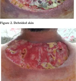
The patient was referred to plastic surgery clinic due to development of a necrotic lesion on the skin measuring 20x10 cm and a subcutaneous cavitary lesion during her follow up (Figure 1). She was considered to have necrotizing fasciitis due to infected appearance of lesion and presence of diabetes mellitus. We planned wound debridement and classical wound care followed by reconstruction with a graft or flap. So necrotic areas in the skin lesion were debrided and biopsy was performed (Figure 2). Histopathological examination revealed a high degree of neutrophilic infiltration and non-specific inflammatory findings. The patient was re-evaluated together with the dermatologist and the infectious disease specialist under the light of these histopathological and clinical findings, and the patient was considered to have an ulcerative type pyoderma gangrenosum. Detailed examinations were done for other underlying reasons; however no abnormal test results were obtained.
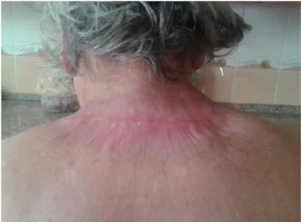
The initial reconstruction plan was abandoned due to the possibility of additional surgical morbidity known as pathergy. An immune-modulatory drug, Dapsone 50 mg bid via per-oral route was recommended by the dermatologist as the patient had diabetes mellitus. The patient was followed up with classical wound care by our plastic surgery clinic. The wound was observed to progressively fill with granulation and eventually completely recovered with scar tissue (Figure 3, 4).
DISCUSSION
PG was first described by Brunsting et al. in 19305. It is a rare non-infectious disease addressed in the spectrum of neutrophilic dermatoses5. The etiology
of PG is not well understood, but it is generally considered to be an aberrant and possibly auto-reactive immune response characterized by a dermal neutrophilic infiltrate2. Classically, lesions begin as
tender papules, papulo-pustules or vesicles, evolving into painful and rapidly enlarging ulcers6. Healing
frequently leaves a cribriform scar, which may lead to considerable disfiguring6. On histology, PG lesions do not display specific features, being typically characterized by a prominent dermal neutrophilic infiltrate with abscess formation, with no signs of infection and usually without significant vasculopathy6.
Figure 2. Debrided skin
Figure 3. Lesion with granulation.
PG has been estimated to occur in three to ten individuals/million worldwide6. PG is most commonly observed in young to middle-aged adults, with women being more affected6. There are four prototypic forms of PG; ulcerative (most common type), pustular, bullous, and vegetative, all of which differ based on their clinical presentations and
Cilt/Volume 43 Yıl/Year 2018 Pyoderma gangrenosum
associated disease5,7. The course of PG can be acute,
relapsing, or chronic7. The onset of PG can be just
as variable as its clinical presentation2. It may arise
spontaneously, or following cutaneous injury (pathergy)1. Some patients present with one or two
slowly growing ulcers while others experience a rapid onset with abrupt appearance of multiple rapidly enlarging ulcerations simultaneously2. In
adults, PG most commonly presents on the lower extremities and trunk7. Other sites of involvement
including breast, hand, head and neck, and peristomal skin have been reported7. In children, the
buttocks, perineum, and head and neck are most commonly affected7.
Figure 4. Recovered scar tissue
The majority of PG cases are associated with underlying diseases and conditions, such as rheumatoid arthritis, inflammatory bowel disease and a broad spectrum of hematological disorders and, rarely, after surgery or minor trauma4. Post-surgical PG is a rare but serious post-operative complication characterized by rapidly enlarging ulcers at the site of surgical incision, occurring on average 7 days after surgery1. The majority of cases
are associated with breast surgery, cardiothoracic surgery, and abdominal surgery1. Various minor procedures involving punctures or excisions may also incite post-surgical PG1.
Diagnosis of PG is challenging as clinical signs closely resemble an infectious process with no diagnostic laboratory or pathognomonic histological findings4. The work-up starts with a cutaneous
biopsy that preferentially includes the border of the ulcer and adjacent skin6. Histology is essential to
exclude vasculitis and malignancy, and special stains and tissue cultures may also be used to rule out
infection6. Complete work-up should include
extensive blood and urine screening and colonoscopy, in order to exclude underlying coexisting conditions6. In order to increase PG recognition and improve diagnosis specificity, diagnostic criteria have been proposed6. Diagnosis
of PG requires the two major criteria (a characteristic PG classical type ulcer, and the exclusion of other cause of cutaneous ulceration), combined with at least two minor criteria (suggestive history pathergy or cribriform scar, presence of a relevant associated disease, compatible histopathological findings, or response to systemic steroid treatment)6. In our case, atypical location of
the lesion and the lack of history of a clear trauma led to some delay in the diagnosis. However, we could rapidly make the diagnosis of PG with presence of a typical ulcer, presence of at least two minor criteria and elimination of other causes. Making an accurate diagnosis of PG and distinguish it from necrotizing fasciitis as early as possible is important for management and outcome of a patient who was initially admitted to a surgery clinic with a rapidly developing cutaneous lesion mimicking necrotizing fasciitis7,8. In necrotizing
fasciitis, the initial clinical manifestation is similar to cellulitis8. The overlying skin is shiny and tense
without clear lines of demarcation8. The associated severe pain is usually out of proportion to clinical lesion8. Unlike PG, the disease process progresses
very rapidly in a matter of hours rather than days8.
As the infection progresses the redness of the skin turns to purple or purple-black with tissue necrosis8.
Eventually frank gangrene sets in and progresses along the fascial planes8. In contrast to PG,
necrotizing fasciitis requires early aggressive management, immediate surgical evaluation, and intravenous antibiotics7.
Similarly to necrotizing fasciitis, suppurative and necrotic appearance of PG often incites surgeons to aggressively debride the wound1. Trauma from
surgery, which induces cytokine release and polymorphonuclear neutrophils chemotaxis, may induce formation of new PG ulcers or cause enlargement of the existing lesion2,4. Surgery alone
has not been shown to be an effective strategy for management of PG2. Any surgical procedure has to
be done as an adjunct to immunosuppression only in patients with stable disease5. Classical wound care
and vacuum-assisted closure has been used successfully to induce granulation tissue in slow
Korkmaz Solmaz et al. Cukurova Medical Journal
healing or large ulcers as long as effective immunosuppression2,5.
Although there is no universally accepted "gold standard", topical and systemic corticosteroids are considered the first immunosuppressive option in patients with PG6. Other anti-inflammatory and
immunosuppressive drugs can be used as steroid-sparing agents or alternative drugs for patients with refractory disease6. In our case, Dapsone was
preferred by the dermatologist for the first step therapy, because our patient had diabetes mellitus. As stated in the literature, classical wound care was performed together with an effective immunosuppressive therapy and rapid wound healing was observed.
In conclusion, surgeons should keep in mind the possibility of pyoderma gangrenosum in a patient who has a cutaneous lesion with suppurative and necrotic appearance mimicking necrotizing fasciitis which requires aggressive surgical debridement, so surgery- induced additional morbidity may be prevented. An integrated work up gains importance in presence of such lesions which are difficult to discriminate and which may be challenging unless diagnosed and treated correctly.
REFERENCES
1. Zuo KJ, Fung E, Tredget EE, Lin AN. A systematic review of post-surgical pyoderma gangrenosum: identification of risk factors and proposed management strategy. J Plast Reconstr Aesthet Surg. 2015;68:295-303.
2. Patel F, Fitzmaurice S, Duong C, He Y, Fergus J, Raychaudhuri SP et al. Effective strategies for the management of pyoderma gangrenosum: a comprehensive review. Acta Derm Venereol. 2015;95:525-31.
3. Tolkachjov SN, Fahy AS, Cerci FB, Wetter DA, Cha SS, Camilleri MJ. Postoperative Pyoderma Gangrenosum: A Clinical Review of Published Cases. Mayo Clin Proc.2016;91:1267-79.
4. Abtahi-Naeini B, Bagheri F, Pourazizi M, Forozeshfard M, Saffaei A. Unusual cause of breast wound: postoperative pyoderma gangrenosum. Int Wound J 2017;14:285-7.
5. Wollina U, Tchernev G. Pyoderma gangrenosum: pathogenetic oriented treatment approaches. Wien Med Wochenschr. 2014;164:263-73.
6. Gameiro A, Pereira N, Cardoso JC, Gonçalo M. Pyoderma gangrenosum: challenges and solutions. Clin Cosmet Investig Dermatol. 2015;8:285-93. 7. Barr KL, Chhatwal HK, Wesson SK, Bhattacharyya
I, Vincek V. Pyoderma gangrenosum masquerading as necrotizing fasciitis. Am J Otolaryngol. 2009;30:273-6.
8. Mahajan AL, Ajmal N, Barry J, Barnes L, Lawlor D. Could your case of necrotisingfascitis be Pyoderma gangrenosum? Br J Plast Surg. 2005;58:409-12.