© 2012 DEÜ
TIP FAKÜLTESİ DERGİSİ CİLT 26, SAYI 3, (ARALIK) 2012, 195 - 199Magnetic Resonance Imaging Findings In A Boy
With Tay-Sachs Disease
TAY-SACHS HASTALIĞI BULUNAN BİR ERKEK ÇOCUĞUNDA MANYETİK REZONANS
GÖRÜNTÜLEME BULGULARI
Erhan BAYRAM
1, Yasemin TOPCU
1, Gülçin AKINCI
1, Handan ÇAKMAKÇI
2, Nur ARSLAN
3,
Semra HIZ KURUL
11Dokuz Eylul University Faculty of Medicine, Department of Pediatric Neurology
2Dokuz Eylul University Faculty of Medicine, Department of Radiology
3Dokuz Eylul University Faculty of Medicine, Department of Pediatric Gastroenterology
Erhan BAYRAM
Dokuz Eylül Üniversitesi Tıp Fakültesi
Çocuk Sağlığı ve Hastalıkları AD Çocuk Nörolojisi BD 35340 İnciraltı, İzmir Tel: (232) 4123624 Faks: (232) 4599723 e-posta: [email protected] SUMMARY
Tay-Sachs is a neurodegenerative lysosomal storage disease that is caused by the mutations in the HEXA gene. Decreased ß-hexosaminidase A activity leads to the accumulation of the GM2 gangliosides in neuron cytoplasms and causes progressive neurologic dysfunction. Magnetic resonance imaging findings drastically change during the progression of the disease. At the early stage of the disease T2 weighted images demonstrate hyperintense lesions in basal ganglia or non-specific findings. In the late phase of the disease cerebral and cerebellar atrophy, and basal ganglia and white matter T2 hyperintensities can be seen. In this paper, we reported a 17 month-old boy with Tay-Sachs disease whose clinical and magnetic resonance imaging findings progressed in 5 months period.
Key words: Tay-Sachs disease, child, magnetic resonance imaging ÖZET
Tay-Sachs HEXA genindeki mutasyonların neden olduğu nörodejeneratif bir lizozomal depo hastalığıdır. ß-heksosaminidaz A aktivitesinin düşüklüğü nedeniyle nöron sitoplazmalarında GM2 gangliozid birikimi ve bunun sonucunda da ilerleyici nörolojik disfonksiyon gelişir. Hastalığın progresyonu ile birlikte beyin manyetik rezonans görüntüleme bulguları da dramatik olarak değişir. Hastalığın erken dönemlerinde ba-zal ganglionlarda T2 ağırlıklı görüntülerde belirgin hiperintens lezyonlar ya da spesifik olmayan bulgular görülebilir. Hastalığı geç dönemlerinde ise serebral ve serebellar atrofi, bazal ganglion ve beyaz cevherde T2 hiperintens lezyonlar görülebilir. Bu makalede 5 aylık bir sürede klinik ve manyetik rezonans görüntüleme bulguları ilerleyen 17 aylık bir TAY-Sachs hastalığı olgusu sunulmuştur.
Anahtar sözcükler: Tay-Sachs hastalığı, çocuk, manyetik rezonans görüntüleme
The GM2 gangliosidoses are a group of rare sphingolipid metabolism disorders, caused by deficiency of lysosomal ß‐hexosaminidase activity. GM2 ganglio‐ sidoses are characterized by the accumulation of GM2
ganglioside in the central nervous system and progressive neurologic deterioration (1). Lysosomal ß‐hexosaminidase consists of two isoenzymes, A and B. Isoenzyme A defici‐ ency causes Tay‐Sachs disease whereas both isoenzymes
A and B deficiency causes Sandhoff disease (2). The incidence of Tay‐Sachs disease in Turkey is 0.54/100000 (3). In Tay‐Sachs and Sandhoff diseases, bilateral thalamic hypodensity have been shown in brain computed tomography studies and that has been considered diagnostic for both disorders (4). Cranial magnetic resonance imaging (MRI) studies with GM2 gangliosidosis patients were reported rarely. Symmetrical T2 high and T1 low intensities in the basal ganglia, and symmetrical T2 high intensities in white matter and cerebral atrophy were described (2).
Here, we presented a case with Tay‐Sachs disease whose cranial MRI findings had changed dramatically in 5 months follow up period.
CASE REPORT
Twelve months of age male patient admitted to hospital with the complaints of unable to speak and walk. He had achieved normal psychomotor development until six months of age and progressive neuromotor deteri‐ oration occured subsequently. At 7 months of age the patient has been unable to sit unaided and 1 year of age unable to speak and walk, and then he showed hypersensitivity to sound. He was the second child of first cousin parents. In the family history, four aunts and one uncle of the patient had died with similar clinical symptoms and signs. On physical examination his weight was 9.8 kg (‐1.43 SDS), height 75 cm (‐1.80 SDS), head circumference 48 cm (0.40 SDS). He had relative macro‐ cephaly and generalized hypotonia. His eye fixation was poor. Cherry red spots were seen in both fundi. Labo‐ ratory tests including complete blood count, biochemistry, creatinine phosphokinase, thyroid function tests were normal. Cranial MR images was obtained at 12 months, and partial agenesis of corpus callosum rostral part was detected (Figure 1). Cerebral and cerebellar white matter signals were normal on MRI. With the possible diagnosis of Tay‐Sachs disease enzyme assay from serum sample was studied. Leukocyte ß‐hexosaminidase A activity was 0.57 nmol/mg protein/h (control range: 87.36‐109.2 nmol/mg protein/h) and total leukocyte ß‐hexosaminidase activity was 1125.89 nmol/mg protein/h (control range: 1161.86‐1657.9 nmol/mg protein/h). The diagnosis of Tay‐
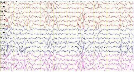
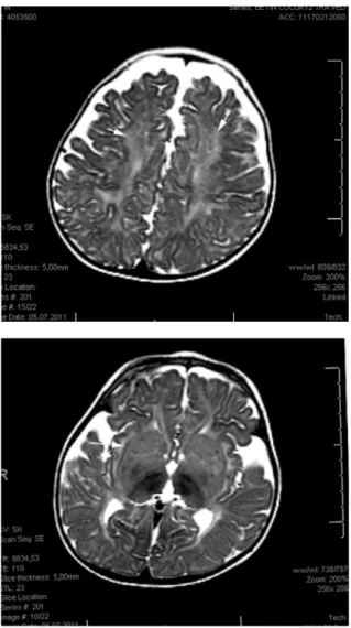
Sachs disease was made with clinical findings and decreased serum ß‐hexosaminidase A activity. Genetic counseling was given. The patient showed slowly progressive clinical symptoms and deterioration. At the age of 16 months a generalized tonic‐clonic convulsion during a febrile period occurred. Electroencephalography showed generalized spike and wave complexes (Figure 2). Follow up cranial MR images of the patient were obtained at the age of 17 months and showed high intensity on T2 weighted image in cerebral and cerebellar white matter. Bilateral, increased volume and signal density were detected in the corpus striatum (Figure 3).
Figure 1. Initial T2 weighted axial MRI (a,b), T1 weighted sagital
MRI (c) of the brain show normal basal ganglia and white matter signal and partial agenesis of the corpus callosum rostral segment(arrow on c).
Figure 2. Generalized spike and wave complexes in EEG of the patient
DISCUSSION
Tay Sachs disease is an autosomal recessive inherited neurodegenerative disorder due to mutations in the HEXA gene localized on chromosome 15q23‐q24, resulting a decreased ß‐hexosaminidase A activity (5,6). As a result of the deficient enzyme activity, the GM2 gangliosidoses accumulate in the cytoplasm of the neurons in central
nervous system and cause neuronal dysfunction. Two types of the disease have been described: acute infantile form and adult onset form (7). The gold standard method for the diagnosis of GM2 gangliosidosis is the measure‐ ment of ß‐hexosaminidase activity in plasma, serum and/ or fibroblasts (1). Our patient’s diagnosis was made by de‐ monstration of deficient enzyme activity of serum sample.
Figure 3. Follow up cranial T2 weighted MRI (a-b) images reveal
high signal intensity on cerebral white matter(arrows on a). Bilateral, increased volume and signal of the corpus striatum (arrow on b).
Several cases have been reported with MRI findings of symmetrical T2 high and T1 low intensities in the caudate nucleus, globus pallidus and putamen. With the prog‐ ression of the disease, cortex, cerebellum and white matter show symmetrical T2 high intensity lesions and later, cerebral atrophy can be seen (2). The accumulation of the GM2 ganglioside in the cytoplasm of the neurons induces neuronal death (8). Maegawa et al reported 21 new cases and reviewed 134 previously reported patients with GM2 gangliosidosis. In their series, the most frequent finding was cerebellar atrophy that was followed by generalized
cerebral atrophy (mean age 1.8‐28.6 years) (1). This result may be resulted from the inclusion of the adult patients in the study. Interestingly, 17.1% of the patients had normal neuroimaging studies (1). In our patient, the first MRI at 12 months of age showed only a partial agenesis of rostral portion of the corpus callosum. Grosso et al and Beck et al reported some GM2 gangliosidosis patients with corpus callosum abnormalities (4,9). Five months later, our patient’s crainal MRI showed high intensity on T2 weighted image in cerebral and cerebellar white matter, and increased volume and signal density were detected in the corpus striatum. These findings were typical for GM2 gangliosidoses (2).
The differential diagnosis with the involvement of the basal ganglia includes Sandhoff disease, Leigh disease, viral encephalitis, Wilson disease, Hallervorden Spatz disease and mitochondrial encephalomyopathy (10). However, the clinical findings, the existence of cherry red spots and also the decreased level of ß‐hexosaminidase A activity confirmed the diagnosis of our patient.
In conclusion, at an early stage of the Tay Sachs disease cranial MRI findings may be non‐specific, but control imagings would provide complementary findings at the diagnosis of the disease.
REFERENCES
1. Maegawa GH, Stockley T, Tropak M, et al. The natural history of juvenile or subacute GM2 gangliosidosis: 21 new cases and literature review of 134 previously repor-ted. Pediatrics 2006;118:1550-1562.
2. Mugikura S, Takahashi S, Higano S, Kurihara N, Kon K, Sakamoto K. MR findings in Tay-Sachs disease. J Comput Asist Tomogr 1996;20:551-555.
3. Ozkara HA, Topcu M. Sphingolipidoses in Turkey. Brain Dev 2004;26:363-366.
4. Grosso S, Farnetani MA, Berardi R, et al. GM2 ganglio-sidosis variant B1 neuroradiological findings. J Neurol 2003;250:17-21.
5. Giraud C, Dussau J, Azouguene E, Feillet F, Puech JP, Caillaud C. Rapid identification of HEXA mutations in Tay-Sachs patients. Biochem Biophys Res Commun 2010;392:599-602.
6. Brismar J, Brismar G, Coates R, Gascon G, Ozand P. Increased density of the thalamus on CT scans in pati-ents with GM2 gangliosidoses. Am J Neuroradiol 1990; 11:125-130.
7. Neudorfer O, Pastores GM, Zeng BJ, Gianutsos J, Zaroff CM, Kolodny EH. Late-onset Tay-Sachs disease: pheno-typic characterization and genopheno-typic correlations in 21 affected patients. Genet Med 2005;7:119-123.
8. Huang JQ, Trasler JM, Igdoura S, Michaud J, Hanal N,
Gravel RA. Apoptotic cell death in mouse models of GM2 gangliosidosis and observations on human Tay-Sachs and Sandhoff diseases. Hum Mol Genet 1997;6:1879-1885. 9. Beck M, Sieber N, Goebel HH. Progressive cerebellar
ataxia in juvenile GM2 gangliosidosis type Sandhoff. Eur J Pediatr 1998;157:866-867.
10. Barcovic SF, Karpati G, Carpenter S, Lang AE. Prog-ressive dystonia with bilateral putaminal hypodensities. Arch Neurol 1987;44:1184-1187.