Molecular Survey of Hepatozoonosis in Natural Infected Dogs: First
Detection and Molecular Characterisation of Hepatozoon canis in
Kyrgyzstan
Kursat ALTAY
1
Mehmet Fatih AYDIN
2Ayperi AYTMIRZAKIZI
3Zarima JUMAKANOVA
3Ayday CUNUSOVA
3Nazir DUMANLI
3,41 Department of Parasitology, Faculty of Veterinary Medicine, University of Sivas Cumhuriyet, TR-58140 Sivas - TURKEY 2 Department of Public Health, Faculty of Health Sciences, University of Karamanoglu Mehmetbey, TR-70200 Karaman
- TURKEY
3 Faculty of Veterinary Medicine, Kyrgyz-Turkish Manas University, 720044, Bishkek, KYRGYZSTAN
4 Department of Parasitology, Faculty of Veterinary Medicine, University of Firat, TR-23119 Elazig - TURKEY Article ID: KVFD-2018-20352 Received: 13.06.2018 Accepted: 26.09.2018 Published Online: 27.09.2018
How to Cite This Article
Altay K, Aydin MF, Aytmirzakizi A, Jumakanova Z, Cunusova A, Dumanli N: Molecular survey of hepatozoonosis in natural infected dogs:
First detection and molecular characterisation of Hepatozoon canis in Kyrgyzstan. Kafkas Univ Vet Fak Derg, 25 (1): 77-81, 2019. DOI: 10.9775/ kvfd.2018.20352
Abstract
Canine hepatozoonosis is a tick-borne protozoan disease and widespread in Europe, Africa, Asia and America. There is not any available data about the presence of Hepatozoon infections in dogs in Kyrgyzstan. In the study we aimed that investigate the presence of Hepatozoon
canis and the prevalance of Hepatozoon infections in dogs from Kyrgyzstan using polymerase chain reaction (PCR) and sequence analysis.
To determine the prevelance of hepatozoonosis in dogs, a total of 170 blood samples were applied to PCR to amplify a fragment of 666 bp found in 18S SSU rRNA gene of Hepatozoon spp. The PCR results shown that Hepatozoon infection rate was 28.8% (49/170) in dogs. Eleven representative positive samples were sequenced to classification of the species. The nucleotide sequences were compared to the H. canis sequences which registered in GenBank using the basic local alignment search tool. Results of sequence analyse of 11 amplicons indicated that 8 were 100% identical and the other 3 sequences shared 99% similarity with H. canis. The sequences were deposited in Genbank with accession numbers from MG917709 to MG917719. It was the first record of H. canis in dogs in Kyrgyzstan.
Keywords: Hepatozoonosis, Hepatozoon canis, Dog, PCR, Sequencing, Kyrgyzstan
Doğal Enfekte Köpeklerde Hepatozoon Enfeksiyonlarının Moleküler
Yöntemlerle Araştırılması: Hepatozoon canis’in Kırgızistan’da İlk Tespiti
Öz
Köpeklerde hepatozoonozis kenelerle nakledilen ve Afrika, Avrupa, Asya ile Amerika’da yaygın olarak görülen protozoan bir hastalıktır. Kırgızistan’da köpeklerde Hepatozoon enfeksiyonlarının varlığına dair bir bilgiye ulaşılamamıştır. Bu çalışmada, polimeraz zincir reaksiyonu (PZR) ve sekans analizi ile Kırgızistan’da köpeklerde Hepatozoon enfeksiyonlarının yaygınlığının ve Hepatozoon canis’in varlığının belirlenmesi amaçlanmıştır. Hepatozoon enfeksiyonlarının prevalansını belirlemek için 170 kan örneğine, Hepatozoon spp. 18S SSU rRNA geninin 666 bp’lik kısımını amplifiye etmek üzere PZR uygulanmıştır. PZR sonucunda Hepatozoon spp. enfeksiyon oranı %28.8 (49/170) olarak ortaya çıkmıştır. On bir PZR pozitif DNA örneğinin DNA dizilimi belirlenmiş ve elde edilen DNA dizilimleri BLAST programı kullanılarak GenBank’ta kayıtlı diğer dizilimlerle karşılaştırılmıştır. Bunlardan 8’inin GenBankasında kayıtlı H. canis DNA dizilimleri ile %100 oranında eşleştiği, diğer 3 dizilimin ise %99 oranında bir benzerliğe sahip olduğu belirlenmiştir. DNA dizilimleri GenBankasına MG917709-MG917719 numaraları altında kaydedilmiştir. Bu çalışma Kırgızistan’da köpeklerde H. canis’in belirlendiği ilk çalışma niteliğindedir.
Anahtar sözcükler: Hepatozoonozis, Hepatozoon canis, köpek, PZR, Sekans analizi, Kırgızistan
INTRODUCTION
Canine hepotozoonosis is a tick-borne protozoan disease
and widespread in Europe, Africa, Asia and America. The first hepatozoonosis case was determined in India, 1905, named as Leukocytozoon canis. After the parasite was
İletişim (Correspondence)
+90 346 2191010 Fax: +90 346 2191812detected in neutrophils, it transferred to the genus of
Hepatozoon [1,2]. There are more than 340 species in
the genus of Hepatozoon and two of them infect dogs,
Hepatozoon canis and Hepatozoon americanum [1,3]. On
the other hand, some new genotypes have been found in dogs with the molecular-genetic based studies in recent years [4,5]. The disease is transmitted by the ingestion of
the vector ticks. Rhipicephalus sanguineus in Asia, Europe, Africa and Brazil, Amblyomma maculatum in South America are known as vector of canine hepatozoonosis [6-8].
Clinical signs of hepatozoonosis in dogs depend on caused species, the nutritional and the others individual factors.
H. canis infections can change from subclinical to severe
and fatal disease. Subclinical and mild disease is common and generally have low parasitemia but 100% of neutrophils can be infected with H. canis in the severe disease which characterised by lethargy, fever and extreme weight loss.
H. americanum infections causes fever, generalised pain
or hyperaesthesia, myositis, muscle atrophy, weakness, depression, reluctance to rise and mucopurulent ocular discharge [2,9,10].
Diagnosis of canine hepatozoonosis may be done with seen of intracytoplasmatic gamonts in neutrophils and monocytes by microscopic examination of the thin blood smears. Secondly the histopathologically meront and mono- soic cysts can be investigated. The biopsy of skeletal muscle is the gold standard for the diagnosis of H. americanum infections in dogs because gamonts of the parasite are rarely seen in thin blood smears. Serological methods such as indirect immunofluorescence and enzyme-linked immunosorbent assay have been used diagnosis of
Hepatozoon infections in dogs. But serological methods
are generally preferred in epidemiological studies to detection of chronic infections [2,9,10].
In the recent years, molecular-genetic based diagnostic methods such as polymerase chain reaction (PCR) and
DNA sequencing have been used for survey of the presence, the characterisation and detection of prevalence of Hepatozoon species in dogs [11-14]. These methods have
been accepted to be more sensitive and specific than microscopic and serological methods for the diagnosis of hepatozoonosis and the other blood parasites [15-22].
We have not found available data about canine hepato-zoonosis in dogs in Kyrgyzstan. In this study we aimed that investigate the presence of H. canis and to determine the prevalence of Hepatozoon infections in dogs from Kyrgyzstan using polymerase chain reaction (PCR) and sequence analysis.
MATERIAL and METHODS
Study Area and Collection of Blood Samples
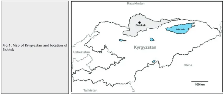
Kyrgyzstan Republic is a Central Asia Union country and it borders Kazakhstan to the north, Uzbekistan to the west, Tajikistan to the southwest and China to the southeast. The country is landlocked and mountainous, located in the Northern Hemisphere in the center of the Eurasian continent, as well as it is far away from large water bodies (the seas and oceans) and close of the desert. Bishkek, formerly Pishpek or Frunze is the largest city and the capital of the country [23] (Fig. 1). The blood samples were collected
from dogs living in a shelter in Bishkek with cooperation with the shelter’s and Kyrgyz-Turkish Manas University Veterinary Teaching Hospital’s staffs.
The sampling was carried out from May 2016-October 2017. The dogs have been accepted without clinical signs with behavioral inspection of the dogs, but particular clinical examination was not conducted. A total of 170 blood samples were obtained. The blood samples were collected in tubes containing ethylendiamine tetraacetic acid (EDTA) and stored -20°C until use for DNA extraction.
Fig 1. Map of Kyrgyzstan and location of
Total DNA Extraction from Blood Samples and Polymerase Chain Reaction (PCR)
Total DNA extraction from blood samples was performed using a PureLink Genomic DNA mini kit (Invitrogen, Carlsbad, USA) following the manufacturer’s instructions. The DNA samples were stored at -20°C until use for PCR. To determine the presence Hepatozoon spp. in the DNA samples, a PCR analysis was carried out using Hep-F (5’-ATACATGAGCAAAATCTCAAC-3’) and Hep-R (5’-CTTATT ATTCCATGCTGCAG-3’) primers which amplify the partial 18S small subunite ribosomal RNA (18S SSU rRNA) gene of Hepatozoon species [24]. Sterile water (DNase, RNase
free) and H. canis positive control DNA were used as negative and positive control in PCR, respectively. H. canis positive control DNA was provided from Department of Parasitology, Faculty of Veterinary Medicine, Selcuk University. The PCR was practiced in a touchdown termo-cycler in a total reaction volume of 25 µL according to Aydin et al.[4].
Eleven PCR positive sample products were purified using QIAquick PCR purification kit (Qiagen, Hilden, Germany). The purified PCR samples were sequenced in a commercial company (Iontek, Istanbul, Turkey). The sequences of the partial 18S SSU rRNA gene of Hepatozoon spp. obtained in the study were submitted to basic local alignment search tool (BLAST) similarity search. After the sequences were identified they have been deposited in GenBank.
RESULTS
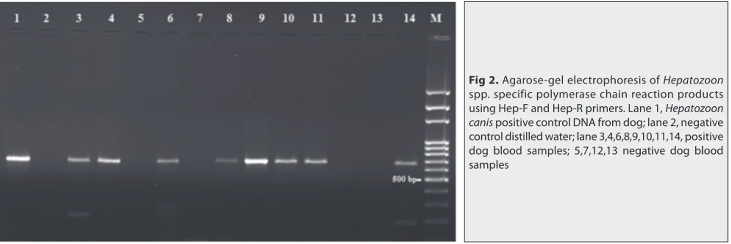
A total of 170 blood samples were analyzed with PCR to amplify a fragment of 666 bp located on 18S SSU rRNA gene of Hepatozoon spp. using Hep-F and Hep-R primer pairs (Fig. 2).
Hepatozoon spp. was detected in 49 blood samples of
investigated 170 dogs with PCR. Out of 170 field samples, 121 were found as negative in terms Hepatozoon spp. To identify and confirm the PCR positive results, randomly selected representative 11 PCR positive samples were
purified and sequenced. A BLAST search performed to compare the sequences alignments with the other
Hepatozoon spp. sequences registered in GenBank under
the accession numbers from MG917709 to MG917719. Nucleotide sequences of partial 18S SSU rRNA gene of
Hepatozoon spp. obtained in the study (MG917709 to
MG917719) were aligned with the GenBank registered 18S SSU rRNA gene sequences of H. canis (KX712126, KX880505, KY197000). Sequence alignment of the PCR positive samples were identified as H. canis. BLAST analyse showed that 8 isolates were shared 100% similarity with
H. canis isolates while the other 3 isolates were shared
99% similarity from Czeck Republic (accesion number: KX712126), Iran (accesion number: KX880505), Turkey (accesion number: KY197000).
DISCUSSION
Canine hepatozoonosis, a worldwide protozoon parasitic infection, has been reported in many countries of Africa, America, Asia and Europe such as Nigeria [25], Italy [14],
Thailand [26], Croatia [27], Brazil [28], Argentina [11], and
Turkey [13]. There are not any available data on presence
Hepatozoon infections in dogs in Kyrgyzstan. The DNA
based molecular diagnostic techniques such as PCR, RLB are widely used for detection and identification of blood parasites from animals. These techniques have superiority in specifity and sensitivity, and they permit to identification new genotypes and/or species [11-22]. Otronto et al.[14]
showed that PCR the most sensitive assay for the detection of H. canis infection in dogs. They suggest that PCR can be used in epidemiological studies as a convenient diagnostic test. Using PCR, this study revealed hepotozoonozis has a high prevalence in dogs in Kyrgyzstan. 49 out of 170 blood samples were found as positive for Hepatozoon spp. with PCR. The result showed that hepatozoonosis has a high prevalence in dogs in Kyrgyzstan.
Hepatozoon canis and H. americanum are primary agents of
canine hepatozoonosis. H. canis is more prevalent species than H. americanum in dogs. H. canis is seen in Europe,
Fig 2. Agarose-gel electrophoresis of Hepatozoon
spp. specific polymerase chain reaction products using Hep-F and Hep-R primers. Lane 1, Hepatozoon
canis positive control DNA from dog; lane 2, negative
control distilled water; lane 3,4,6,8,9,10,11,14, positive dog blood samples; 5,7,12,13 negative dog blood samples
Asia, Africa and also America while H. americanum is limited in America continent [1-3,29]. In this study 49 samples
were detected positive with Hepatozoon-genus specific PCR. The partial sequences were determined of 11 PCR positive samples. The positive samples were identified as
H. canis according to sequence alignments. BLAST analysis
of the sequences showed that 8 isolates were shared 100% similarity with H. canis isolates while the other 3 isolates were shared 99% similarity from Czeck Republic (accesion number: KX712126), Iran (accesion number: KX880505), Turkey (accesion number: KY197000). It was the first detection of H. canis in dogs from Kyrgyzstan using molecular methods. There is need more comprehensive studies to detection of the infection status in Kyrgyzstan. Currently, there are no protective vaccines for the prevention of canine hepatozoonosis. Therefore, the only powerful method to prevent the canine hepatozoonosis is the control of vector ticks. On the other hands wild carnivores such as jackals have been found naturally infected with H. canis [30]. This situation enhances the
importance of epidemiological knowledge of Hepatozoon infections in terms of the development and application of appropriate control strategies. This survey reveals the high prevalence of canine hepatozoonosis in dogs in Kyrgyzstan, and H. canis is the primary agent of the diseases in the country. There is still need more prevalent epidemiological studies on canine hepatozoonosis in the country.
A
cknowledmentsWe thank Dr. Ferda Sevinc (Selcuk University) for Hepatozoon
canis positive control DNA animal shelter staffs and
Kyrgyz-Turkish Manas University Veterinary Teaching Hospital staffs for their assistance in sample collection.
REFERENCES
1. Baneth G: Perspectives on canine and feline hepatozoonosis. Vet
Parasitol, 181, 3-11, 2011. DOI: 10.1016/j.vetpar.2011.04.015
2. Dantas-Tores F, Otranto D: Hepatozoonosis. In, Marcondes CB (Ed):
Arthropod Borne Diseases, Springer International Publishing Switzerland, 363-368, 2017. DOI: 10.1007/978-3-319-13884-8
3. Smith TG: The genus Hepatozoon (Apicomplexa: Adeleina). J Parasitol,
82, 565-585, 1996. DOI: 10.2307/3283781
4. Aydin MF, Sevinc F, Sevinc M: Molecular detection and characterization
of Hepatozoon spp. in dogs from central part of Turkey. Ticks Tick Borne
Dis, 6, 388-392, 2015. DOI: 10.1016/j.ttbdis.2015.03.004
5. Orkun Ö, Koç N, Sürsal N, Çakmak A, Nalbantoğlu S, Karaer Z:
Molecular characterization of tick-borne blood protozoa in stray dogs from central Anatolia region of Turkey with a high-rate Hepatozoon infection. Kafkas Univ Vet Fak Derg, 24 (2): 227-232, 2018. DOI: 10.9775/ kvfd.2017.18678
6. Baneth G, Samish M, Alekseev E, Aroch I, Shkap V: Transmission of
Hepatozoon canis to dogs by naturally fed or percutaneously injected Rhipicephalus sanguineus ticks. J Parasitol, 87, 606-611, 2001. DOI:
10.1645/0022-3395(2001)087[0606:TOHCTD]2.0.CO;2
7. Baneth G, Samish M, Shkap V: Life cycle of Hepatozoon canis
(Apicomplexa: Adeleorina: Hepatozoidae) in the ticks Rhipicephalus
299, 2007. DOI: 10.1645/GE-494R.1
8. Mathew JS, Ewing SA, Panciera RJ, Woods JP: Experimental
transmission of Hepatozoon americanum Vincent- Johnson et al., 1997 to dogs by the Gulf Coast tick, Amblyomma maculatum Koch. Vet Parasitol, 80, 1-14, 1998. DOI: 10.1016/S0304-4017(98)00189-7
9. Taylor MA, Coop RL, Wall RL, Parasites of dogs and cats. Veterinary
Parasitology, Blackwell Publishing, 409-433, 2007.
10. Baneth G, Vicent-Jhonson N: Hepatozoonosis. In, Shaw SE, Day MJ
(Eds): Arthropod Borne Infectious Diseases of the Dog and Cat. 78-88, Manson Publishing Ltd, London, 2005.
11. Eiras DF, Basabe J, Scodellaro CF, Banach DB, Matos ML, Krimer A, Baneth G: First molecular characterization of canine
hepato-zoonosis in Argentina: Evaluation of asymptomatic Hepatozoon canis infection in dogs from Buenos Aires. Vet Parasitol, 149, 275-279, 2007. DOI: 10.1016/j.vetpar.2007.07.010
12. Aktas M, Ozubek S, Altay K, Sayın Ipek ND, Balkaya I, Utuk AE, Kirbas A, Simsek S, Dumanli N: Molecular detection of tick-borne
rickettsial and protozoan pathogens in domestic dogs from Turkey.
Parasit Vectors, 8:157, 2015. DOI: 10.1186/s13071-015-0763-z
13. Aktas M, Ozubek S, Altay K, Balkaya I, Utuk AE, Kirbas A, Simsek S, Dumanli N: A molecular and parasitological survey of Hepatozoon
canis in domestic dogs in Turkey. Vet Parasitol, 209, 264-267, 2015. DOI:
10.1016/j.vetpar.2015.02.015
14. Otranto D, Dantas-Torres F, Weigl S, Latrofa MS, Stanneck D, de Caprariis D, Capelli G, Baneth G: Diagnosis of Hepatozoon canis in young
dogs by cytology and PCR. Parasit Vectors, 4:55, 2011. DOI: 10.1186/1756-3305-4-55
15. Altay K, Dumanlı N, Aktaş M, Özübek S: Survey of Anaplasma
infections in small ruminants from East part of Turkey. Kafkas Univ Vet Fak
Derg, 20 (1): 1-4, 2014. DOI: 10.9775/kvfd.2013.9189
16. Altay K, Ataş AD, Özkan E: Sivas yöresi koyun keçi ve kenelerde
Theileria ve Babesia türlerinin moleküler yöntemlerle araştırılması. Manas J Agr Vet Life Sci, 7 (1): 30-39, 2017.
17. Altay K, Dumanlı N, Holman PJ, Aktaş M: Detection of Theileria ovis
in naturally infected sheep by nested PCR. Vet Parasitol, 127 (2): 99-104, 2005. DOI: 10.1016/j.vetpar.2004.09.012
18. Altay K, Aktaş M, Dumanlı N: Doğu ve Güneydoğu Anadolu
Bölgelerinde küçük ruminantlarda Theileria enfeksiyonları. Türkiye Parazitol
Derg, 31 (4): 268-271, 2007.
19. Dumanlı N, Aktaş M, Çetinkaya B, Çakmak A, Köroğlu E, Saki CE, Erdoğmuş Z, Nalbantoğlu S, Öngör H, Şimşek S, Karahan M, Altay K:
Prevalence and distribution of tropical theileriosis in eastern Turkey. Vet
Parasitol, 127 (1): 9-15, 2005. DOI: 10.1016/j.vetpar.2004.08.006
20. Karagenc TI, Pasa S, Kirli G, Hosgor M, Bilgic HB, Ozon YH, Atasoy A, Eren H: A parasitological, molecular and serological survey of
Hepatozoon canis infection in dogs around the Aegean coast of Turkey. Vet Parasitol, 135, 113-119, 2006. DOI: 10.1016/j.vetpar.2005.08.007
21. Pasa S, Kiral F, Karagenc T, Atasoy A, Seyrek K: Description of dogs
naturally infected with Hepatozoon canis in the Aegean region of Turkey.
Turk J Vet Anim Sci, 33 (4): 287 289-295, 2009. DOI: 10.3906/vet-0801-11
22. Altay K, Dumanlı N, Aktaş M: A study on ovine tick-borne
hemoprotozoan parasites (Theileria and Babesia) in the East Black Sea Region of Turkey. Parasitol Res, 111 (1): 149-153, 2012. DOI: 10.1007/ s00436-011-2811-8
23. Ilyasov S, Zabenko O, Gaydamak N, Kirilenko A, Myrsaliev N, Shevchenko V, Penkina L: Climate profile of the Kyrgyz Republic, The
State Agency for Environmental Protection and Forestry under the Government of the Kyrgyz Republic, The United Nations Development Programme,Bishkek, 2013. http://www.kg.undp.org/content/dam/ kyrgyzstan/Publications/env-energy/kgz_Kyrgyzstan%20Climate%20 profile_ENG_for%20web-opt.pdf; Accessed: 17.05.2018.
24. Inokuma H, Okuda M, Ohno K, Shimoda K, Onishi T: Analysis of
the 18S rRNA gene sequence of a Hepatozoon detected in two Japanese dogs. Vet Parasitol, 106, 265-271, 2002. DOI: 10.1016/S0304-4017 (02)00065-1
25. Sasaki M, Omobowale O, Ohta K, Tozuka M, Matsuu A, Hirata H, Nottidge HO, Ikadai H, Oyamada T: A PCR-based epidemiological
survey of Hepatozoon canis in dogs in Nigeria. J Vet Med Sci, 70 (7): 743-745, 2008. DOI: 10.1292/jvms.70.743
26. Jittapalapong S, Rungphisutthipongse O, Maruyama S, Schaefer JJ, Stich RW: Detection of Hepatozoon canis in stray dogs and cats in
Bangkok Thailand. Ann N Y Acad Sci, 1081, 479-488, 2006. DOI: 10.1196/ annals.1373.071
27. Vojta L, Mrljak V, Curkovic S, Zivicnjak T, Marinculic A, Beck R: Molecular epizootiology of canine hepatozoonosis in Croatia. Int J
Parasitol, 39, 1129-1136, 2009. DOI: 10.1016/j.ijpara.2009.02.007
28. Rubini AS, dos Santos Paduan K, Von Ah Lopes V, O’Dwyer LH:
Molecular and parasitological survey of Hepatozoon canis (Apicomplexa: Hepatozoidae) in dogs from rural area of Sao Paulo state, Brazil. Parasitol
Res, 102 (5): 895-899, 2008. DOI: 10.1007/s00436-007-0846-7
29. Ivanov A, Tsachev I: Hepatozoon canis and hepatozoonosis in the
dog. Trakia J Sci, 6 (2): 27-35, 2008.
30. Penzhorn BL, Netherlands EC, Cook CA, Smit NJ, Vorster I, Harrison-White RF, Oosthuizen MC: Occurrence of Hepatozoon canis (Adeleorina:
Hepatozoidae) and Anaplasma spp. (Rickettsiales: Anaplasmataceae) in black-backed jackals (Canis mesomelas) in South Africa. Parasit Vectors, 11:158, 2018. DOI: 10.1186/s13071-018-2714-y