85
Isolated left ventricular diverticulum in an adult:
dynamic contrast-enhanced MRI findings
Eriflkinde izole sol ventrikül-divertikülüm: dinamik kontrast MR bulgular›
Cihan Duran, MD, Mecit Kantarc›*,MD, Kutlay Karaman, MD
Department of Radiology, Florence Nightingale Hospital, Kadir Has University School of Medicine, Istanbul, Turkey *Department of Radiology, GATA Haydarpasa Education Hospital ‹stanbul, Turkey
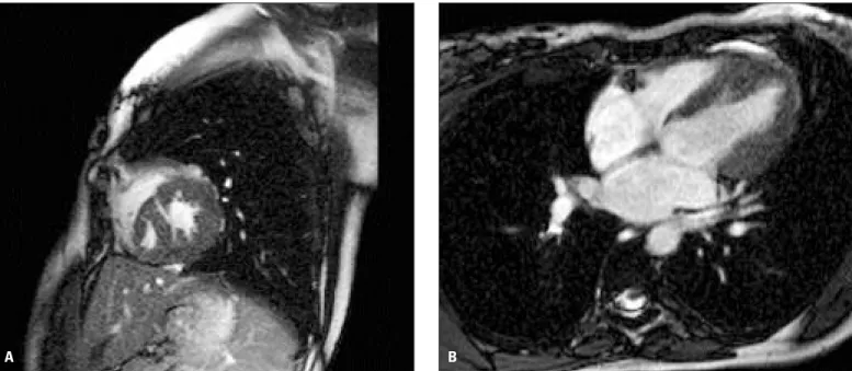
Congenital ventricular diverticulum is a rare cardiac mal-formation initially described in 1838 (1). Congenital aneurysm and diverticulum of the left ventricle are rare findings that can be detected by both echocardiography and/or left ventricular angiography. Ventricular aneurysm and diverticulum can be differentiated by several criteria. Contractility is the only reli-able parameter: aneurysm expands, whereas diverticulum contracts during ventricular systole. A 19-year-old woman, presenting with upper abdominal discomfort and palpitation, and sustained wide QRS tachycardia, was referred to our hos-pital. A 2D echocardiogram revealed left ventricular hypertro-phy and an apical diverticulum. Dynamic cardiac magnetic resonance imaging (MRI) demonstrated a diverticulum origi-nating at the apex of the left ventricle, near the interventricular
septum (Figure 1A, B). The ventricular diverticulum was attached by a narrow neck to the rest the left ventricle and was clearly visualized by MRI. The ostium of the diverticulum was opened at the anteroseptal wall, and the diverticulum itself did show active systolic contraction (Figure 2A, B). Although car-diac catheterization with left ventriculography can show the diverticulum, non-invasive dynamic cardiac MRI makes it eas-ier to see the difference between the aneurysm and diverticu-lum. Congenital diverticulum is a rare lesion and is discovered during adulthood, and usually asymptomatic (2). Our case with congenital ventricular diverticulum that has been demonstrat-ed by dynamic cardiac MRI, and the images suggest that dynamic cardiac MRI can be used for definition of this cardiac pathology.
Address for Correspondence: Mecit Kantarc›, MD, Department of Radiology, GATA Haydarpasa Education Hospital Istanbul, Turkey
Tel: +90 533 568 66 25 (GSM), +90 442 2361212-1521 (work), Fax: +90(442) 2361301, E-mail: [email protected]
Fig.1. Dynamic cardiac MR images in sagittal (A) and axial (B) views shows the diverticulum originating at the apex of the left ventricle, near the interventricular septum (D: diverticulum; LV: left ventricle; MR: magnetic resonance, arrows: interventricular septum).
A B
References
1. Gruberg L, Goldstein SA, Pfister AJ, Monsein LH, Evans DM, Leon MB. Cantrell's syndrome: left ventricular diverticulum in an adult
patient. Circulation 2000;101:109-10.
2. Wu JM, Yu CY. Isolated congenital left ventricular diverticulum. Pediatr Cardiol 1996;17:254-6.
Anadolu Kardiyol Derg 2005;5: 85-6 Duran et al.
Isolated left ventricular diverticulum in an adult
86
Fig. 2. Systolic phase dynamic MR images in sagittal (A) and axial (B) views show the diverticulum itself with active systolic contraction. (MR: magnetic resonance)