Incidence, risk factors and severity of retinopathy of
prematurity in Turkey (TR-ROP study): a prospective,
multicentre study in 69 neonatal intensive care units
Ahmet Yagmur Bas,
1Nihal Demirel,
1Esin Koc,
2Dilek Ulubas Isik,
3İbrahim Murat Hirfanoglu,
2Turan Tunc,
4on behalf of the TR-ROP Study Group
To cite: Bas AY, Demirel N, Koc E, et al. Br J Ophthalmol 2018;102:1711–1716. 1Department of Neonatology, Yildirim Beyazit University Faculty of Medicine, Ankara, Turkey
2Department of Neonatology, Gazi University Faculty of Medicine, Ankara, Turkey 3Department of Neonatology, Etlik Zubeyde Hanim Women’s Health Teaching and Research Hospital, University of Health Sciences, Ankara, Turkey 4Neonatology Division, Memorial Hospital, Istanbul, Turkey
Correspondence to Professor Ahmet Yagmur Bas, Department of Neonatology, Etlik Zubeyde Hanim Women’s Health Teaching and Research Hospital, Ankara 06010, Turkey; yagmur32@ yahoo. com Received 22 December 2017 Revised 23 January 2018 Accepted 16 February 2018 Published Online First 8 March 2018
AbsTrACT
background To evaluate the prevalence, risk factors and treatment of retinopathy of prematurity (ROP) in Turkey and to establish screening criteria for this condition.
Methods A prospective cohort study (TR-ROP) was performed between 1 April 2016 and 30 April 2017 in 69 neonatal intensive care units (NICUs). Infants with a birth weight (BW)≤1500 g or gestational age (GA)≤32 weeks and those with a BW>1500 g or GA>32 weeks with an unstable clinical course were included in the study. Predictors for the development of ROP were determined by logistic regression analyses.
results The TR-ROP study included 6115 infants: 4964 (81%) with a GA≤32 weeks and 1151 (19%) with a GA>32 weeks. Overall, 27% had any stage of ROP and 6.7% had severe ROP. A lower BW, smaller GA, total days on oxygen, late-onset sepsis, frequency of red blood cell transfusions and relative weight gain were identified as independent risk factors for severe ROP in infants with a BW≤1500 g. Of all infants, 414 needed treatment and 395 (95.4%) of the treated infants had a BW≤1500 g. Sixty-six (16%) of the treated infants did not fulfil the Early Treatment for Retinopathy of Prematurity requirements for treatment.
Conclusions Screening of infants with a GA≤34 weeks or a BW<1700 g appears to be appropriate in Turkey. Monitoring standards of neonatal care and conducting quality improvement projects across the country are recommended to improve neonatal outcomes in Turkish NICUs.
Trial registration number NCT02814929, Results.
InTroduCTIon
Retinopathy of prematurity (ROP), a vasoprolifer-ative disorder of the immature retina in premature infants, is a significant cause of blindness in many middle-income countries. The prevalence of ROP is lower in high-income countries, where risk factors such as oxygen administration and blood oxygen saturation are strictly monitored.1 Severe ROP is
typically found in infants with a very low gesta-tional age (GA) at birth in developed countries.1 2
Heavier and more mature babies can also develop ROP in developing countries, because there is insuf-ficient awareness of the risk factors of the disease process, a shortage of skilled professionals and/or a shortage of essential equipment to care for infants.3
In recent years, Turkey has been developing programmes to improve neonatal health. This study
(TR-ROP) determined the prevalence and treatment modalities of infants with ROP in Turkey and was the first multicentre study to analyse risk factors for ROP development in the country. Based on data obtained from infants, criteria for ROP screening in Turkey were evaluated. Because Turkey has received many refugees in recent years, this study also evalu-ated the prevalence of ROP in preterm infants born to refugees.
MeThods
The TR-ROP study was promoted by the Turkish Neonatology Society and included preterm infants screened for ROP between 1 April 2016 and 30 April 2017. In Turkey, the total number of neonatal intensive care units (NICUs) including neonatologists on the medical staff is 134 (22 private, 40 university and 72 state hospitals). In total, 69 NICUs (8 private, 39 university and 22 state hospitals) agreed to take part in the study (51% of all). Heads of the NICUs and directors of hospitals gave written consent to participate in the research. It was approved by the ethics committee and informed consent was obtained from the parents before the initial screening.
study population
This prospective cohort study evaluated the inci-dence and severity of ROP in relation to GA, birth weight (BW) and treatment modalities. The independent risk factors for the development of severe ROP in infants with a BW≤1500 g and for any ROP in infants with a BW>1500 g were assessed.
Infants with a BW≤1500 g or GA≤32 weeks and those with a BW>1500 g or GA>32 weeks, who were determined by the attending clini-cian to be at risk for ROP development, were screened. Then the medical records of retinal examinations of preterm infants who met the screening criteria were evaluated. The data on refugee infants were also recorded. Exam-inations took place in the NICU or outpatient facility (for discharged infants). Eligible infants who were discharged before the first screening and missed or did not complete all screening sessions were excluded from the study. The data are restricted to all babies who underwent all the screening sessions. Infants with congen-ital anomalies, chromosomal abnormalities and
on February 5, 2020 by guest. Protected by copyright.
http://bjo.bmj.com/
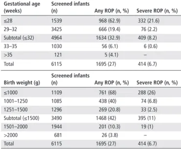
Table 1 ROP in relation to gestational age and birth weight
Gestational age (weeks)
screened infants
(n) Any roP (n, %) severe roP (n, %)
≤28 1539 968 (62.9) 332 (21.6) 29–32 3425 666 (19.4) 76 (2.2) Subtotal (≤32) 4964 1634 (32.9) 409 (8.2) 33–35 1030 56 (6.1) 6 (0.6) >35 121 5 (4.1) – Total 6115 1695 (27) 414 (6.7) birth weight (g) screened infants
(n) Any roP (n, %) severe roP (n, %)
≤1000 1109 761 (68) 288 (26) 1001–1250 1085 438 (40) 74 (6.8) 1251–1500 1296 269 (20.8) 33 (2.5) Subtotal (≤1500) 3490 1468 (42) 395 (11) 1501–2000 1944 201 (10.3) 19 (1) >2000 681 26 (3.8) – Total 6115 1695 (27) 414 (6.7)
ROP, retinopathy of prematurity.
those who died before the first ophthalmic examination were excluded from the study.
dataset
Neonatologists who agreed to participate in this study provided data regarding ROP in their NICUs. A case report form (CRF) for each enrolled patient was completed by the participating neonatologist. Data were collected through an online data entry system via a special network named the ‘Trials-Network’. All the questions in the CRF were required to be answered. The data entry system did not allow the collaborator to proceed and submit the data if no response was received for any question in the CRF. Anonymous data were entered into password protected database to maintain confidentiality. The records from 69 NICUs were pooled together and analysed at the end of the study. Clinical characteristics
Antenatal, natal and postnatal risk factors for the development of ROP including maternal age, use of antenatal corticosteroids, preeclampsia/eclampsia, infants of diabetic mothers, chorio-amnionitis (clinical or histopathological), in vitro fertilisation, multiple births, mode of delivery, sex, GA, BW, small for gesta-tional age (SGA; 10th percentile),4 resuscitation in the delivery
room, respiratory distress syndrome (RDS), surfactant treatment, duration of invasive/noninvasive mechanical ventilation and oxygen therapy, intracranial haemorrhage >Grade II according to Papile staging,5 haemodynamically significant patent ductus
arteriosus (PDA), early/late neonatal sepsis (clinically proven or culture positive), necrotising enterocolitis (NEC)≥Stage II in accordance with the modified Bell criteria,6 the number of red
blood cell (RBC) transfusions (15 mL/kg for each transfusion), bronchopulmonary dysplasia (BPD), oxygen requirement at 36 weeks postmenstrual age, relative weight gain and breastfeeding were recorded on the CRF for each patient.
ophthalmic examinations
The International Classification of ROP guidelines were used to record the stage of the disorder, location by zone and signs of plus disease.7 All infants meeting the screening criteria were
scheduled to have their first examination at between 4 and 6 weeks of life. Ophthalmic examinations were continued until full retinal vascularisation and the maximum stage of ROP for each infant was reported. The data were analysed for the most advanced stage of ROP in the eye with the most severe disease.
Severe ROP was defined as ROP needing treatment. Criteria for treatment of ROP were based on the Early Treatment for Retinopathy of Prematurity (ETROP);8 however, not all treated
patients met this criteria and were defined as the ‘unclassified’ group. The study also investigated the need for laser photocoag-ulation, intravitreal bevacizumab (IVB) and vitreoretinal surgery for ROP.
The NICUs having no treatment options transferred the infants to other facilities where ROP treatment is available. The referring neonatologists completed the CRF forms for these patients after being in contact with the receiving facilities. statistical analyses
Statistical analyses were conducted using SPSS statistical soft-ware for Windows, V.21.0 (SPSS, Chicago, Illinois, USA). The results are presented as numbers (n), frequencies (%), means with the respective SDs and medians with their IQRs. Para-metric tests were used to analyse variables. The χ2 test was used to compare categorical variables. A two-tailed value of p≤0.05
was considered statistically significant. Multiple logistic regres-sion analyses were used to evaluate risk factors for any degree of ROP (BW>1500 g) and severe ROP in infants (BW≤1500 g), using the selection of factors associated (p≤0.05) with ROP determined by univariate analyses. In the model, no ROP versus severe ROP (BW≤1500 g) and no ROP versus any degree of ROP (BW>1500 g) were compared. Variables with a p≤0.05 using logistic regression analyses were accepted as indepen-dent risk factors. The OR and 95% CI for each risk factor were determined. The one-way analysis of variance was performed to determine the statistical significance for GA and BW among NICUs in university, state and private hospitals.
resulTs
During the study period, data from 69 centres including NICUs of 39 university hospitals (2823 infants), 22 state hospitals (2605 infants) and 8 private hospitals (687 infants) were obtained. All of the participating centres had ophthalmology units for ROP screening, but only 41/69 performed laser photocoagulation and/or antivascular endothelial growth factor (anti-VEGF) treat-ments and 5/69 centres performed vitreoretinal surgery.
The TR-ROP study included 6115 preterm infants: 4964 (81%) with a GA≤32 weeks and 1151 (19%) with a GA>32 weeks. The mean BW and GA for the total cohort were 1,457±479 g and 28.9±6.3 weeks, respectively. There were 3163 (51.7%) females and 2952 (48.3%) males in the study group. The mean postnatal day and postmenstrual age at the initial diagnosis of ROP were 49.2±16 days and 33.8±2.9 weeks, respectively. Overall, 27% of the patients were found to have any stage of ROP and 6.7% had severe ROP. The incidences of ROP and severe ROP in rela-tion to GA and BW are shown in table 1. The majority (96%) of infants with any stage of ROP had a GA≤32 weeks and 80% of the infants with severe ROP had a GA≤28 weeks.
Of the total study cohort, a total of 551 infants (9%) were born to refugees. There were no statistically significant differ-ences in any degree of ROP and severe ROP between very low birth weight (VLBW) infants of citizens (n=3193) and refugees (n=297).
Univariate analyses identified several risk factors as potential markers. Table 2 shows the relationships between severe ROP and risk factors in infants with a BW≤1500 g.
on February 5, 2020 by guest. Protected by copyright.
Table 2 Univariate analyses of covariates for severe ROP development in infants with a BW≤1500 g
Covariates
Infants bW≤1500 g
univariate analysis (severe roP vs no roP)
no roP (n=2022) severe roP (n=395) P value 95% CI or
Maternal age (years)* 28.9±6.4 28.7±6.2 0.565 0.979 to 1.012 0.995
Antenatal steroid, two doses 870 (43%) 145 (36.7 %) 0.02‡ 0.614 to 0.959 0.767
Preeclampsia 544 (26 %) 83 (21%) 0.015‡ 0.556 to 0.938 0.722 Gestational diabetes 106 (5 %) 23 (5.8 %) 0.640 0.702 to 1.777 1.117 Chorioamnionitis 165 (8 %) 56 (14 %) <0.001‡ 1.343 to 2.570 1.858 IVF pregnancy 247 (12%) 41 (10 %) 0.832 0.586 to 1.180 0.832 Multiple births Twins 424 (21%) 80 (20.3 %) 0.728 0.729 to 1.248 0.953 Triplets 76 (3.8 %) 14 (3.5%) 0.810 0.519 to 1.668 0.931 Vaginal delivery 236 (12 %) 83 (21 %) <0.001‡ 1.524 to 2.656 2.012
Gestational age (weeks)* 29.8±2.2 26.5±1.9 <0.001‡ 0.441 to 0.511 0.475
BW (g)* 1215±215 888±228 <0.001‡ 0.994 to 0.995 0.994 Male gender 934 (46 %) 207 (52 %) 0.028‡ 1.035 to 1.593 1.284 SGA 520 (25.7 %) 50 (12.7 %) <0.001‡ 0.306 to 0.572 0.418 Resuscitation at birth 853 (42 %) 306 (77 %) <0.001‡ 3.667 to 6.070 4.717 RDS 1228 (83 %) 361 (91 %) <0.001 4.820 to 9.957 6.928 Surfactant treatment 959 (47 %) 340 (86 %) <0.001‡ 5.092 to 9.240 6.859
Duration of invasive mechanical ventilation (days)† 0±2 (0–148) 17±40 (0–308) <0.001‡ 1.063 to 1.080 1.071 Duration of noninvasive ventilation (days)† 3±7 (0–87) 18±22 (0–120) <0.001‡ 1.079 to 1.100 1.090
Total days on oxygen† 10±23 (0–171) 65±53 (0–308) <0.001‡ 1.047 to 1.057 1.052
PDA requiring treatment 349 (36 %) 210 (53 %) <0.001‡ 4.326 to 6.837 5.438
Intracranial haemorrhage (>Grade II) 73 (3.6 %) 70 (17.7 %) <0.001‡ 4.057 to 8.142 5.748
Early-onset neonatal sepsis 433 (21 %) 167 (42 %) <0.001‡ 2.149 to 3.378 2.694
Late-onset neonatal sepsis 677 (33 %) 294 (74 %) <0.001‡ 4.537 to 7.394 5.792
NEC (≥Stage II) 142 (7 %) 84 (21 %) <0.001‡ 2.680 to 4.840 3.601
BPD 273 (13 %) 266 (67 %) <0.001 10.324 to 16.884 13.203
Frequency of RBC transfusions
Once 426 (21 %) 41 (10%) <0.001‡ 2.637 to 7.503 4.448
Twice and more 532 (26 %) 331 (83%) <0.001‡ 18.607 to 44.438 28.756
Breastfeeding more than 80% of feeding at PN 28 days, (n, %)
1347 (66 %) 162 (41 %) <0.001‡ 0.279 to 0.434 0.348
Age of regain BW (days)* 11.3±5.1 14.5±6 <0.001‡ 1.080 to 1.121 1.100
Relative weight gain at 28 days (g)* 382±180 229±135 <0.001‡ 0.993 to 0.995 0.994
*The values are presented as mean±SD.
†The values are presented as median±IQR, min-max values are given in parentheses. ‡The variables that were put in the logistic regression model.
BPD, bronchopulmonary dysplasia; BW, birth weight; IVF, in vitro fertilisation; n, number of patients; NEC, necrotising enterocolitis; p, significant difference between patients with severe ROP versus those with no ROP, defined as p<0.05; PDA, patent ductus arteriosus; PN, postnatal; RBC, red blood cell ; RDS, respiratory distress syndrome; ROP, retinopathy of prematurity; SGA, small for gestational age.
All risk factors found to be significant were analysed using a multivariate logistic regression model. Table 3 shows the inde-pendent risk factors for severe ROP in VLBW infants.
Using multivariate logistic regression analyses, the following were independent risk factors for any ROP in infants with BW>1500 g: GA (for every 100 g) (OR, 0.863; 95% CI 0.775 to 0.960; p=0.007), BW (for every week) (OR, 0.997; 95% CI 0.996 to 0.998; p<0.001), RBC transfusion (≥once) (OR, 1.545; CI 1.067 to 2.237; P=0.021) and total days on oxygen (for each day on oxygen) (OR, 1.023; CI 1.014 to 1.032; p<0.001).
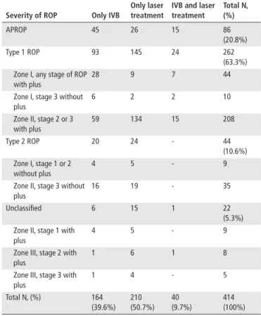
Of all of the infants screened for ROP, 414 (6.7%) needed treatment. A total of 395 (95.4%) of the treated infants had a BW≤1500 g and treatment was performed in 19 infants with a BW of 1501–2000 g. Severe ROP was diagnosed in five babies with BW>1500 g and GA>32 weeks who required treatment. Treatment was applied bilaterally in 385 patients and was
performed in one eye in 29 cases. Five infants with a GA≤28 weeks underwent vitreoretinal surgery. Table 4 lists the severities and treatment modalities of ROP in the treated patients.
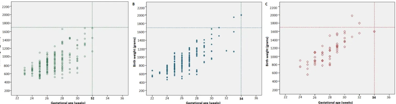
The incidence of severe ROP in university hospitals, in state hospitals and in private hospitals was 6.2%, 6.8% and 8.4% respectively. Mean GA and mean BW of infants with severe ROP varied between different types of NICU (table 5).
Appropriate criteria for screening for the NICUs in university hospitals should be <1700 g or ≤32 weeks and for the NICUs in state hospitals and private hospitals should be <1700 g or ≤34 weeks (figure 1).
dIsCussIon
ROP is a serious morbidity of prematurity, whose incidence and severity increase with decreasing GA and BW. Studies conducted in high-income countries have shown that infants born at ≥32
on February 5, 2020 by guest. Protected by copyright.
http://bjo.bmj.com/
Table 3 Independent risk factors for severe retinopathy of prematurity in infants with a birth weight≤1500 g
Adjusted or 95% CI P value
Gestational age (weeks)* 0.812 0.726 to 0.910 <0.001 Birth weight (g)† 0.998 0.997 to 0.999 <0.001 Small for gestational age 0.471 0.277 to 0.799 0.005 Total days on oxygen‡ 1.025 1.019 to 1.031 <0.001
Late-onset sepsis 1.423 1.016 to 1.994 0.04
Frequency of red blood cell transfusions≥twice
2.384 1.389 to 4.092 0.002
Relative weight gain at 28 days (grams)
0.998 0.997 to 0.999 <0.001 P<0.05 represents statistical significance.
*OR for every week. †OR for every 1 g. ‡OR for each day on oxygen.
Table 4 Severity and treatment modalities of ROP in treated patients
severity of roP only IVb
only laser treatment
IVb and laser treatment Total n, (%) APROP 45 26 15 86 (20.8%) Type 1 ROP 93 145 24 262 (63.3%) Zone I, any stage of ROP
with plus
28 9 7 44
Zone I, stage 3 without plus
6 2 2 10
Zone II, stage 2 or 3 with plus 59 134 15 208 Type 2 ROP 20 24 - 44 (10.6%) Zone I, stage 1 or 2 without plus 4 5 - 9
Zone II, stage 3 without plus
16 19 - 35
Unclassified 6 15 1 22
(5.3%) Zone II, stage 1 with
plus
4 5 - 9
Zone III, stage 2 with plus
1 6 1 8
Zone III, stage 3 with plus 1 4 - 5 Total N, (%) 164 (39.6%) 210 (50.7%) 40 (9.7%) 414 (100%) APROP, aggressive posterior retinopathy of prematurity; IVB, intravitreal bevacizumab; N, number of patients; ROP, retinopathy of prematurity.
Table 5 Mean BWs and GAs of infants with severe ROP according to types of units university hospitals (n=192) state hospitals (n=164) Private hospitals (n=58) P value* Mean GA (weeks) 26.3±2 26.7±2.2 27.9±2.1 <0.05 Mean BW (g) 878±250 905±273 1128±299 <0.05
*The values are significantly higher in private hospitals than in university and state hospitals.
BW, birth weight; GA, gestational age; n, number of patients; ROP, retinopathy of prematurity.
weeks are not at risk for developing ROP and most infants born at >28 weeks who develop ROP have a mild disease that spon-taneously regressed without treatment.9 The findings of the
TR-ROP study were comparable to those from other developing countries and showed that more mature and heavier babies were at risk for severe ROP.10
There were no differences in any ROP and severe ROP devel-opment between VLBW infants of refugees and citizens in our study. The Ministry of Health of Turkey has been involved in direct healthcare services in the refugee camps and through the referral of refugees to Turkish hospitals. The 2015 report of the Turkish Neonatology Society reported a mortality rate of
26% for babies with a BW<1500 g, according to data obtained from 59 NICUs.11 However, there were insufficient data on the
neonatal mortality of refugees in this report.
GA, BW and oxygen therapy are well-known major risk factors in the development of ROP.12 In this study, a lower BW,
shorter GA and total days on oxygen were found as independent risk factors for severe ROP in infants with a BW≤1500 g and for any ROP in infants with a BW>1500 g.
Some previous studies have reported that the prevalence of ROP was higher in SGA infants compared with appropriate for GA preterms, while SGA was not found to be a risk factor for ROP in other reports.13 14 Factors that are considered an
increased risk for severe ROP in SGA babies include chronic uterine hypoxia, abnormal growth factor levels, nutrient restriction and antioxidant deficiency.15 However, in our study,
SGA was surprisingly associated with a decreased incidence of severe ROP in VLBW infants when using a multivariate logistic regression model.
There was a relationship between poor postnatal weight gain and an increased risk for ROP.16 Poor postnatal weight
gain was also found as an independent risk factor for severe ROP in infants with a BW≤1500 g in our study. Using univar-iate analyses, several risk factors including RDS, respiratory support, sepsis, NEC, PDA, intracranial haemorrhage and BPD were significantly associated with severe ROP in VLBW infants in our cohort. These perinatal morbidities may have decreased postnatal weight gains.
This study showed that RBC transfusions had strong effects on the development of ROP. Transfusions may increase oxygen delivery to the retina because of the lower oxygen affinity of adult haemoglobin in packed red cells. Repeated transfusions may also cause free iron accumulation, which may result in increased production of free hydroxyl radicals as assessed by the Fenton reaction, resulting in damage to the retina.17
Although the role of blood transfusions as a risk factor for ROP was suggested by numerous reports,18 19 several studies have
reported that a transfusion limitation policy did not reduce the prevalence of ROP.20 Our data suggested that limiting blood
transfusion in regards to threshold haemoglobin values in guidelines could contribute to reducing the prevalence of ROP.
Multiple studies have reported the role of neonatal sepsis in the development of ROP.21 22 In this study, late onset sepsis was
an independent risk factor for severe ROP in VLBW infants. Sepsis may act through cytokines and endotoxins, which directly affect retinal angiogenesis. This process is frequently accompanied by hypotension, which can cause tissue perfusion impairment and retinal ischaemia.23
Treatment was performed in 6.7% of the infants screened for ROP in the current study. In nearly half of the infants with severe ROP, the treatment modality involved laser photocoagula-tion and IVB was performed in the other half as the first choice.
on February 5, 2020 by guest. Protected by copyright.
Figure 1 Plots of BW versus GA for infants treated for ROP in three different types of NICU. (A) University hospitals. (B) State hospitals. (C) Private hospitals. BW, birth weight; GA, gestational age; NICU, neonatal intensive care unit; ROP, retinopathy of prematurity.
A nationwide population-based study from the UK reported that diode laser photocoagulation was performed in 90.5% of infants requiring treatment.1 The higher usage of IVB in our study may be
due to ease of administration (typically at the bedside). In addition, paediatric anaesthesia for performing laser photocoagulation was not available in some NICUs in our study.
Notably, 66 (16%) of 414 infants were treated earlier than type 1 ROP and did not fulfil the ETROP requirements for treatment in our study. Twenty-six of these 66 infants were treated with IVB. The popularity of anti-VEGF agents is increasing in Turkey; however, the long-term safety and efficacy of these agents are still not definitively known. The risk of progression to retinal detach-ment in type 1 ROP is around 15%, but is much lower with less severe disease.8 Evidence-based data are not available to confirm
a favourable risk–benefit ratio of IVB usage in cases earlier than type 1 ROP.
In our study, the incidence of severe ROP varied between the three types of NICU which reflects the differences in neonatal care. The rates of severe ROP were lower in university hospital NICUs, where practices for newborn care are likely to be better than non-university NICUs. Based on the results of present study, the screening criteria for ROP need to be wider in state and private hospitals than applied in the university hospitals. ROP programmes in Turkey should adopt the criteria of <1700 g or ≤34 weeks to capture all babies requiring treatment.
The strength of the TR-ROP study was that it was a large multi-centre cohort study that allowed us to prospectively obtain data via a special network. However, the neonatologists did not go through any training in order to standardise definitions of potential risk factors before the study started. Similarly, the participating ophthal-mologists also did not undergo any processes to standardise how they graded ROP. These situations are the limitations of the study.
In conclusion, screening criteria for ROP in Turkey needs to be wider than developed countries. The high incidence of infants with ROP in our study emphasised the need for aggressive measures for prevention and control of the disease. The safe implementation of oxygen therapy with appropriate monitoring, better antenatal and neonatal care, meticulous attention to hygienic procedures and control of sepsis may reduce the prevalence of ROP. There-fore, monitoring standards of neonatal care and conducting quality improvement projects across the country are essential for improving neonatal outcomes in Turkish NICUs.
Correction notice This article has been corrected since it published Online First. Typos in several of the Collaborator names have been corrected.
Acknowledgements We thank Bulent Celik from the Department of Statistics, Gazi University, Ankara, for statistical analysis.
Collaborators TR-ROP Study Group Collaborators (Neonatology): Fatma Nur Sari (Dr Zekai Tahir Burak Women’s Health Education and Research Hospital, University of Health Sciences, Ankara); Guner Karatekin (Zeynep Kamil Maternity and Children’s Training and Research Hospital, University of Health Sciences, Istanbul); Esad Koklu (Megapark Hospital, Kahramanmaras); Huseyin Altunhan (Necmettin Erbakan University, Meram Faculty of Medicine, Konya); Hatice Turgut (Inonu University Faculty of Medicine, Malatya); Fatma Narter (Kartal Lutfi Kirdar Education and Research Hospital, University of Health Sciences, Istanbul); Nuriye Tarakci (Dr Faruk Sukan Maternity and Children’s Hospital, Konya); Kadir Serafettin Tekgunduz (Ataturk University Faculty of Medicine, Erzurum); Servet Ozkiraz (Medicalpark Hospital, Gaziantep); Cumhur Aydemir (Bulent Ecevit University Faculty of Medicine, Zonguldak); Ahmet Ozdemir (Erciyes University Faculty of Medicine, Kayseri); Bilin Cetinkaya (Baskent University Faculty of Medicine, Adana); Ebru Kazanci (Kanuni Sultan Suleyman Training and Research Hospital, University of Health Sciences, Istanbul); Ayhan Tastekin (Medipol University, Istanbul); Sebnem Calkavur (Dr Behcet Uz Children’s Hospital, University of Health Sciences, Izmir); Banu Mutlu Ozyurt (Mersin State Hospital, Mersin); Yasar Demirelli (Erzurum Nenehatun Maternity Hospital, Erzurum); Huseyin Selim Asker (NCR International Hospital, Gaziantep); Birgul Mutlu (Doruk Yildirim Private Hospital, Bursa); Ozgun Uygur (Tepecik Training and Research Hospital, University of Health Sciences, Izmir); Hilal Ozkan (Uludag University Faculty of Medicine, Bursa); Didem Armangil (Yuksek Ihtisas University Faculty of Medicine, Ankara); Ferda Ozlu (Cukurova University Faculty of Medicine, Adana); Mustafa Kurthan Mert (Numune Training and Education Hospital, University of Health Sciences, Adana); Hacer Ergin (Pamukkale University Faculty of Medicine, Denizli); Beyza Ozcan (Konya Education and Research Hospital, University of Health Sciences, Konya); Evrim Kiray Bas (Sisli Hamidiye Etfal Education and Research Hospital, University of Health Sciences, Istanbul); Emel Okulu (Ankara University Faculty of Medicine, Ankara); Betul Acunas (Trakya University Faculty of Medicine, Edirne); Ulker Celik (Denizli State Hospital, Denizli); Sait Ilker Uslu (Ondokuz Mayıs University, Faculty of Medicine, Samsun); Mehmet Mutlu (Karadeniz Technical University Faculty of Medicine, Trabzon); Nihat Demir (Yuzuncu Yil University Faculty of Medicine, Van); Funda Eroglu (Ankara Guven Hospital, Ankara); Zeynel Gokmen (Baskent University Faculty of Medicine, Konya); Serdar Beken (Acibadem University Faculty of Medicine, Istanbul); Bilge Tanyeri Bayraktar (Bezmialem University Faculty of Medicine, Istanbul); Nilay Hakan (Mugla Sitki Kocman University Faculty of Medicine, Mugla); Kazım Kucuktasci (Denizli Private Saglik Hospital, Denizli); Aysen Orman (Firat University Faculty of Medicine, Elazig); Serdar Comert (Suleymaniye Maternity, Research & Training Hospital, University of Health Sciences, Istanbul); Sabahattin Ertugrul (Dicle University Faculty of Medicine, Diyarbakir); Nuran Ustun (Medeniyet University Faculty of Medicine, Istanbul); Ozlem Sahin (Umraniye Education and Research Hospital, University of Health Sciences, Istanbul); Demet Terek (Ege University Faculty of Medicine, Izmir); Yusuf Kale (Cengiz Gokcek Maternity and Children’s Hospital, Gaziantep); Murat Konak (Konya Selcuk University Faculty of Medicine, Konya); Sadık Yurttutan (Kahramanmaras Sutcu Imam University Faculty of Medicine, Kahramanmaras); Ozge Aydemir (Osmangazi University Faculty of Medicine, Eskisehir); Aysegul Zenciroglu (Dr Sami Ulus Maternity and Children’s Hospital, University of Health Sciences, Ankara); Dilek Sarici (Kecioren Education and Research Hospital, University of Health Sciences, Ankara); Nilufer Guzoglu (Kirikkale University Faculty of Medicine, Kirikkale); Sahin Hamilcikan (Bagcilar Education and Research Hospital, University of Health Sciences, Istanbul); Tugba Gursoy (Koc University Faculty of Medicine, Istanbul); Funda Tuzun (Dokuz Eylul University Faculty of Medicine, Izmir); Rahmi Ors (Medova Hospital, Konya); Selda Arslan (Mustafa Kemal University Faculty of Medicine, Hatay); Arzu Akdag (Bursa Dortcelik Children’s Hospital, Bursa); Asli Memisoglu (Marmara University Faculty of Medicine, Istanbul); Beril Yasa (Istanbul University, Istanbul Faculty of Medicine, Istanbul); Berna Hekimoglu (Trabzon Kanuni Education and Research Hospital, University of Health Sciences, Trabzon); Ozden Turan (Baskent University Faculty of Medicine, Ankara);
on February 5, 2020 by guest. Protected by copyright.
http://bjo.bmj.com/
Hakan Aylanc (Onsekizmart University Faculty of Medicine, Canakkale); Sahin Takci (Gaziosmanpasa University Faculty of Medicine, Tokat); Tolga Celik (Hacettepe University Faculty of Medicine, Ankara); Suzan Sahin (Adnan Menderes University Faculty of Medicine, Aydin); Ilknur Kilic (Atasehir Kadikoy Sifa Hospital, Istanbul). TR-ROP Study Group Collaborators (Ophthalmology): Caner Kara (Etlik Zubeyde Hanim Women’s Health Teaching and Research Hospital, University of Health Sciences, Ankara); Zuhal Ozen Tunay (Dr Zekai Tahir Burak Women’s Health Education and Research Hospital, University of Health Sciences, Ankara); Gokhan Celik (Zeynep Kamil Maternity and Children’s Training and Research Hospital, University of Health Sciences, Istanbul); Ibrahim Gozen (Megapark Hospital, Kahramanmaras); Gunhal Satirtav (Necmettin Erbakan University, Meram Faculty of Medicine, Konya); Nihat Polat (Inonu University Faculty of Medicine, Malatya); Ayse Yesim Oral (Kartal Lutfi Kirdar Education and Research Hospital, University of Health Sciences, Istanbul); Mine Tokgoz (Konya Numune Hospital, Konya); Sadullah Keles (Ataturk University Faculty of Medicine, Erzurum); Burak Bilgin (Medicalpark Hospital, Gaziantep); Silay Canturk Ugurbas (Bulent Ecevit University Faculty of Medicine, Zonguldak); Cagatay Karaca (Erciyes University Faculty of Medicine, Kayseri); Nedime Sahinoglu Keskek (Baskent University Faculty of Medicine, Adana); Dilbade Yildiz Ekinci (Kanuni Sultan Suleyman Training and Research Hospital, University of Health Sciences, Istanbul); Ozlem Balci (Istanbul Medipol University, Istanbul); Emir Volkan Altan (Dr Behcet Uz Children’s Hospital, University of Health Sciences, Izmir); Sevda Bakbak(Mersin State Hospital, Mersin); Nihan Aksu Ceylan (Erzurum Regional Education and Research Hospital, University of Health Sciences, Erzurum); Sabit Kimyon (Gaziantep University Faculty of Medicine, Gaziantep); Gunay Alyamac (Retina Eye Hospital, Bursa); Gamze Ture (Tepecik Training and Research Hospital, University of Health Sciences, Izmir); Meral Yildiz (Uludag University Faculty of Medicine, Bursa); Feyza Calis (Yuksek Ihtisas University Faculty of Medicine, Ankara); Selcuk Sizmaz (Cukurova University Faculty of Medicine, Adana); Emine Sukgen (Numune Training and Education Hospital, University of Health Sciences, Adana); Ebru Nevin Cetin (Pamukkale University Faculty of Medicine, Denizli); Muammer Ozcimen, (Konya Education and Research Hospital, University of Health Sciences, Konya); Semra Tiryaki Demir (Sisli Hamidiye Etfal Education and Research Hospital, University of Health Sciences, Istanbul); Huban Atila (Ankara University Faculty of Medicine, Ankara); Altan Ozal (Trakya University Faculty of Medicine, Edirne); Gokhan Tufaner (Denizli State Hospital, Denizli); Ozlem Eski Yucel (Ondokuz Mayıs University Faculty of Medicine, Samsun); Mehmet Kola (Karadeniz Technical University Faculty of Medicine, Trabzon); Erbil Seven (Yuzuncu Yil University Faculty of Medicine, Van); Sengul Ozdek (Gazi University Faculty of Medicine, Ankara); Ali Hakan Durukan (Ankara Guven Hospital, Ankara); Ali Kal (Baskent University Faculty of Medicine, Konya); Ali Riza Cenk Celebi (Acibadem University Faculty of Medicine; Istanbul); Ibrahim Arif Koytak (Bezmialem University Faculty of Medicine, Istanbul); Goksu Alacamli (Mugla Sitki Kocman University Faculty of Medicine, Mugla); Arif Esme (Denizli Private Saglik Hospital; Denizli); Onur Catak (Firat University Faculty of Medicine, Elazig); Irfan Perente (Suleymaniye Maternity, Research & Training Hospital, University of Health Sciences, Istanbul); Alparslan Sahin (Dicle University Faculty of Medicine, Diyarbakir); Aylin Ardagil Akcakaya (Istanbul Medeniyet University Faculty of Medicine Istanbul); Gulunay Kiray (Umraniye Education and Research Hospital, University of Health Sciences, Istanbul); Serhat Nalcaci (Ege University Faculty of Medicine, Izmir); Umit Aksoy (Cengiz Gokcek Maternity and Children’s Hospital, Gaziantep); Berker Bakbak (Konya Selcuk University Faculty of Medicine, Konya); Aysegul Comez
(Kahramanmaras Sutcu Imam University Faculty of Medicine, Kahramanmaras); Huseyin Gursoy (Osmangazi University Faculty of Medicine, Eskisehir); Emrah Utku Kabatas (Dr Sami Ulus Maternity and Children’s Hospital, University of Health Sciences, Ankara); Ikbal Seza Petricli (Etlik Zubeyde Hanim Women’s Health Teaching and Research Hospital, University of Health Sciences, Ankara); Mehmet Erhan Yumusak (Kirikkale University Faculty of Medicine, Kirikkale); Ahmet Kirgiz (Bagcilar Education and Research Hospital, University of Health Sciences, Istanbul); Gunay Uludag (Koc University Faculty of Medicine, Istanbul); Aylin Yaman (Dokuz Eylul University Faculty of Medicine, Izmir); Zeynep Dadaci (Medova Hospital, Konya);Ali Karatas (Bursa Dortcelik Children’s Hospital, Bursa); Hande Celiker (Marmara University Faculty of Medicine, Istanbul);Zafer Cebeci (Istanbul University, Istanbul Faculty of Medicine, Istanbul); Mahmut Cenap Esenulku (Trabzon Kanuni Education and Research Hospital, University of Health Sciences, Trabzon); Imren Akkoyun (Baskent University, Faculty of Medicine, Ankara); Ismail Ersan (Onsekizmart University Faculty of Medicine, Canakkale); Selim Demir (Gaziosmanpasa University Faculty of Medicine, Tokat); Sibel Kadayifcilar (Hacettepe University Faculty of Medicine, Ankara); Ayse Ipek Akyuz Unsal (Adnan Menderes University Faculty of Medicine, Aydin); Mumin Hocaoglu (Istanbul Retina Institute, Istanbul). Contributors All authors made substantial contributions to conception, design, analysis and interpretation of data and contributed to writing the article and approved the current version.
Funding This research received no specific grant from any funding agency in the public, commercial or not-for-profit sectors
Competing interests None declared. Patient consent Guardian consent obtained.
ethics approval The study was approved by the ethical review committee of Gulhane Faculty of Medicine.
Provenance and peer review Not commissioned; externally peer reviewed. open access This is an open access article distributed in accordance with the Creative Commons Attribution Non Commercial (CC BY-NC 4.0) license, which permits others to distribute, remix, adapt, build upon this work non-commercially, and license their derivative works on different terms, provided the original work is properly cited and the use is non-commercial. See: http:// creativecommons. org/ licenses/ by- nc/ 4. 0/
© Article author(s) (or their employer(s) unless otherwise stated in the text of the article) 2018. All rights reserved. No commercial use is permitted unless otherwise expressly granted.
RefeRenCes
1 Adams GGW, Bunce C, Xing W, et al. Treatment trends for retinopathy of prematurity in the UK. BMJ Open 2017;21:7.
2 Gilbert C. Retinopathy of prematurity: a global perspective of the epidemics, population of babies at risk and implications for control. Early Hum Dev 2008;84:77–82.
3 Chaudhry TA, Hashmi FK, Salat MS, et al. Retinopathy of prematurity: an evaluation of existing screening criteria in Pakistan. Br J Ophthalmol 2014;98:298–301. 4 Mikolajczyk RT, Zhang J, Betran AP, et al. A global reference for fetal-weight and
birthweight percentiles. Lancet 2011;377:1855–61.
5 Papile LA, Burstein J, Burstein R, et al. Incidence and evolution of subependymal and intraventricular hemorrhage: a study of infants with birth weights less than 1,500 gm. J Pediatr 1978;92:529–34.
6 Bell MJ, Ternberg JL, Feigin RD, et al. Neonatal necrotizing enterocolitis. Therapeutic decisions based upon clinical staging. Ann Surg 1978;187:1–7.
7 International Committee for the Classification of Retinopathy of Prematurity. The International Classification of Retinopathy of Prematurity revisited. Arch Ophthalmol 2005;123:991–9.
8 Early Treatment For Retinopathy Of Prematurity Cooperative Group. Revised indications for the treatment of retinopathy of prematurity: results of the early treatment for retinopathy of prematurity randomized trial. Arch Ophthalmol 2003;121:1684–9. 9 Holmström G, Hellström A, Jakobsson P, et al. Evaluation of new guidelines for ROP
screening in Sweden using SWEDROP - a national quality register. Acta Ophthalmol 2015;93:265–8.
10 Tabarez-Carvajal AC, Montes-Cantillo M, Unkrich KH, et al. Retinopathy of prematurity: screening and treatment in Costa Rica. Br J Ophthalmol 2017;101:1709–13. 11 Society TN. Mortality rates in Turkish NICUs in 2016. Turk Neonatol Soc Bull
2017;29:32–4. Article in Turkish.
12 Phelps DL. Retinopathy of prematurity. In: Martin RJ, Fanaroff AA, Walsh MC, eds. Fanaroff and Martin’s neonatal perinatal medicine: diseases of the fetus and infant. Philadelphia: Elsevier Saunders, 2015:1767–74.
13 Bernstein IM, Horbar JD, Badger GJ, et al. Morbidity and mortality among very-low-birth-weight neonates with intrauterine growth restriction. The vermont Oxford Network. Am J Obstet Gynecol 2000;182:198–206.
14 Filho JBF, Valiatti FB, Eckert GU, et al. Is being small for gestational age a risk factor for retinopathy of prematurity? A study with 345 very low birth weight preterm infants. J Pediatr 2009;85:48–54.
15 Kavurt S, Özcan B, Aydemir O, et al. Risk of retinopathy of prematurity in small for gestational age premature infants. Indian Pediatr 2014;51:804–6.
16 Hellström A, Hård AL, Engström E, et al. Early weight gain predicts retinopathy in preterm infants: new, simple, efficient approach to screening. Pediatrics 2009;123:e638–e645. 17 Wardle SP, et al. Effect of blood transfusion on lipid peroxidation in preterm infants.
Arch Dis Child Fetal Neonatal Ed 2002;86:46F–8.
18 Hesse L, Eberl W, Schlaud M, et al. Blood transfusion. Iron load and retinopathy of prematurity. Eur J Pediatr 1997;156:465–70.
19 Dani C, Reali MF, Bertini G, et al. The role of blood transfusions and iron intake on retinopathy of prematurity. Early Hum Dev 2001;62:57–63.
20 Kirpalani H, Whyte RK, Andersen C, et al. The Premature Infants in Need of Transfusion (PINT) study: a randomized, controlled trial of a restrictive (low) versus liberal (high) transfusion threshold for extremely low birth weight infants. J Pediatr 2006;149:301–7. 21 Tolsma KW, Allred EN, Chen ML, et al. Neonatal bacteremia and retinopathy of
prematurity: the ELGAN study. Arch Ophthalmol 2011;129:155–63. 22 Stoll BJ, Hansen NI, Adams-Chapman I, et al. Neurodevelopmental and growth
impairment among extremely low-birth-weight infants with neonatal infection. JAMA 2004;292:2357–65.
23 Lee J, Dammann O. Perinatal infection, inflammation, and retinopathy of prematurity. Semin Fetal Neonatal Med 2012;17:26–9.
on February 5, 2020 by guest. Protected by copyright.