1222 Journal of Gastroenterology and Hepatology 21 (2006) 1221–1226 © 2006 Journal of Gastroenterology and Hepatology Foundation and Blackwell Publishing Asia Pty Ltd
Letters to the Editor
Blackwell Publishing AsiaMelbourne, AustraliaJGHJournal of Gastroenterology and Hepatology0815-93192006 Blackwell Publishing Asia Pty Ltd July 200621712211226Letter to the EditorLetters to the EditorLetters to the Editor
L E T T E R S T O T H E E D I T O R
A true fundic diverticulum of the
gallbladder
To the Editor,
True diverticulum of the gallbladder is an extremely rare congeni-tal anomaly of the gastrointestinal system. All previously reported cases in the current literature were diagnosed after surgical resec-tion.1–5
The patient presented here is the first true gallbladder diverticulum case demonstrated well by magnetic resonance cho-langiopancreatography (MRCP) before any surgical interventions. A 44 year-old man was referred to our hospital after his initial management of recurrent pancreatitis attacks. He described abdominal pain with nausea which started 4 days before his admission. Laboratory investigation was performed in a previous hospital and revealed elevated serum amylase level of 396 IU/L (normal <95 IU/L) and lipase level of 706 IU/L (normal <195 IU/L).
The patient had a 5-year history of recurrent pancreatitis attacks. The first attack occurred in 1999 and the second attack in 2001. Previous abdominal ultrasound and computerized tomogra-phy scan had revealed signs of pancreatitis and a pancreatic cyst with a dimension of 4.2 cm × 3.2 cm in the tail of the pancreatic gland. He had no prior history of jaundice, hepatitis, parasitic infection or abdominal trauma. The patient consumed more than 80 g a day of alcohol between 1982 and 1995 and continued to drink alcohol after this but in decreased amounts. He also smoked 30 cigarettes a day for 20 years. There was no ingestion of any herbal medicine or a particular diet and he had not undergone a blood transfusion or a surgical procedure during the last 6 months. There was no family history of hepatitis, parasitic infection and gallstones, except that both his mother and aunt had passed away due to gastric cancer.
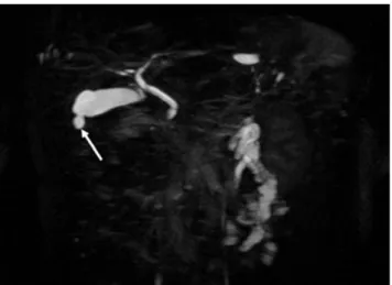
On physical examination, the patient was dehydrated and lean with a body mass index (BMI) of 21. There was mild epigastric and right upper quadrant tenderness. Laboratory evaluation was repeated and revealed elevated amylase and lipase values with normal CA 19–9 and CEA values. Upright abdominal X-ray showed no evidence of abnormal calcification. The ultrasound of the abdomen revealed enlargement of the pancreas and dilatation of the intrahepatic biliary ducts and common bile duct. Magnetic resonance imaging demonstrated signs of pancreatitis in both pan-creatic corpus and tail with a cyst. As demonstrated in Figure 1, MRCP images showed a solitary, true gallbladder diverticulum with a diameter of 10 mm, and very little biliary sludge in the lumen of the gallbladder. A hepatobiliary iminodiacetic acid (HIDA) scan of the gallbladder was performed and showed no abnormality on the whole gallbladder.
Pseudodiverticulum (false diverticulum) is the most common type of diverticulum of the gallbladder. It is characterized by incomplete muscular wall herniation and usually occurs due to acquired conditions such as cholelithiasis and cholecystitis.2,3
On the contrary, true diverticulum is a rare congenital anomaly of the gallbladder and includes all three layers of the gallbladder wall. The incidence of true diverticulum was reported to be 0.06% in a series of congenital anomalies of the gallbladder, and 0.0008% in the resected gallbladders.
Although true diverticula were demonstrated well as grossly visible outpouchings of the entire gallbladder wall during the pathological examinations in previous studies, they have not been
demonstrated by non-invasive imaging tests before surgical resec-tion. In other words, they were found incidentally. In this regard, our patient is the first true gallbladder diverticulum case uniquely demonstrated by MRCP before any surgical interventions.
Although there has been no satisfactory information regarding the nature of the true diverticulum of the gallbladder, it is always described as a non-harmful condition in previous reports. Addi-tionally, recurrent episodes of epigastrium and right upper quad-rant abdominal pain, perforation of the gallbladder diverticulum, and carcinoma in a true diverticulum of the gallbladder were also reported by the same authors.1–5
We believe that the frequency of the true diverticulum would increase by considerable usage of MRCP and increasing awareness. Then, our knowledge could be improved about the real frequency and the clinical importance of this condition.
The MRCP pictures were showing biliary sludge in the lumen of the gallbladder. Albeit, the amount of the sludge was very little, it is also possible that the biliary sludge may have played a role in the recurrence of pancreatitis in our patient.6
We would have liked to have analyzed the bile and the sludge, but the patient did not allow to us to do an endoscopic retrograde cholangiopancreatogra-phy. Likewise, the patient did not accept the gallbladder resection. Thus, we could not obtain histopathological confirmation of the diverticulum.
Metin Basaranoglu* and Numan Cem Balci†
*Internal Medicine Department, Kadir Has University Hospital, Istanbul, and †Radiology Department, Kocaeli University, Kocaeli, Turkey
References
1 Nadeau OE. Diverticulum of the gallbladder perforating the liver: report of a case. Med. Surg. 1917; 1: 73.
2 Gross RE. Congenital anomalies of the gallbladder: a review of one
hundred and forty eight cases with report of a double gallbladder. Arch.
Surg. 1936; 32: 131–62.
Figure 1 Coronal MRCP image of the abdomen. The T2-weighted fat
saturated projection MRCP image reveals a true diverticulum as a pearl or a tear drop at the bottom of the gallbladder (arrow).
Letters to the Editor
Journal of Gastroenterology and Hepatology 21 (2006) 1221–1226 1223
© 2006 Journal of Gastroenterology and Hepatology Foundation and Blackwell Publishing Asia Pty Ltd
3 Shackelford RT. Diagnosis of Surgical Disease, Vol. 2. Philadelphia:
W.B. Saunders, 1969; 947.
4 Chin NW, Chapman I. Carcinoma in a true diverticulum of the gallblad-der. Am. J. Gastroenterol. 1988; 83: 667–9.
5 Kramer AJ, Bregman A, Zeddies CA, Guynn VL. Gallbladder diverticulum: a case report and review of the literature. Am. Surg.
1998; 64: 298–301.
6 Venneman NG, Renooij W, Rehfeld JF et al. Small gallstones,
pre-served gallbladder motility, and fast crystallization are associated with pancreatitis. Hepatology 2005; 41: 738–46.
Blackwell Publishing AsiaMelbourne, AustraliaJGHJournal of Gastroenterology and Hepatology0815-93192006 Blackwell Publishing Asia Pty Ltd 20062112211226Letter to the EditorLetters to the EditorLetters to the Editor
L E T T E R S T O T H E E D I T O R
Prolonged hepatitis E in an
immunocompromised patient
To the Editor,Acute hepatitis E is usually followed by complete recovery and chronic hepatitis does not occur. Viral shedding usually lasts up to a month and has never been reported after 4 months. We report the case of a patient with prolonged hepatitis E virus (HEV). HEV RNA was detected in stools or serum over a 10-month period, associated with flares of elevated transaminases.
A 45-year-old male (Mr F) was diagnosed with a stage IV CD30+, ALK-, anaplastic large T-cell lymphoma. He was treated with four cycles of the ACVBP chemotherapy regimen, from August to October 2003, followed by autologous stem-cell trans-plantion in January 2004. The patient had a complete response 8 months after transplantation.
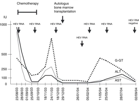
In November 2003, Mr F was referred to our unit for acute hepatitis. The liver tests were: aspartate aminotransferase (AST) 239 IU/l, alanine aminotransferase (ALT) 171 IU/l, alkaline phosphatase 802 IU/l, gamma-glutamyl transpeptidase (GGT)
299 IU/L and bilirubin 91 µmol/L (Fig. 1). HEV RNA was detected in the stools using real-time reverse transcription-polymerase chain reaction (RT-PCR) described elsewhere.1,2
Other causes of acute hepatitis were ruled out. Liver biopsy showed moderate lobular hepatitis with spotty hepatocyte necrosis and polymorph inflammation in the lobule.
Serum taken at initial hospitalization in August was checked retrospectively and was positive for HEV. Overall, HEV RNA was detected in stools (four times) and/or in serum (five times) over a 10-month period between August 2003 and May 2004. Liver enzymes fluctuated with three flare-ups. On 6 September 2004, liver tests were normal and serum and stools were negative for HEV RNA. The sequence comparison, genetic distances and phy-logenetic analysis of the genomic amplification products indicated that the HEV strain from the patient belonged to genotype 3.
The mode of contamination of HEV is unclear. The patient lives in a rural community and hunts partridge, but he has never eaten boar or deer. He had not traveled outside of France in the year preceding the onset of acute hepatitis. The majority of the cases of hepatitis E in our region have an unknown source of infection.2
This observation depicts prolonged hepatitis E in an immuno-compromised patient. During acute hepatitis E, HEV RNA can be detected in the stools at the onset of the icteric phase and typically persists from approximately 10 days to 1 month. HEV RNA in the serum usually becomes undetectable after the peak in aminotrans-ferase activity. In a study where HEV was experimentally deliv-ered to a volunteer, serum HEV RNA disappeared on day 16 after the onset.3
Aggarwal et al. found that the maximum duration after the onset of first symptoms at which a stool sample was positive was 30 days.4
However, viral shedding for as long as 52 days after the onset of icterus and protracted periods of viremia of 45– 112 days’ duration have also exceptionally been described.5
This is the first description of viral shedding for more than 4 months. The RT PCR we used is only quantitative and it is therefore impossible to quantify viral shedding. The patient was
Figure 1 Evolution of the liver tests over the
study period. Arrows depict treatment regi-men and hepatitis E virus (HEV) RNA detec-tion in the serum or the stools (all HEV RNA positive samples are not listed to simplify the figure). 1000 500 250 100 2 3 /0 8 /0 3 28/08/03 01 /0 9 /0 3 2 5 /0 9 /0 3 2 2 /1 0 /0 3 1 8 /1 1/0 3 2 4 /1 1 /0 3 1 8 /1 2 /0 3 2 6 /0 1 /0 4 05 /0 2 /0 4 1 1 /0 5 /0 4 2 5 /0 5 /0 4 2 8 /0 7 /0 4 2 9 /1 2 /0 3 0 AST ALT G-GT Autologus bone marrow transplantation IU 0 2 /0 9 /0 4 Chemotherapy HEV RNA HEV RNA HEV RNA HEV RNA HEV RNA HEV RNA HEV RNA negative