Full Terms & Conditions of access and use can be found at
https://www.tandfonline.com/action/journalInformation?journalCode=iard20
Acta Radiologica
ISSN: 0284-1851 (Print) 1600-0455 (Online) Journal homepage: https://www.tandfonline.com/loi/iard20
Myositis Ossificans of Psoas Muscle: Magnetic
Resonance Imaging Findings
M. Sirvanci, A. K. Ganiyusufoglu, K. Karaman, M. Tezer & A. Hamzaoglu
To cite this article: M. Sirvanci, A. K. Ganiyusufoglu, K. Karaman, M. Tezer & A. Hamzaoglu (2004) Myositis Ossificans of Psoas Muscle: Magnetic Resonance Imaging Findings, Acta Radiologica, 45:5, 523-525
To link to this article: https://doi.org/10.1080/02841850410005697
Published online: 09 Jul 2009.
Submit your article to this journal
Article views: 600
CASE REPORT ACTA RADIOLOGICA
Myositis Ossificans of Psoas Muscle: Magnetic Resonance Imaging
Findings
M. SIRVANCI, A. K. GANIYUSUFOGLU, K. KARAMAN, M. TEZER & A. HAMZAOGLU
Department of Radiology, University of Kadir Has, Faculty of Medicine, Istanbul, Turkey; Department of Orthopedics, Florence Nightingale Hospital, Istanbul, Turkey
Sirvanci M, Ganiyusufoglu AK, Karaman K, Tezer M, Hamzaoglu A. Myositis ossificans of psoas muscle: magnetic resonance imaging findings. Acta Radiol 2004;45: 523–525.
Myositis ossificans is a benign, localized, self-limiting ossifying soft tissue mass with unknown pathogenesis. It may be confused with a malignant tumor of bone or soft tissues, especially in the early stages of the disease. In this report, we present a case of myositis ossificans affecting unilateral psoas muscle, which to our knowledge is a very uncommon location. There was no history of trauma. We describe the imaging findings and clues to early diagnosis of myositis ossificans.
Key words: Myositis ossificans; magnetic resonance imaging; psoas muscle
Mustafa Sirvanci, Velioglu sokak No: 9/7 Husnufirat apartmani, Ayazma, 81160 Uskudar, Istanbul, Turkey (fax. z0090 216 343 2762, e-mail. [email protected])
Myositis ossificans denotes a benign and frequently self-limiting process with an ossifying soft tissue mass in the skeletal muscle. In 75% of cases, a
history of direct major trauma or repeated minor injuries is clearly established. However, in some patients no history of antecedent trauma is present (3). In this situation, it may be confused with, and requires differentiation from a malignant tumor of bone or soft tissues, especially in the early stages of the disease. In this report, we present a case of myositis ossificans affecting unilateral psoas muscle, which, to our knowledge, is a very uncommon location. There was no history of trauma.
Case Report
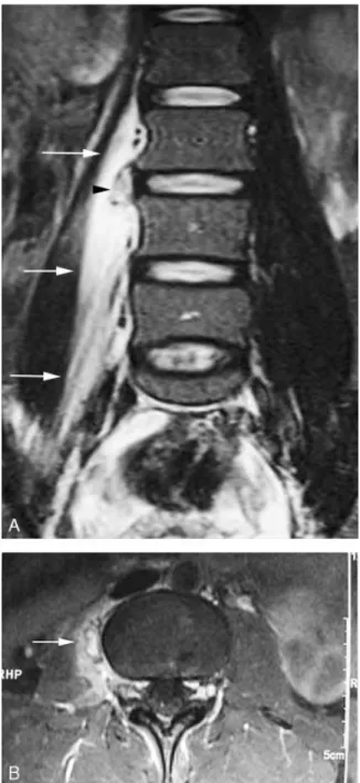
A 31-year-old woman presented with a 4-week history of right-sided lower back pain. Her past medical history was unremarkable except for a successful delivery 3 months previously. She denied any specific trauma. The patient was afebrile and laboratory values were normal except for an elevated erythrocyte sedimentation rate. Lumbar X-ray examination did not show any pathological findings. With a presumed diagnosis of spondylo-discitis or lumbar discal pathology, MRI examina-tion was done with a 1.5-T scanner (Sonata; Siemens, Erlangen, Germany). On coronal STIR images, the lesion was readily apparent with well-defined borders and hyperintense central
region surrounded by a hypointense rim (Fig. 1A). There was also an excessive, diffuse hyperintensity consistent with edema in the psoas muscle sur-rounding the lesion. Both the lesion (except for its hypointense rim) and surrounding edema showed marked enhancement after IV Gd-DTPA injection (Fig. 1B). There were no pathological signal changes or contrast enhancement of nearby intervertebral disk and vertebral bodies. These findings were thought to represent myositis ossificans, but an infection, or a sarcoma, were considered in the differential diagnosis. In order to further image the lesion with peripheral hypointense rim, a CT scan was performed with a spiral CT scanner (Prospeed SX Power; G. E. Medical Systems, Milwaukee, Wisc., USA). The CT scan documented a 8620 mm ellipsoid lesion with a well-demarcated ossified peripheral rim surrounding a central non-ossified region of low attenuation (Fig. 2). The diagnosis of myositis ossificans was concluded by these CT findings and conservative management was adopted. The patient’s symptoms resolved 2 months later and a follow-up MRI examination was per-formed which showed near complete regression of the surrounding perilesional edema.
Discussion
Myositis ossificans is a benign, localized, self-limiting ossifying soft tissue mass with unknown pathogenesis. It is characterized by non-neoplastic
heterotopic bone and cartilage formation in soft tissues in or adjacent to muscle and in proximity to bone. The lesion commonly affects large muscles of extremities, such as upper arm and thigh (1, 4). Pathophysiology of this disorder is thought to be posttraumatic inflammatory changes in skeletal muscle, although the exact etiology is still uncertain.
During the acute inflammatory phase, the erythro-cyte sedimentation rate may be elevated, as in our case, which may suggest an infection or tumor.
Radiologic findings parallel the histologic pattern of maturation. By the third week, calcification appears in the periphery of the lesion. After 4–6 weeks, the lesion shows a rim of curvilinear peripheral ossification like a shell around the lesion (characteristic zoning phenomenon). In the final healing stage, peripheral calcification pro-gresses centrifugally to form cancellous bone. Maturation is completed in about 5–6 months (1, 4).
The most characteristic radiological findings are radiological involution and a peripheral rim-like calcification/ossification, reflecting zoning phenom-enon (1, 4, 5). The diagnosis may be established by observation of the characteristic zoning phenom-enon via CT scan. CT is the most sensitive imaging method for detecting heterotopic mineralization, zoning phenomenon (4, 5).
The MRI appearance of myositis ossificans also correlates with the stage of the disease (3, 4). On MRI, diffuse and sometimes extensive surrounding edema disproportionate to the central lesion is a prominent finding in the early stage of the lesions, when imaged within 8 weeks of the onset of symptoms (4). This feature is unusual for a neoplastic lesion. In the acute phase, MRI can show the lesion before the appearance of calcifica-tions (4). Curvilinear and irregular areas of decreased signal intensity surrounding and coursing through the lesions can be seen especially in intermediate type lesions. After administration of IV Gd-DTPA, marked enhancement is seen especially in early lesions. Mature lesions are well-defined and inhomogeneous masses with a signal intensity
Fig. 1. MRI demonstrating myositis ossificans of the right psoas muscle located near to L3–L4 intervertebral disk. A. Coronal STIR image shows a well-demarcated hyperintense lesion with a hypointense rim (arrowhead). Note also extensive surrounding edema along the adjacent psoas muscle (arrows). B. Axial post-contrast T1-weighted image with fat saturation shows marked enhancement of the lesion and the surrounding edema. The hypointense rim of the lesion can be seen clearly (arrow).
Fig. 2. CT scan obtained after MRI showing the characteristic features of myositis ossificans. Axial CT scan shows an ellipsoid mass with a peripheral ring of new bone formation (arrow) surrounding a central non-ossified region, reflecting zoning phenomenon.
524 M. Sirvanci et al.
approximating that of fatty bone marrow on both T1 and T2-weighted images without associated edema and enhancement. On all pulse sequences, decreased signal changes related to bone trabeculae are seen surrounding and within the lesion (2, 5).
Early-intermediate myositis ossificans lesions may be confused with soft tissue sarcomas or infections especially when there is no history of trauma (3). In myositis ossificans of psoas muscle, as in our case, the main differential diagnostic considerations are sarcoma and tuberculous spondylitis with extension to psoas muscle. In addition to the lack of osseous pathological signal changes, detection of a hypoin-tense rim around the lesion led us to regard myositis ossificans as the most likely differential diagnostic consideration.
In conclusion, although there are varying appear-ances of myositis ossificans related to the histologic changes as the disorder progresses, there are highly suggestive MRI findings for early detection of
myositis ossificans even in uncommon sites, as in our case. When excessive, diffuse surrounding edema suggesting inflammation around a soft tissue mass is detected, myositis ossificans should be considered among the differential diagnosis.
References
1. Ehara S, Shiraishi H, Abe M, Mizutani H. Reactive heterotopic ossification. Its patterns on MRI. Clin Imaging 1998;22:292–6.
2. De Smet AA, Norris MA, Fisher DR. Magnetic resonance imaging of myositis ossificans: analysis of seven cases. Skeletal Radiol 1992;21:503–7.
3. Palmer WE, Kuong SJ, Elmadbouh HM. MR imaging of myotendinous strain. Am J Roentgenol 1999;173:703–9. 4. Parikh J, Hyare H, Saifuddin A. The imaging features of
post-traumatic myositis ossificans, with emphasis on MRI. Clin Radiol 2002;57:1058–66.
5. Siegel MJ. Magnetic resonance imaging of musculoske-letal soft tissue masses. Radiol Clin North Am 2001; 39:701–20.
Myositis Ossificans of Psoas Muscle 525