Clinical Note
Juxtapapillary Diverticulum: Findings on MRI
N. Cem Balci, MD,
1* Tara Noone, MD,
2Elif Aku¨n, MD,
3Ahmet Akinci, MD,
3and
Hans-Ulrich Klo¨r, MD
3The purpose of our study was to describe the imaging findings of juxtapapillary diverticulum on magnetic reso-nance imaging (MRI). The MRI and magnetic resoreso-nance cholangiopancreatography (MRCP) examinations of 14 pa-tients with juxtapapillary diverticula that were diagnosed on endoscopic retrograde cholangiopancreatography (ERCP) (N⫽ 8) or endoscopy (N ⫽ 6) were retrospectively evaluated. T1-weighted spoiled gradient-echo, T2-weighted half Fourier single shot fast spin-echo (HASTE), and T2-weighted True FISP (fast imaging with steady state preces-sion) images and thin-slice MRCP images were obtained on all patients. In five patients, diluted gadolinium DPTA (1/ 100) was used as an oral contrast. T2-weighted True FISP and HASTE images demonstrated air-fluid levels within all diverticula. Hyperintense oral contrast on T1-weighted spoiled gradient-echo images aided detection of the smaller diverticula. MRCP images obtained in the coronal plane best demonstrated the relationship of the diverticula to the papilla. MRI with the use of HASTE, True FISP, and oral contrast-enhanced T1-weighted sequences was able to de-pict juxtapapillary diverticula in our series.
Key Words: papilla; diverticulum; MRI; MRCP; ERCP;
du-odenum
J. Magn. Reson. Imaging 2003;17:487– 492. © 2003 Wiley-Liss, Inc.
A JUXTAPAPILLARY DIVERTICULUM is defined as a duodenal diverticulum within close proximity to the duodenal papilla. Some authors hypothesize that a mo-tility disorder of the sphincter of Oddi is related to juxtapapillary diverticulum formation (1– 6). They are commonly associated with concomitant biliary or pan-creatic disease, including choledocholithiasis, common bile duct dilatation, cholelithiasis, and pancreatitis. The majority of juxtapapillary diverticula are discovered
incidentally on barium studies or on endoscopic exam-inations.
Patients with juxtapapillary diverticula may undergo magnetic resonance imaging (MRI) of the upper abdo-men and magnetic resonance cholangiopancreatogra-phy (MRCP) to explore their related pancreaticobiliary disease symptoms. The majority of these patients have imaging findings consistent with choledocholithiasis, cholelithiasis, and/or chronic pancreatitis, which may result from the presence of the juxtapapillary divertic-ulum (3,5,8,9). Juxtapapillary diverticula can alter can-nulation of the papilla at ERCP; therefore, prior detec-tion with MRI and MRCP can aid management planning (7–9). Patients who do not undergo ERCP or endoscopic examination may benefit from the ability of MRI and MRCP to detect these lesions, enabling determination of the etiology of clinical symptomatology and guidance for appropriate clinical management (5,6).
The purpose of our retrospective study was to de-scribe the imaging findings of the juxtapapillary diver-ticulum on MRI and MRCP. To our knowledge, these MRI findings have not been described previously.
MATERIALS AND METHODS
From January 2001 to May 2002, a total of 30 consec-utive patients (14 male, 16 female; age range 63–78 years, mean 68.3 years) were identified and underwent MRI of the upper abdomen and MRCP, in conjunction with ERCP or endoscopic examination of the duode-num. All of these patients were referred for MRI to rule out biliary and pancreatic disease. Fourteen patients were diagnosed with juxtapapillary diverticula by ERCP (N⫽ 8) or endoscopy (N ⫽ 6), and their MRI examina-tions were retrospectively evaluated.
MRI was performed for the following reasons: eleva-tion of biliary and/or pancreatic enzymes (N⫽ 8), ele-vated liver enzymes (N⫽ 7), clinical suspicion of chole-cystitis (N⫽ 2), clinical suspicion of malignancy (N ⫽ 3), and failed cannulation of the papilla at ERCP (N⫽ 2). A total of eight patients underwent ERCP examination after the MRI examination. In four patients, ERCP was performed to further evaluate clinically suspected sphincter of Oddi dysfunction (SOD) or stenosis as the cause of pancreatitis, and in four patients, ERCP was performed to remove sludge from the common bile duct (CBD). Two patients underwent ERCP as the initial ex-amination, but were then examined by MRCP because
1Department of Radiology, Florence Nightingale Hospital, Istanbul,
Turkey.
2Department of Radiology, Medical University of South Carolina,
Charleston, South Carolina.
3Department of Internal Medicine, Kadir Has University, Istanbul,
Tur-key.
*Address reprint request to: N.C.B., Bog˘azici Sitesi, A Blok, No: 13, Daire: 20, 80860, Istinye, Istanbul, Turkey.
E-mail: [email protected]
Received August 27, 2002; Accepted December 16, 2002. DOI 10.1002/jmri.10281
Published online in Wiley InterScience (www.interscience.wiley.com).
cannulation of the papilla was not successful at ERCP. Six patients underwent endoscopy to evaluate duode-nal disease. ERCP in our institution was performed in standard fashion with selective cannulation and injec-tion of the common bile and pancreatic ducts with con-trast material.
MRI was performed on a 1.5-Tesla MRI system (Mag-netom Sonata; Siemens Medical Systems, Erlangen, Germany) equipped with high performance gradients (40 mT/m maximum gradient strength and 200 sec rise time). MRI of the upper abdomen was performed with the use of a phased-array body coil. Patients had a fasting period of six hours before the examination, and one half hour before the MRI examination, five patients received an oral solution of 5 mL of Gd-DTPA added to 500 mL of tap water. The following imaging sequences were obtained in all patients: breath-hold, T1-weighted spoiled gradient echo, in phase and out of phase (rep-etition time [TR]⫽ 150–170 msec, echo time [TE] ⫽ 2–4 msec, flip angle [FA] ⫽ 70°); T1-weighted fat sup-pressed, breath-hold, in-phase gradient-echo images (TR ⫽ 180–190 msec, TE ⫽ 4.6 msec, FA ⫽ 70°); T2-weighted half Fourier single shot fast spin echo (HASTE) with and without fat suppression (TR ⫽ ⬁, TE ⫽ 90 msec) in axial and coronal planes; and T2-weighted True-FISP (fast imaging with steady state pre-cession; TR⫽ 3.6 msec, TE ⫽ 1.8 msec, FA ⫽ 70°) in axial and coronal planes. MRCP was performed using a thin slice HASTE (TR ⫽ ⬁, TE ⫽ 90 msec) in multiple paracoronal planes with 4-mm section thickness, ac-quiring 14 –16 sections total. Post-gadolinium images were obtained using a T1-weighted spoiled gradient-echo sequence with the same parameters as the pre-contrast scan. Gadolinium chelates (Magnevist, Scher-ing, Berlin, Germany) were administered at a dose of 0.1 mmol/kg and serial contrast-enhanced images were obtained. All imaging sequences, except for the MRCP, were acquired using 14 –16 sections and a sec-tion thickness of 6 – 8 mm. The matrix for all scans was 128 –145⫻ 256 (phase encoding x frequency encoding). MRI and MRCP images were reviewed retrospectively by two radiologists and by two gastroenterologists. A total of 14 juxtapapillary diverticula were visualized and retrospectively evaluated on MRI. On the cross sectional images, the location, size, and content of the diverticulum, as well as the morphology and signal in-tensity of the pancreatic parenchyma and biliary tract, were evaluated. For signal intensity assessment, paraspinal muscles and liver parenchyma were used as references on fat saturated images.
RESULTS
All juxtapapillary diverticula were located in the medial wall of the second portion of the duodenum. The diam-eters of the diverticula ranged from 1– 4 cm (mean, 1.7 cm). T2-weighted images demonstrated air-fluid levels, identified by the presence of hyperintense fluid layering dependently relative to signal void air in all patients. In patients receiving oral gadolinium, diverticula were not visualized on HASTE and T2-weighted fast spin-echo images secondary to the T2 shortening effect of the oral contrast. On T2-weighted True FISP images, air-fluid
levels were observed, without the signal loss of the fluid contents in patients administered oral contrast (Fig. 1). T2-weighted HASTE images and True FISP images best demonstrated the wall of the diverticulum and its rela-tionship to the papilla (Figs. 1 and 2). The relarela-tionship of the papilla to the wall of the diverticulum was eval-uated on the T2-weighted HASTE and true FISP images in the coronal and axial planes. Coronal MRCP images demonstrated the relationship between the diverticu-lum and the papilla (Fig. 2). In twelve patients, the communication between the duodenal wall and the di-verticulum was clearly shown (Fig. 2). On T1-weighted images, air-fluid levels were visualized as hypointense fluid layering dependently relative to the signal void air. In patients with oral contrast in the duodenum, hyper-intense fluid within the diverticula observed on T1-weighted images was helpful to identify two small di-verticula (Fig. 1).
In three patients, an atrophic pancreatic gland was observed. In six patients, diminished signal of the pan-creatic gland was observed on T1-weighted fat sup-pressed images as compared to the liver parenchyma. In two patients, the CBD was dilated with mild intra-hepatic biliary dilatation, but no stone formation in the CBD. In three patients, multiple gallstones were de-tected (Fig. 1). In three patients, no associated pathol-ogy was present on MRI.
Delayed outflow of contrast was observed on ERCP in three patients on late fluoroscopic images, consistent with SOD (Fig. 2). Three patients underwent diverticu-lectomy. The remainder of the patients did not undergo surgical treatment.
DISCUSSION
Duodenal diverticula are outpouchings of the mucosa and muscularis mucosa through the intestinal wall. Juxtapapillary diverticula arise within a radius of 2–3 cm from the papilla of Vater (2,5). The prevalence of juxtapapillary diverticula increases with age and is re-ported to be as high as 27% (5). In our small study population, the prevalence was higher because we se-lected patients either with a known indication for ERCP or in whom pancreatic pathology was likely.
Juxtapapillary diverticula are usually depicted either at ERCP or incidentally on barium studies of the duo-denum. The computed tomography (CT) findings of jux-tapapillary diverticula have been described as part of larger series and case reports. The MRI findings of this entity have not been described previously. Our findings on MRI correlate well with previously described CT find-ings, which included detection of an air-fluid level (10,11). On MRI, both HASTE and True-FISP tech-niques demonstrated air-fluid levels. Previous studies have demonstrated the usefulness of these techniques for the assessment of bowel pathology. HASTE is a motion-insensitive technique and provides clear defini-tion of the bowel wall (12–14). Imaging findings of duo-denal diverticula were reviewed within a series report-ing the MRI findreport-ings of a range of duodenal pathology; this study reported imaging findings similar to those of the juxtapapillary duodenal diverticula observed in this study (13). In our study, juxtapapillary diverticula were
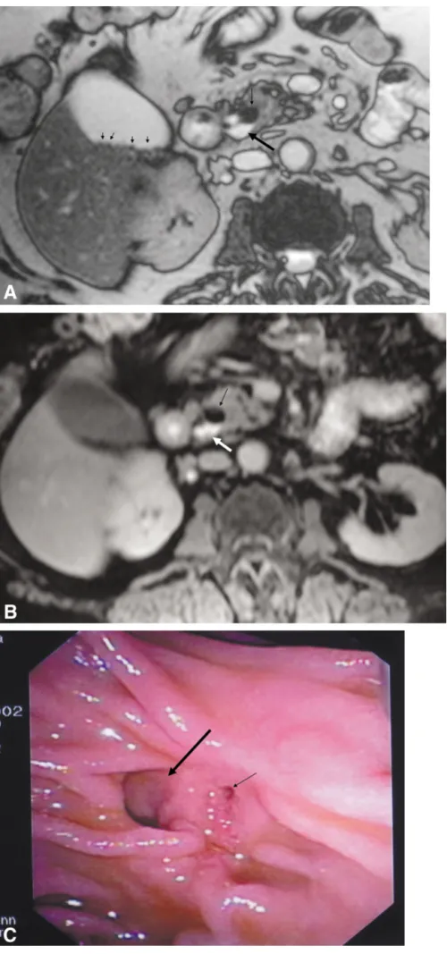
Figure 1. Small juxtapapillary
divertic-ulum in a 67-year-old patient with mul-tiple gallstones. T2-weighted True FISP image in the axial plane (TR⫽ 3.6 msec, TE ⫽ 1.8 msec, FA ⫽ 70°) (a) reveals hyperintense fluid (long arrow) and a signal void air level (short arrow) within the diverticulum. Multiple small gall-stones are observed in the gallbladder (small arrows). T1-weighted post-con-trast fat suppressed spoiled gradient-echo image (TR⫽ 165 msec, TE ⫽ 2.3 msec, FA ⫽ 70°) (b) in the axial plane reveals hyperintense fluid due to the oral contrast (white arrow), with a signal void air level above (black arrow). The corre-sponding endoscopic image during ERCP (c) demonstrates the diverticulum (long arrow) and its relationship to the papilla (short arrow).
demonstrated on both axial and coronal sections with the HASTE technique. With the use of the strong gra-dient system of our scanner, a True FISP sequence with a short TR and short TE was performed, decreasing susceptibility to bowel and respiratory motion artifacts. True FISP is a fast imaging technique with steady-state precession, which provides high signal of fluid-contain-ing structures (15). In our study, this technique en-abled better delineation of the diverticular wall. It has been shown that the use of either of these techniques may be useful in the demonstration of bowel pathology (16,17).
Although the majority of juxtapapillary diverticula are asymptomatic and discovered incidentally, there are reported associations with pancreaticobiliary dis-eases. The most commonly associated biliary pathology is gallstone formation. This may be explained either by biliary stasis, resulting from compression of the distal CBD by the diverticulum, or by an incompetent
sphinc-ter of Oddi which causes reflux of duodenal contents. Accumulation of beta-glucuronidase producing organ-isms leads to formation of pigment bile stones in the CBD in patients with juxtapapillary diverticula (1,2,4). In our study, the cases associated with dilatation of the CBD revealed sludge, rather than stone formation, in the distal CBD. Three patients had multiple calculi in the gallbladder. Another disorder associated with the juxtapapillary diverticulum is pancreatitis in a chronic or acute relapsing form. This is attributed to the stasis of pancreatic secretions (1,2). Six of our patients had laboratory and imaging findings consistent with chronic pancreatitis. MRI demonstrated parenchymal signal decreases consistent with the presence of pan-creatitis. Diminished signal of the pancreatic gland on T1-weighted fat saturated images associated with chronic pancreatitis has been described previously (18,19). Pancreatic cystic neoplasms and pseudocysts can be considered in the differential diagnosis with the
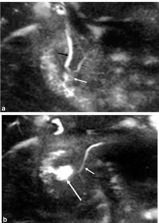
Figure 2. Juxtapapillary diverticulum
in a 70-year-old patient. MRCP with the use of HASTE sequence (TR ⫽ ⬁, TE⫽ 90 msec) reveals the distal com-mon bile duct (thick black arrow) and pancreatic duct (thin black arrow) en-tering into the diverticulum (white ar-row) (a). In the next section posteriorly, (b) the diverticulum (long arrow) and the pancreatic duct (short arrow) are demonstrated. On the corresponding ERCP images (c,d), catheterization of the papilla within the diverticulum is demonstrated (c). On the late (10-minute) phase ERCP image (d), there is contrast in the CBD (thick arrow) and in the pancreatic duct (thin arrow), in-dicating impaired biliary outflow sec-ondary to SOD. The juxtapapillary di-verticulum is marked with a white arrow.
juxtapapillary diverticulum if the former are located in close proximity to the papilla (20). The air-fluid level in the diverticular sac and the demonstration of the com-munication between the duodenum and diverticulum are MRI features that can distinguish the diverticula from pancreatic pseudocysts and cystic neoplasms. Treatment of juxtapapillary diverticula is surgical. Ei-ther trans-abdominal or laparoscopic diverticulectomy may be performed (21). Endoscopic sphincterotomy has been used as an alternative approach for the relief of CBD stasis (6). In three of our cases, diverticulectomy was performed. As the result of short-term follow-up, resultant relief of symptoms and long-term effects of this procedure need to be investigated in larger series. In conclusion, we described the MRI findings of jux-tapapillary diverticula. T2-weighted True FISP and HASTE images best demonstrated the diverticula. Di-lute oral contrast aided detection of the smaller
diver-ticula. The associated imaging findings of pancreatitis, cholelithiasis, and dilatation of the CBD also were eval-uated effectively by MRI.
REFERENCES
1. Hagege H, Berson A, Pelletier G, et al. Association of juxtapapillary diverticula with choledocholithiasis but not with cholecystolithia-sis. Endoscopy 1992;24:248 –251.
2. Egawa N, Kanisawa T, Tu Y, Sasaki N, Tsuruta K, Okamoto A. The role of juxtapapillary duodenal diverticulum in the formation of gallbladder stones. Hepatogastroenterology 1998;45:917–920. 3. Uomo G, Manes G, Ragozzino A, Cavellera A, Rabitti PG.
Periamp-ullary extraluminal duodenal diverticula and acute pancreatitis: an underestimated etiological association. Am J Gastroenterol 1996; 91:1186 –1188.
4. Ponce J, Garrigues V, Sala T, Pertejo V, Val A, Hoyos M. Motor pattern of the sphincter of Oddi in patients with juxtapapillary diverticula. J Clin Gastroenterol 1990;12:162–165.
5. Lobo DN, Balfour TW, Iftikhar SY, Rowlands BJ. Periampullary diverticula and pancreaticobiliary disease. Br J Surg 1999;86:588 – 597.
6. Eapen T, Reddy KR. Cholangitis and pancreatitis due to a juxta-papillary duodenal diverticulum— endoscopic sphincterotomy is the other alternative in selected cases. Am J Gastroenterol 1982; 77:303–304.
7. Leivonen MK, Halttunen JAA, Kivilaakso EO. Duodenal diverticu-lum at endoscopic retrograde cholangiopancreatography: analysis of 123 patients. Hepatogastroenterology 1996;43:961–966. 8. Fujita N, Noda Y, Kobayashi G, Kimura K, Yogo A. ERCP for
intradi-verticular papilla: two-devices-in-one-channel method. Gastroin-test Endosc 1998;48:517–520.
9. Toth E, Lindstrom E, Fork F-T. An alternative approach to the inac-cessible intradiverticular papilla. Endoscopy 1999;31:554 –556. 10. Stone EE, Brant WE, Smith GB. Computed tomography of
duode-nal diverticula. J Comput Assist Tomogr 1989;13:61– 63. 11. De Rai P, Castoldi L, Tiberio G. Intraluminal duodenal diverticulum
causing acute pancreatitis: CT scan and review of the literature. Dig Surg 2000;17:288 –292.
12. Semelka RC, Kelekis NL, Thomasson D, Brown MA, Laub GA. HASTE MR Imaging: description of technique and preliminary re-sults in the abdomen. J Magn Reson Imaging 1996;6:698 – 699. 13. Marcos HB, Semelka RC, Noone TC, Woosley JT, Lee JK. MRI of
normal and abnormal duodenum using half-Fourier single-shot RARE and gadolinium-enhanced spoiled gradient echo sequences. Magn Reson Imaging 1999;17:869 – 880.
14. Lee JK, Marcos HB, Semelka RC. MR imaging of the small bowel using the HASTE sequence. AJR Am J Roentgenol 1998;170:1457– 1463.
15. Ross JS. Newer sequences for spinal MR imaging: smorgasbord or succotash for acronyms? AJNR Am J Neuroradiolgy 1999;20:361– 373.
16. Gourtsoyiannis N, Papanikolaou N, Grammatikakis J, Maris T, Prassopoulos P. MR imaging of the small bowel with a true-FISP sequence after enteroclysis with water solution. Invest Radiol 2000; 35:707–711.
17. Gourtsoyiannis N, Papanikolaou N, Grammatikakis J, Maris T, Prassopoulos P. MR enteroclysis protocol optimization: comparison between 3D FLASH with fat saturation after intravenous gadolin-ium injection and true FISP sequences. Eur Radiol 2001;11:908 – 913.
18. Semelka RC, Kroeker MA, Shoenut JP, Kroeker R, Yaffe CS, Micf-likier AB. Pancreatic disease: prospective comparison of CT, ERCP, and 1.5 Tesla MR imaging with dynamic gadolinium enhancement and fat suppression. Radiology 1991;181:785–791.
19. Semelka RC, Shoenut JP, Kroeker MA, Micflikier AB. Chronic pan-creatitis: MR imaging features before and after administration of gadopentetate dimeglumine. J Magn Reson Imaging 1993;3:79 – 82.
20. Balci NC, Semelka RC. Radiologic features of cystic, endocrine and pancreatic neoplasms. Eur J Radiol 2001;38:113–119.
21. Togaya N, Shimoda M, Hamada K, Ishikawa K, Kogure H. Laparo-scopic duodenal diverticulectomy. Surg Endosc 2000;14:592–593.