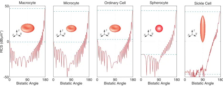
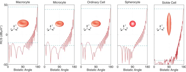
Rigorous solutions of scattering problems involving red blood cells
Tam metin
Şekil
Benzer Belgeler
Recent studies have pointed out that the maximum allowable loading ratio or derating factor (DF) of the IMs have various values for several combinations of the magnitude and angle
Sonuç olarak, Avrupa Dijital Yeterlilik Çerçevesine göre hazırlanan Dijital Okuryazarlık Beceri formunda öğretmen adaylarının giriş düzeyinde bilmesi gereken
In this study, in vitro biomineralization of calcium phosphate (CaP) crystals was controlled in a truly biomimetic system composed of ALP, OCN and OPN, which are the main
The work in [40] has detailed a job scheduling and file replication mechanism for a batch of data intensive jobs that exhibit batch-shared I/O behavior. Batch shared I/O behavior
Ong’un “birincil sözlü kültür” ve “ikincil sözlü kültür” tanımlarından hareketle ikincil sözlü kültür ürünü olarak adlandırabilecek olan Çağan Irmak’ın
Very recently, a deep sub-wavelength (sub λ/100) nanocavity design has been proposed to achieve perfect narrowband light absorption in the mid-to-far infrared range [ 43 ].. Here,
In addition to the estimation of channel coefficients, this subsystems removes the cyclic prefix interval from the received samples and sends the remaining samples of the OFDM
In a study appearing on page 1071, the GME Variome Consortium collected whole-exome data from populations across the GME region, including Northwest and Northeast Africa,
