Biomimicry of multifunctional
nanostructures in the neck feathers of
mallard (Anas platyrhynchos L.) drakes
Tural Khudiyev
1, Tamer Dogan
1,3& Mehmet Bayindir
1,2,31UNAM-National Nanotechnology Research Center, Bilkent University, 06800 Ankara, Turkey,2Institute of Materials Science and Nanotechnology, Bilkent University, 06800 Ankara, Turkey,3Department of Physics, Bilkent University, 06800 Ankara, Turkey.
Biological systems serve as fundamental sources of inspiration for the development of artificially colored
devices, and their investigation provides a great number of photonic design opportunities. While several
successful biomimetic designs have been detailed in the literature, conventional fabrication techniques
nonetheless remain inferior to their natural counterparts in complexity, ease of production and material
economy. Here, we investigate the iridescent neck feathers of
Anas platyrhynchos drakes, show that they
feature an unusual arrangement of two-dimensional (2D) photonic crystals and further exhibit a
superhydrophobic surface, and mimic this multifunctional structure using a nanostructure composite
fabricated by a recently developed top-down iterative size reduction method, which avoids the
above-mentioned fabrication challenges, provides macroscale control and enhances hydrophobicity
through the surface structure. Our 2D solid core photonic crystal fibres strongly resemble drake neck
plumage in structure and fully polymeric material composition, and can be produced in wide array of colors
by minor alterations during the size reduction process.
N
ature offers a great diversity of well-optimized photonic engineering designs, which often represent a
compromise between several potentially conflicting purposes and can maintain such a diverse array of
functions as mate attraction, UV protection, water repulsion, camouflage and sensory enhancement
1,2.
During the last decade, advancements in nanoscience and modern fabrication methods have enabled the detailed
investigation and functional mimicry of these natural designs, especially with regards to the structural coloration
effects found in biological systems
3–5. The physical phenomena responsible for structural coloration have also
received considerable attention
6,7, as these structures derive their colors from a number of optical effects, such as
interference, scattering, photonic crystal effects, or a combination thereof
8. Photonic crystals in living systems are
especially notable for their iridescence and exceptionally bright coloration
9, which may assist in camouflage
10,11,
communication
12, sensing
13–15and other purposes that still remain largely unexplored. The imitation of these
nanostructures represents significant advances
16in the area of nano-optics, and promotes the design of novel
photonic configurations
17,18. However, current fabrication methods cannot satisfactorily mimic the architectural
complexity
19found in natural systems without greatly compromising control capacity, ease of fabrication and/or
production costs. In particular, while nature-inspired 1D and 3D photonic crystals can be fabricated
20,21by
self-assembly or deposition techniques, functional imitations of 2D photonic crystals observed in nature
22–24are yet to
be produced.
Here we investigate and successfully imitate a peculiar 2D photonic scheme observed on the neck feathers of
mallard drakes. Our bioinspired 2D photonic crystals successfully replicate not only the optical properties, but
also the material features and the architectural complexity of the original structure. A novel top-down approach,
called iterative size reduction (ISR)
25–27, is used to avoid potential issues associated with the fabrication of solid
core 2D photonic crystals and to produce a biomimetic design that displays the same structural complexity and
functionality as mallard feather barbules with low fabrication costs, short processing times and minimal labor
intensity. In addition to its optical properties, the barbule surface is also shown to be strongly hydrophobic, and
this property is also successfully replicated in our bio-inspired 2D photonic crystals. The present work represents
the first successful fabrication of an all-polymer 2D solid-core photonic crystal capable of functioning at optical
frequencies. We further demonstrate that biomimetic fiber arrays displaying a great range of colors can be
obtained by minor alterations in a single fabrication step, without necessitating individual process optimization
procedures for each desired color.
OPEN
SUBJECT AREAS:
PHOTONIC CRYSTALS SYNTHESIS AND PROCESSING NANOWIRESReceived
13 January 2014
Accepted
28 March 2014
Published
22 April 2014
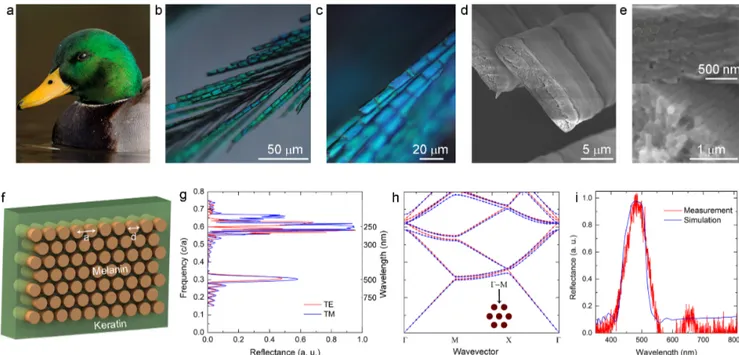
Correspondence and requests for materials should be addressed to M.B. (bayindir@nano. org.tr)Neck feathers of mallard (A. platyrhynchos) drakes (Figure 1a) are
investigated to determine the phenomena responsible for their bright
green coloration. Optical microscopy characterization suggests that
neck feathers are strongly iridescent, as the green hue observed under
a low angle of incidence (x20 magnification, Figure 1b) changes into
a distinctive blue color when the angle of illumination is increased
(x50 magnification, Figure 1c). The barbule morphology possesses a
non-circular cross-section, which may serve to maximize the
func-tional surface areas of the barbule elements responsible for iridescent
coloration (Figure 1d). Well-aligned, hexagonally distributed rods
are observed to occur on barbule edges in five- or six-layer arrays
(Figure 1e, Figure S1) and represent an unusual form of 2D photonic
crystal
28. Barbules are primarily composed of melanin pigments
(rods) embedded in a keratin matrix (filling material), and display
real refractive indices of 2.00 and 1.56, values of which fluctuate
slightly over the visible spectrum
29. Complex refractive index values
of these materials play a minor role in coloration by the 2D photonic
crystal effect, as they mainly alter the reflected intensity. These
fluc-tuations assumed to be negligible in our calculations.
A moderate refractive index contrast, such as that found in
mal-lard feathers, allows the reflection of a narrow band of wavelengths,
the position of which shifts depending on the viewing angle.
Consequently, optical simulation techniques were utilized to
deter-mine the location and properties of the reflection band associated
with barbule elements. Design parameters (Figure 1f) obtained from
SEM images of neck feather barbules are used in FDTD and MPB
simulations
30, and the resulting directional band-gap diagram and
reflectance spectrum are both found to overlap in the green and
ultraviolet regions of the electromagnetic spectrum (Figure 1g–h).
The consistency of our simulation and measurement results (Figure
S2) further support the notion that the presence of 2D photonic
crystals is responsible for the green color displayed by feather
bar-bules (Figure 1i).
The structural orientation of the barbule elements is then utilized
in the fabrication of a novel material for use in 2D photonic crystal
based devices. Iterative size reduction is utilized in order to mimic the
structural properties observed in mallard feathers, and the size and
material data taken from feather barbules are used to simplify the
design process. Polycarbonate (PC) and polyvinyldifluoride (PVDF)
are selected as the material components due to their thermal
com-patibility, which is required for the thermal drawing process, and low
refractive index contrast (n
PC51.58 and n
PVDF51.41), which closely
approximates the original configuration (Figure S3). The first step
macroscopic preform (Figure 2a, Figure S4a, b) is prepared by
suc-cessively wrapping PVDF and PC polymers around a PC rod, which
is then thermally consolidated in a furnace (see Methods). The final
macrostructure measures several centimeters in diameter, and is
thermally drawn in a custom-built fibre tower system to effect the
iterative reduction of structure dimensions (see Ref. 25 for details).
Fabricated step I fibres (Figure 2d) possess an excellent
cross-sec-tional regularity that is preserved along the length of the fibre.
Microstructures obtained after step I are cut and prepared for the
second step of the fabrication (Figure 2b).
Composite fibres with outer diameters of several hundred microns
are embedded in a polymer sheath (i.e. a rectangular hollow preform
prepared for the insertion of first step microfibres) (Figure S4c, d) in
self-arranged hexagonal arrays and redrawn to produce indefinitely
long 2D photonic crystal microstructures. Microwire arrays present
within the PVDF matrix of step II fibres preserve their initial
struc-ture profile, as can be observed from their cross-section images
(Figure 2e). A third step, similar in procedure to step II but utilizing
only one step II fibre, can then be performed to produce step III fibres
with nano- to microscale lattice constants (Figure 2c).
Microstruc-tures with rectangular cross-sections composed of hexagonal arrays
of nanorods are obtained after three consecutive drawing steps, and
bear a great resemblance to the fine structure of mallard feathers. 2D
photonic crystal structures obtained as a result of step II and step III
fabrications span a wide range of lattice sizes (50 mm-100 nm).
SEM images of step III fibres are relatively low-quality due to the
low contrast between the photonic crystal components (Figure 2f).
Figure 1|
Investigation ofA. platyrhynchos feathers. (a), Mallard neck feathers are investigated for their structural properties (Photographed by Jacob S. Spendelow). Optical microscopy images of mallard neck feathers reveal that higher magnifications result in a change of coloration from (b), green to (c), blue, which suggests the presence of iridescence. Hexagonal arrays of rods are observed from (d), longitudinal and (e), cross-sectional SEM images of feather barbules to be distributed throughout the barbule periphery. (f), Scheme representing the distribution of melanin rods embedded in the keratin matrix. The associated lattice constants (a) and diameters (d) are observed to be around 150 nm and 130 nm, respectively. (g), Reflection simulation of neck feather barbules is performed using the above-mentioned values. (h), Band structure calculations show that directional band-gaps exist in the green and UV regions of the spectrum. (i), Experimental and simulation results associated with the optical features of mallard feathers are in good agreement.Minor deformations observed in rod structures may slightly alter the
local hue, but do not significantly change the overall color.
Our multiple size reduction process can therefore be utilized to
convert the white-colored macro-prototype into structurally colored
photonic crystal fibres (Figure 3a). The green hue observed in optical
microscope images of 2D all-polymer photonic crystal structures
(Figure 3b) shifts into blue under higher magnifications as a result
of its iridescence (Figure 3c). Longitudinal SEM images reveal that
the PVDF shell acts as an embedding matrix for PC nanorods
(Figure 3d). Roughnesses are observed to exist on the photonic
crys-tal surface, but are too small to display Mie scattering or thin film
interference, and are therefore expected to play an insignificant role
in coloration
26,31. Lattice constants and rod diameters are measured
to be around 200 nm and 175 nm for green-colored 2D photonic
crystal fibres. The reflection spectra and band structure behavior of
produced fibres can be simulated accurately by using these values
(Figure 3f, g). Our measurement and unpolarized light simulation
data are also in good agreement for green-colored fibres (Figure 3h).
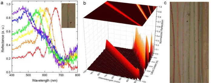
Our bioinspired solid-core 2D photonic crystal fibres are also used
for the production of a diverse array of colored fibres spanning the
visible spectrum. Decreasing lattice constants and rod diameters
gradually change the hues observed on a tapered part of photonic
crystal fibre (Figure 4a). A 3D map of the photonic crystal effect
clearly demonstrates band-gap positions associated with specific
lat-tice constants, and can assist in the design of colored photonic crystal
fibres (Figure 4b). Narrow reflection bands allow the clear
obser-vation of each color. The reduction factor can be altered during the
third step of the drawing process to modify the lattice constant of the
final product and to facilitate the single-step fabrication of arbitrarily
long, structurally colored photonic crystal fibres (Figure 4c).
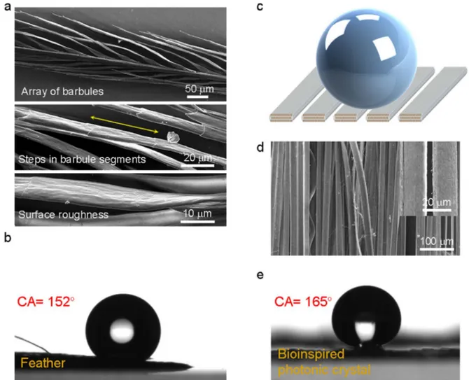
Mallard neck feathers (unlike flight feathers
32) and our
bio-inspired photonic 2D crystal fibres are both found to be strongly
hydrophobic due to their similar surface architectures, though
dif-ferent effects are responsible for their enhanced hydrophobicities
(Figure 5). SEM images of feather barbules suggest hat three surface
effects contribute to feather hydrophobicity (CA5152u 6 2u): (1)
The gaps present within the barbule arrays, (2) the interlocking steps
between each barbule cluster and (3) the nanoscale roughness of each
barbule surface (Figure 5a,b)
33. Similarly, the surface nanopattern
and arrangement of our photonic crystal fibres, as well as the
intrins-ically hydrophobic nature of the PVDF polymer shell, contribute to
the superhydrophobicity of our bio-inspired photonic crystals
(Figure 5c,d). PVDF is a fluoride-containing polymer, and
fluori-nated residues are well-known to promote hydrophobicity
34(Figure S5a). However, our all-polymer step III photonic crystal
structures (CA5160u 6 5u) are more hydrophobic than PVDF
poly-mer films (CA591u), suggesting that, in addition to material
com-position, surface hierarchy also contributes significantly to the
superhydrophobicity of the fibrous photonic crystal surfaces
(Figure 5e). Higher hydrophobicities are also observed for step II
fibres (CA5124u), which possess microscale surface patterns, as
opposed to the nanoscale features of step III fibres (Figure S5b).
The polymeric composition of our 2D photonic crystal design also
confers additional features such as biocompatibility, elasticity and
Figure 2|
Functional biomimetics of mallard drake feathers. A low temperature, multimaterial fibre drawing method is used for the iterative reduction of a macroscopic preform down to microscale photonic crystal. (a), The first step preform is obtained by successively wrapping PVDF and PC polymer films around a PC rod. The outer layer of step I fibres removed prior to the second step of fabrication. (b), In the second step, a rectangular formation is used in order to increase the aspect ratio of the final result, so as to resemble the design observed in A. platyrhynchos feathers. PVDF embedded PC microwires with lattice constants of several microns are produced at the end of step II. (c), Step III is performed by repeating previous fabrication procedures, and permits the precise control of the lattice constant in the 100-300 nm region. (d–f), Cross-sectional SEM images of produced fibers after steps I, II and III, respectively.pliability to structurally colored fibres
35–38. These fibres can also be
produced in millimeter to nanometer scales, and can therefore be
utilized on different operation wavelengths (i.e., UV, visible and IR).
In addition, our fabrication method allows the precise control of the
fibre production process, and therefore can effectively facilitate the
fabrication of a broad range of complex nanoarchitectures. The
fabrication of hard-to-produce 2D photonic crystal structures with
distinct material compositions can also be accomplished by choosing
thermally compatible raw materials
22,23. Such photonic crystal fibers
are promising for use in applications relating to filtering
39, colored
fabric woven
40,41and their superhydrophobic character is particularly
useful in anisotropic wettable fibers
42.
Figure 3
|
Characterization of biomimetic all-polymer photonic crystal structures. (a), A diverse array of 2D photonic crystal nanostructures can be engineered from a unique macroscale preform. (b), The vivid green hue observed under light microcopy confirms the presence of photonic crystal effects. (c), Biomimetic photonic crystal fibres appear iridescent as the magnification of light microscope is increased. The green color is transformed into blue as a result of the photonic crystal effect. (d–e), SEM images reveal the ribbonlike photonic crystal formation and hexagonal distribution of PC rods inside the PVDF matrix, with lattice constants around 200 nm. (f), Reflection and (g), bandstructure calculations are performed using the optical and size parameters of the material components. (h), Reflection measurements from green colored photonic crystal structures are consistent with FDTD modelling results.Figure 4
|
Band-gap tunable all-polymer colorful photonic crystal structures. (a), A tapered photonic crystal structure exhibits lattice constant-dependent changes in color along 200 mm of its length. Measurements are taken from each hue observed on the tapered photonic crystal (inset). (b), A map of bandstructure calculations can be used in the design of any desired color. (c), A large array of colors can also be obtained during the third fabrication step by altering the reduction factor. Millimeter-scale structures displaying seven distinct colors are shown as a proof-of-concept.Methods
Macroscopic composite preparation, consolidation and thermal size reduction. Photonic crystals are produced using three iterative fabrication steps. A single step I preform is utilized throughout the fabrication process, and uses a PC rod as a core component, which in turn is prepared using a hollow PC preform with an initial diameter of 30 mm. Briefly, the PC preform is cut into four equal pieces, and a turning machine is used to shape a single quadrant into a cylindrical rod with a diameter of 10 mm. A PVDF polymer film is then wrapped around this rod until a total diameter of 12 mm is reached, and a PC film is wrapped around the PC/PVDF core until the total diameter exceeds 20 mm (the complete preform therefore has a diameter ratio of 105258 for PC5PVDF5PC).
The macroscopic preform is then consolidated above the glass transition temper-ature of its components (185uC for 10 minutes, under a 1023Torr vacuum), and
thermally drawn in a custom-built fibre tower, specifications of which can be found in Ref 25. Briefly, the macroscopic structure is heated and drawn into step I microwires in a vertical two-zone furnace, with a top zone temperature of 224uC. Drawing parameters, such as drawing speed, down feed speed and temperature, are controlled throughout the fabrication process. Fibres with outside diameters of 400 mm are obtained as a result of step I fabrication, and the outer coat of PC is stripped prior to the subsequent steps (the non-adhesive layer between PVDF and PC facilitates the convenient removal of PC).
Functional biomimicry of the rectangular photonic crystal structures observed in mallard feathers is accomplished using a rectangular step II preform (step II preforms are hollow PC structures into which step I fibres are inserted). The rectangular step II preform is prepared by wrapping PC film around a bare glass substrate with the dimensions of 20 mm 3 1.5 mm 3 7 cm, to a final thickness of 35 mm 3 15 mm 3 7 cm. This preform is then thermally consolidated in a consolidation furnace, with an extended heating time (.2 h) to ensure that the outer PC layer conforms fully to the shape of the glass rod. The glass rod is then dissolved using 48% HF, leaving behind the rectangular preform. A 4cm-long array of 500 step I fibres is inserted into this step
II preform and thermally drawn at a temperature of 230uC in our custom fibre tower system (see also Ref. 25 for other parameters used in the fibre drawing process, such as capstan and down-feed speed). Fibres with outside diameters of 1.5 mm are obtained at the end of the second step. Step III is identical to step II, except that step III preforms are filled with a single step II fibre. Arbitrarily long photonic crystals can be produced with a broad crystal parameter range (including lattice constant . 100 nm) after step III, and the process can be repeated to obtain smaller structure diameters. The overall reduction factor at the end of step III is 105.
Sample preparation and imaging.Dichloromethane (DCM) (Carlo Erba) is utilized to etch the PC sheath covering the photonic crystal structures, exposing the latter for longitudinal imaging by electron and optical microscopy. Exposed nanostructures are rinsed gently with DCM for the removal of residual polymer. PVDF shells of photonic crystal structures are highly resistant to DCM etching, and are not damaged during the nanostructure extraction process. Cross-sectional images of mallard feathers and bio-inspired photonic crystals were obtained by embedding both structures in epoxy resin and sectioning them by ultramicrotome prior to imaging. Cross-sectional images are difficult to obtain due to the low contrast between the material components of both feather barbules and our bio-inspired fibres. SEM imaging is performed under both high pressure and low pressure modes to minimize this problem.
FDTD simulations and MPB calculations.FDTD simulations are performed by using commercial finite-difference time-domain software (Lumerical Solutions Inc.). A planewave source is used for illumination. FDTD simulations are calculated in two dimensional simulation regions. Frequency-domain power monitors are used to collect reflected light from periodic layers of rods embedded in host material. Refractive indices of PC and PVDF are assumed to be constant in the visible spectrum, and taken as 1.58 and 1.41 (see Figure S3).
Figure 5
|
Enhanced hydrophobicity in feather and feather-inspired photonic crystal structure. (a), Neck feathers exhibit superhydrophobicity due to presence of structural hierarchy. (b), Contact angle measurement of a single mallard feather. (c), Our photonic crystal surface enhances the intrinsic hydrophobicity of PVDF film (see Figure S5) by increasing surface roughnesses. (d), The nanoscale surface pattern observed on step III structures and the alignment of the fiber arrays both contribute to surface superhydrophobicity. (e), The water contact angle is measured to exceed 165u for this particular array.(MIT Photonic-Bands) MPB is an open-source code developed by MIT for the computation of photonic band structures. For our work, triangular lattice config-urations were used for the calculations involving both feather barbules and barbule-mimetic photonic crystal structures, radius to lattice (r/a) ratios of which were measured from SEM images to be 0.4330 and 0.4375, respectively.
1. Liu, K. & Jiang, L. Bio-inspired design of multiscale structures for function integration. Nano Today 6, 155–175 (2011).
2. Duprat, C., Protie`re, S., Beebe, A. Y. & Stone, H. A. Wetting of flexible fibre arrays. Nature 482, 510–513 (2012).
3. Fudouzi, H. Tunable structural color in organisms and photonic materials for design of bioinspired materials. Sci. Technol. Adv. Mater. 12, 064704 (2011). 4. Finnemore, A. et al. Biomimetic layer-by-layer assembly of artificial nacre. Nature
Commun. 3, 966 (2012).
5. Kolle, M. et al. Mimicking the colourful wing scale structure of the Papilio blumei butterfly. Nature Nanotechnol. 5, 511–515 (2010).
6. Vukusic, P., Sambles, J. R. & Lawrence, C. R. Structural colour: Colour mixing in wing scales of a butterfly. Nature 404, 457 (2000).
7. Whitney, H. M. et al. Floral iridescence, produced by diffractive optics, acts as a cue for animal pollinators. Science 323, 130–133 (2009).
8. Kinoshita, S. & Yoshioka, S. Structural colors in nature: the role of regularity and irregularity in the structure. ChemPhysChem 6, 1442–1459 (2005).
9. Vukusic, P., Sambles, J. R., Lawrence, C. R. & Wootton, R. J. Structural colour: Now you see it —now you don’t. Nature 410, 36 (2001).
10. Wiersma, D. S. Disordered photonics. Nature Photon. 7, 188–196 (2013). 11. Wilts, B. D. et al. Brilliant camouflage: photonic crystals in the diamond weevil,
Entimus imperialis. Proc. R. Soc. B 279, 2524–2530 (2012).
12. Ghiradella, H. Light and color on the wing: Structural colors in butterflies and moths. Appl. Opt. 30, 3492–3500 (1991).
13. Potyrailo, R. A. et al. Morpho butterfly wing scales demonstrate highly selective vapour response. Nature Photon. 1, 123–128 (2007).
14. Kim, J. H., Moon, J. H., Lee, S. Y. & Park, J. Biologically inspired humidity sensor based on three-dimensional photonic crystals. Appl. Phys. Lett. 97, 103701 (2010). 15. Sambles, J. R. Biophotonics: Blue butterflies feel the heat. Nature Photon. 6,
141–142 (2012).
16. Parker, A. R. & Townley, H. E. Biomimetics of photonic nanostructures. Nature Nanotechnol. 2, 347–353 (2007).
17. Pris, A. D. et al. Towards high-speed imaging of infrared photons with bio-inspired nanoarchitectures. Nature Photon. 6, 195–200 (2012).
18. Walish, J. J., Kang, Y., Mickiewicz, R. A. & Thomas, E. L. Bioinspired electrochemically tunable block copolymer full color pixels. Adv. Mater. 21, 3078–3081 (2009).
19. Vukusic, P. & Sambles, J. R. Photonic structures in biology. Nature 424, 852–855 (2003).
20. Diao, Y. Y. & Liu, X. Y. Controlled colloidal assembly: Experimental modeling of general crystallization and biomimicking of structural color. Adv. Func. Mater. 22, 1354–1375 (2012).
21. Kolle, M. et al. Bio-inspired band-gap tunable elastic optical multilayer fibres. Adv. Mater. 25, 2239–2245 (2013).
22. Zi, J. et al. Coloration strategies in peacock feathers. Proc. Natl. Acad. Sci. 100, 12576–12578 (2003).
23. Vigneron, J. P. et al. Structural origin of the colored reflections from the black-billed magpie feathers. Phys. Rev. E 73, 021914 (2006).
24. Eliason, C. M. & Shawkey, M. D. A photonic heterostructure produces diverse iridescent colours in duck wing patches. J. R. Soc. Interface 9, 2279–2289 (2012). 25. Yaman, M. et al. Arrays of indefinitely long, uniform nanowires and nanotubes.
Nature Mater. 10, 494–501 (2011).
26. Khudiyev, T., Ozgur, E., Yaman, M. & Bayindir, M. Structural coloring in large scale core–shell nanowires. Nano Lett. 11, 4661–4665 (2011).
27. Ozgur, E., Aktas, O., Kanik, M., Yaman, M. & Bayindir, M. Macroscopic assembly of indefinitely long and parallel nanowires into large area photodetection circuitry. Nano Lett. 12, 2483–2487 (2012).
28. Joannopoulos, J. D., Johnson, S. G., Winn, J. N. & Meade, R. D. Photonic crystals: Molding the flow of light. Princeton University Press. (2011).
29. Brink, D. J. & van der Berg, N. G. Structural colours from the feathers of the bird Bostrychia hagedash. J. Phys. D: Appl. Phys. 37, 813–818 (2004).
30. Johnson, S. G. & Joannopoulos, J. D. Block-iterative frequency-domain methods for Maxwell’s equations in a planewave basis. Opt. Express 8, 173–190 (2001). 31. Khudiyev, T., Huseyinoglu, E. & Bayindir, M. Non-resonant Mie scattering:
Emergent optical properties of core-shell polymer nanowires. Sci. Rep. 4, 4607 (2014).
32. Eliason, C. M. & Shawkey, M. D. Decreased hydrophobicity of iridescent feathers: a potential cost of shiny plumage. J. Exp. Biol. 214, 2157–2163 (2011). 33. Oner, D. & McCarthy, T. J. Ultrahydrophobic surfaces. Effects of topography
length scales on wettability. Langmuir, 16, 7777–7782 (2000).
34. Biffinger, J. C., Kim, H. W. & DiMango, S. G. The polar hydrophobicity of fluorinated compounds. ChemBioChem 5, 622–627 (2004).
35. Qi, Y. & McAlpine, M. C. Nanotechnology-enabled flexible and biocompatible energy harvesting. Energ. Environ. Sci. 3, 1275–1285 (2010).
36. Holmes-Siedle, A. G., Wilson, P. D. & Verrall, A. P. PVdF: An electronically-active polymer for industry. Mater. Design. 4, 910–918 (1984).
37. Nakazawa, M., Shi, Y. W., Matsuura, Y., Iwai, K., & Miyagi, M. Hollow polycarbonate fiber for Er: YAG laser light delivery. Opt. Lett. 31, 1373–1375 (2006).
38. Han, M., Hyun, D. H., Park, H. H., Lee, S. S., Kim, C. H., & Kim, C. A novel fabrication process for out-of-plane microneedle sheets of biocompatible polymer. J. Micromech. Microeng. 17, 1184–1191 (2007).
39. Stecher, M. et al. Polymeric THz 2D photonic crystal filters fabricated by fiber drawing. IEEE Trans. Terahertz Sci. Technol. 2, 203–207 (2012).
40. Abouraddy, A. F. & Bayindir, M. et al. Towards multimaterial multifunctional fibres that see, hear, sense and communicate. Nature Mater. 6, 336–347 (2007). 41. Gauvreau, B. et al. Color-changing and color-tunable photonic bandgap fiber
textiles. Opt. Express 16, 15677–15693 (2008).
42. Wu, H. et al. Biomimetic nanofiber patterns with controlled wettability. Soft Matter. 4, 2429–2433 (2008).
Acknowledgments
We would like to thank to Ekin O. Ozgur for her help in obtaining SEM images of mallard drake feathers, and Alper D. Ozkan for his critical reading of the manuscript and fruitful discussions. This work is supported by TUBITAK under the Project Nos. 110M412 and 111A015. The research leading to these results has received funding from the European Research Council under the European Union’s Seventh Framework Programme (FP/ 2007-2013)/ERC Grant Agreement n. 307357. M.B. acknowledges partial support from the Turkish Academy of Sciences (TUBA).
Author contributions
T.K., T.D. and M.B. designed and carried out research, analyzed data and wrote the paper.
Additional information
Supplementary informationaccompanies this paper at http://www.nature.com/ scientificreports
Competing financial interests:The authors declare no competing financial interests. How to cite this article:Khudiyev, T., Dogan, T. & Bayindir, M. Biomimicry of multifunctional nanostructures in the neck feathers of mallard (Anas platyrhynchos L.) drakes. Sci. Rep. 4, 4718; DOI:10.1038/srep04718 (2014).
This work is licensed under a Creative Commons Attribution-NonCommercial-ShareAlike 3.0 Unported License. The images in this article are included in the article’s Creative Commons license, unless indicated otherwise in the image credit; if the image is not included under the Creative Commons license, users will need to obtain permission from the license holder in order to reproduce the image. To view a copy of this license, visit http://creativecommons.org/licenses/by-nc-sa/3.0/