©2012 Landes
Bioscience.
Do not distribute
www.landesbioscience.com Bioengineered 343
Bioengineered 3:6, 343-346; November/December 2012; © 2012 Landes Bioscience
ArticLe ADDeNDA ArticLe ADDeNDum
Addendum to: Cakmak T, Angun P, Demiray YE, Ozkan AD, Elibol Z, Tekinay T. Differential effects of nitrogen and sulfur deprivation on growth and biodiesel feedstock production of Chlamydomonas reinhardtii. Biotechnol Bioeng 2012; 109:1947-57; PMID:22383222; http://dx.doi. org/10.1002/bit.24474
Keywords: Chlamydomonas reinhardtii,
biodiesel, nitrogen, sulfur, triacylglycerol Submitted: 04/17/12
Revised: 07/07/12 Accepted: 07/10/12
http://dx.doi.org/10.4161/bioe.21427
*Correspondence to: Turgay Tekinay; Email: [email protected]
N
itrogen (N) and sulfur (S) have inter-related and distinct impacts on microalgal metabolism; with N star-vation having previously been reported to induce elevated levels of the biodiesel feedstock material triacylglycerol (TAG), while S deprivation is extensively studied for its effects on biohydrogen produc-tion in microalgae.1,2 We have previouslydemonstrated that N- and S-starved cells of Chlamydomonas reinhardtii display different metabolic trends, suggesting that different response mechanisms exist to compensate for the absence of those two elements.3 We used C. reinhardtii
CC-124 mt(-) and CC-125 mt(+) strains to test possible metabolic changes related to TAG accumulation in response to N and S deprivation, considering that gamete differentiation in this organism is mainly regulated by N.4 Our
find-ings contribute to the understanding of microalgal response to element depriva-tion and potential use of element depri-vation for biodiesel feedstock production using microalgae, but much remains to be elucidated on the precise contribution of both N and S starvation on microalgal metabolism.
Due to high bioenergy outturn and the synthesis of high value added products associated with this group, microalgae are frequently investigated as a poten-tial means for various biotechnological applications.5 Biofuel production from
microalgae has emerged as a promising way of partially remediating the depen-dency of global energy demand on fossil
Nitrogen and sulfur deprivation differentiate lipid accumulation targets
of Chlamydomonas reinhardtii
Turgay Cakmak,1,2 Pinar Angun,1 Alper D. Ozkan,1 Zeynep Cakmak,1,3 Tolga T. Olmez1 and Turgay Tekinay1,*
1Laboratory of Sustainable Technologies; UNAM Institute of Materials Science and Nanotechnology; Bilkent University; Ankara, Turkey; 2Department of
Molecular Biology and Genetics; Faculty of Science; Istanbul Medeniyet University; Istanbul, Turkey; 3Department of Biology; Faculty of Arts and Sciences;
Kırıkkale University; Kırıkkale, Turkey
fuels. Compared with fossil fuels, biofuel production from microalgae is currently not cost-effective; however, continued increases in oil prices, together with a potential decrease in the cost of biodiesel from microalgae, are expected to make biofuels a viable alternative in the near future. A great variety of microalgae show distinct metabolic properties and are able to switch their metabolic output levels in response to different abiotic stress fac-tors. Levels of TAG, a principal biodiesel feedstock, vary widely across microalgae, depending on their species-specific nature and environmental factors such as changes in element concentration or presence, light intensity and temperature.6,7
N vs. S Deprivation: Differential Survival Strategy of Microalgae
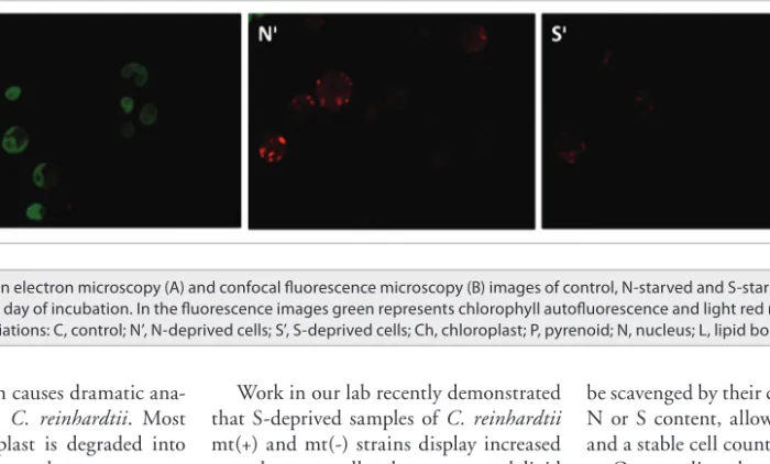
As a macroelement, N has a profound importance for microalgal metabolism and the limitation of this element is com-pensated by radical changes in several key metabolic pathways. In the process of acclimation to N deficiency, microalgae have been reported to degrade ribosomes and decrease enzyme activities involved in photosynthesis, glyoxylate cycle, gluco-neogenesis and photosynthetic carbon fix-ation cycle while simultaneously inducing carotenoid production to protect against oxidative stress, increasing the expression levels of TAG synthesis related genes in significant quantities, and differentiating into gametes, considered a potential sur-vival strategy since zygotes can withstand adverse conditions.8,9 Figure 1 shows that
©2012 Landes
Bioscience.
Do not distribute
344 Bioengineered Volume 3 issue 6
be scavenged by their conspecifics for their N or S content, allowing limited growth and a stable cell count.
Our studies showed that N starva-tion generally yielded similar effects as S starvation, but the negative impacts on cell count, total protein and chlorophyll levels were much more severe (Table 1). This result is likely caused by the rela-tive importance and abundance of N compared with S, such that while S can be salvaged from dead cells or obtained from intracellular stores, N must be sup-plied constantly for adequate growth. N content of dry C. reinhardtii biomass is known to be over 10-fold greater than the S content.2 As such, a much greater mass
of N is necessary for C. reinhardtii, while a comparatively lesser amount of S, such as that found in the initial cells inoculated into the S-free medium, may be enough to partially facilitate growth. Compared with S-starved cells, N-starved C.
rein-hardtii cells also displayed a lower amount
of enlargement, which may also be cor-related with the greater metabolic stress N-starved samples undergo (Table 1). We have observed that cellular functions are affected more rapidly in N-starved Work in our lab recently demonstrated
that S-deprived samples of C. reinhardtii mt(+) and mt(-) strains display increased growth rates, cell volumes, neutral lipid and TAG accumulation compared with their N-deprived equivalents, while a more rapid decrease in chlorophyll content was observed in C. reinhardtii cultures under N deprivation (Table 1). Furthermore, the metabolic changes associated with nutrient starvation occurred in a time-dependent manner, generally reaching a maximum on the fourth and fifth days of starvation and decreasing or remain-ing stable afterwards.3 This trend may
indicate that vegetative cells of C.
rein-hardtii can mitigate the effects of N and S
starvation for four to five days before the stress associated with long-term nutrient deprivation leads to autophagy to recycle part of the cytoplasm including organ-elles. N deficiency was reported to induce autophagy, which is a self-degrading pro-cess common in eukaryotes that provides needed energy and raw materials for cellu-lar repair, in many organisms14 including
C. reinhardtii.15 Longer starvation period
that autophagy response is insufficient would lead cell death. Dead cells may then nitrogen deprivation causes dramatic
ana-tomical changes in C. reinhardtii. Most notably the chloroplast is degraded into smaller sphere-like sub-compartments and cytoplasmic lipid droplets are formed (Fig. 1A). Besides these, chloroplast deg-radation is not as fast as N deprivation in S-deprived cells (Fig. 1B) supporting the data reported previously.3 Microalgae have
been reported to synthesize arylsulphatase to recover SO4-2 from SO
4-2 esters,
upregu-late acyltransferases and ATP sulfury-lase expression, downregulate proteins involved in translation and folding, and decrease chloroplast ribosomal polypep-tides greatly.10,11 Furthermore, cessation of
cell cycle, upregulation of the enzymes of the oxidative pentose phosphate pathway, N- or S- scavenging proteins, activation of various mechanisms for reactive oxygen species removal, repression of Calvin cycle enzymes and decrease of photosynthetic activity are observed during both types of deprivations.12,13 Such changes represent
attempts to survive the increased oxidative stress associated with nutrient deprivation and to recover N or S from the environ-ment by highly selective uptake processes for those elements.
Figure 1. transmission electron microscopy (A) and confocal fluorescence microscopy (B) images of control, N-starved and S-starved C. reinhardtii cells sampled on fifth day of incubation. in the fluorescence images green represents chlorophyll autofluorescence and light red represents Nile red fluorescence. Abbreviations: c, control; N’, N-deprived cells; S’, S-deprived cells; ch, chloroplast; P, pyrenoid; N, nucleus; L, lipid bodies.
©2012 Landes
Bioscience.
Do not distribute
www.landesbioscience.com Bioengineered 345
that the disturption of starch metabolism may increase lipid production capacity, as has been suggested previously.2,21 While
both metabolites were found to increase greatly upon starvation with no apparent antagonistic effects, this is likely the result of cell enlargement caused by S depriva-tion instead of a true lack of competidepriva-tion between lipid and starch synthesis. Our results suggest that a global shutdown in energetic functions may occur upon S deprivation. Flagella are almost always lost after third day of S deprivation and chlo-rophyll levels drop considerably, leading to the conclusion that anabolic reactions are severely reduced in that particular cell. Herein, we propose that upon reduction of energy consumption, the trend of metabo-lism favors storage of energetic denser molecules. Lipids are highly energetic molecules having a higher energy yield per gram than sugars, while starch is known to be the densest form of sugars that is usually used for storage in plants.
Records detailing the use of N starva-tion to increase lipid producstarva-tion for bio-diesel production exist in literature,2 our
study suggests that S starvation is the preferable approach due to the lack of ade-quate cell growth and biovolume attain-ment upon N exposure.
the latter may afford a measure of control over their levels.16 As such, the decrease
in chlorophyll synthesis is interpreted to be a part of the alteration, deactivation and disassembly of photosynthetic com-plexes as a response to oxidative stress resulting from S starvation. Likewise, N deprivation is closely associated with the degradation of ribulose-1,5-bisphosphate carboxylase oxygenase to recycle the lat-ter’s N content17 and the depletion of
this protein may necessitate alterations in the mechanism of photosynthesis, leading to the decrease in chlorophyll content observed in N-deficient C.
rein-hardtii.18,19 An increase in carotenoid
con-tent, observed in both N- and S-starved samples, is a response to the stress condi-tions brought about by nutrient deficiency and is consistent with previous stud-ies.20 As such, their accumulation may
be a stress response intended to prevent oxidative damage. Our recent investiga-tion showed that both starch and neutral lipids greatly accumulate in S-deprived
C. reinhardtii and that those increases
correspond to the rapid decrease in pro-tein levels observed during the first day of starvation. Production of starch took pri-ority over lipid synthesis, suggesting that the two metabolites may compete and microalgal cells. N starvation leads to an
almost instantaneous cell growth arrest due to the obligatory presence of N in every protein and most metabolites can-not be compensated by autophagy or other recycling pathways. On the other hand, S deprivation leads to less sudden but again severe responses in overall metabolism and cellular functions. This temporal delay in response probably corresponds to a period of cellular recycling by autophagy and better accumulation of stress marker mol-ecules (carotenoid, TAG, etc.).
S Deprivation May Be Used as a Potential Means for TAG Production from Microalgae
As previously reported,3 chlorophyll
con-tent decreased rapidly upon both S and N starvation, while a corresponding increase in carotenoid content was also observed.
C. reinhardtii is known to restructure its
photosynthetic machinery upon S depri-vation, resulting in a decrease in the expression of many of the proteins mak-ing up the photosystem complexes I and II within 24 h.1 Such adjustments occur
to minimize oxidative stress, as reactive oxygen species (ROS) are generated dur-ing photosynthesis and the shutdown of
Table 1. changes in growth and biochemical parameters in wild type C. reinhardtii cc-124 and cc-125 strains after four days of N or S deprivation
Parameters tested C. reinhardtii CC-124 (mt -) C. reinhardtii CC-125 (mt +)
N deprivation S deprivation N deprivation S deprivation
cell Growth 83% decrease 65% decrease 66% decrease 49% decrease
total biovolume 62.6% decrease 220% increase 54.6% decrease 310% increase
relative dry weight 32% decrease 27% decrease 23% decrease 20% decrease
Protein level 88% decrease 89% decrease 87% decrease 89% decrease
chlorophyll content 61% decrease 26% decrease 89% decrease 74% decrease
carotenoid content 3.6-fold increase 2.8-fold increase 1.9-fold increase 2.3-fold increase
cell biovolume 2.9-fold increase 6.1-fold increase 1.7-fold increase 5.8-fold increase
Starch level 2.3-fold increase 3.4-fold increase 4.3-fold increase 4.7-fold increase
relative polisaccharide level 8.1-fold increase 9.9-fold increase 13.1-fold increase 8.6-fold increase total neutral lipid level 2.4-fold increase 2.6-fold increase 1.7-fold increase 3-fold increase
relative tAG level 6.9-fold increase 15.3-fold increase 29.1-fold increase 16.5-fold increase
©2012 Landes
Bioscience.
Do not distribute
346 Bioengineered Volume 3 issue 6
15. Pérez-Pérez ME, Florencio FJ, Crespo JL. Inhibition of target of rapamycin signaling and stress activate autophagy in Chlamydomonas reinhardtii. Plant Physiol 2010; 152:1874-88; PMID:20107021; http:// dx.doi.org/10.1104/pp.109.152520.
16. Nishiyama Y, Yamamoto H, Allakhverdiev SI, Inaba M, Yokota A, Murata N. Oxidative stress inhibits the repair of photodamage to the photosynthetic machin-ery. EMBO J 2001; 20:5587-94; PMID:11598002; http://dx.doi.org/10.1093/emboj/20.20.5587. 17. García-Ferris C, Moreno J. Redox regulation of
enzymatic-activity and proteolytic susceptibility of ribulose-1,5-bisphosphate carboxylase oxygenase from Euglena gracilis. Photosynth Res 1993; 35:55-66; http://dx.doi.org/10.1007/BF02185411. 18. Plumley FG, Schmidt GW. Nitrogen-dependent
reg-ulation of photosynthetic gene expression. Proc Natl Acad Sci U S A 1989; 86:2678-82; PMID:16594026; http://dx.doi.org/10.1073/pnas.86.8.2678. 19. Siaut M, Cuine S, Cagnon C, Fessler B, Nguyen
M, Carrier P, et al. Oil accumulation in the model green alga Chlamydomonas reinhardtii: character-ization, variability between common laboratory strains and relationship with starch reserves. BMC Biotechnol 2011; 11:11; PMID:21281462; http:// dx.doi.org/10.1186/1472-6750-11-7.
20. Salguero A, de la Morena B, Vigara J, Vega JM, Vilchez C, León R. Carotenoids as protective response against oxidative damage in Dunaliella bardawil. Biomol Eng 2003; 20:249-53; PMID:12919805; http://dx.doi.org/10.1016/S1389-0344(03)00065-0. 21. Li Y, Han D, Hu G, Sommerfeld M, Hu Q. Inhibition
of starch synthesis results in overproduction of lipids in Chlamydomonas reinhardtii. Biotechnol Bioeng 2010; 107:258-68; PMID:20506159; http://dx.doi. org/10.1002/bit.22807.
7. Deng X, Fei X, Li Y. The effects of nutrition-al restriction on neutrnutrition-al lipid accumulation in Chlamydomonas and Chlorella. Afr J Microbiol Res 2011; 5:260-70.
8. Dean AP, Sigee DC, Estrada B, Pittman JK. Using FTIR spectroscopy for rapid determination of lipid accumulation in response to nitrogen limitation in freshwater microalgae. Bioresour Technol 2010; 101:4499-507; PMID:20153176; http://dx.doi. org/10.1016/j.biortech.2010.01.065.
9. Beck C, Haring M. Gametic differentiation of Chlamydomonas. Int Rev Cytol 1996; 168:259-302; http://dx.doi.org/10.1016/S0074-7696(08)60886-4. 10. Zhang L, Happe T, Melis A. Biochemical and
morphological characterization of sulfur-deprived and H2-producing Chlamydomonas reinhardtii (green alga). Planta 2002; 214:552-61; PMID:11925039; http://dx.doi.org/10.1007/s004250100660. 11. Yıldız FH, Davies JP, Grossman AR. Characterization
of sulfate transport in Chlamydomonas reinhardtii during sulfur-limited and sulfur-sufficient growth. Plant Physiol 1994; 104:981-7; PMID:12232142. 12. Grossman AR, Croft M, Gladyshev VN, Merchant
SS, Posewitz MC, Prochnik S, et al. Novel metab-olism in Chlamydomonas through the lens of genomics. Curr Opin Plant Biol 2007; 10:190-8; PMID:17291820; http://dx.doi.org/10.1016/j. pbi.2007.01.012.
13. Toepel J, Albaum SP, Arvidsson S, Goesmann A, la Russa M, Rogge K, et al. Construction and evalua-tion of a whole genome microarray of Chlamydomonas
reinhardtii. BMC Genomics 2011; 12:579;
PMID:22118351; http://dx.doi.org/10.1186/1471-2164-12-579.
14. Yorimitsu T, Klionsky DJ. Autophagy: molecular machinery for self-eating. Cell Death Differ 2005; 12(Suppl 2):1542-52; PMID:16247502; http:// dx.doi.org/10.1038/sj.cdd.4401765.
References
1. Zhang Z, Shrager J, Jain M, Chang CW, Vallon O, Grossman AR. Insights into the survival of Chlamydomonas reinhardtii during sulfur starva-tion based on microarray analysis of gene expression. Eukaryot Cell 2004; 3:1331-48; PMID:15470261; http://dx.doi.org/10.1128/EC.3.5.1331-1348.2004. 2. Wang ZT, Ullrich N, Joo S, Waffenschmidt S,
Goodenough U. Algal lipid bodies: stress induction, purification, and biochemical characterization in wild-type and starchless Chlamydomonas reinhardtii. Eukaryot Cell 2009; 8:1856-68; PMID:19880756; http://dx.doi.org/10.1128/EC.00272-09.
3. Cakmak T, Angun P, Demiray YE, Ozkan AD, Elibol Z, Tekinay T. Differential effects of nitro-gen and sulfur deprivation on growth and bio-diesel feedstock production of Chlamydomonas reinhardtii. Biotechnol Bioeng 2012; 109:1947-57; PMID:22383222; http://dx.doi.org/10.1002/ bit.24474.
4. Sager RGS, Granick S. Nutritional control of sexu-ality in Chlamydomonas reinhardi. J Gen Physiol 1954; 37:729-42; PMID:13174779; http://dx.doi. org/10.1085/jgp.37.6.729.
5. Spolaore P, Joannis-Cassan C, Duran E, Isambert A. Commercial applications of microalgae. J Biosci Bioeng 2006; 101:87-96; PMID:16569602; http:// dx.doi.org/10.1263/jbb.101.87.
6. Converti A, Casazza AA, Ortiz EY, Perego P, Del Borghi M. Effect of temperature and nitro-gen concentration on the growth and lipid con-tent of Nannochloropsis oculata and Chlorella vul-garis for biodiesel production. Chem Eng Process 2009; 48:1146-51; http://dx.doi.org/10.1016/j. cep.2009.03.006.