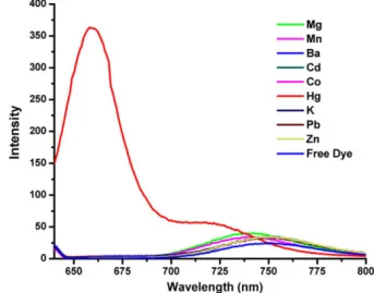
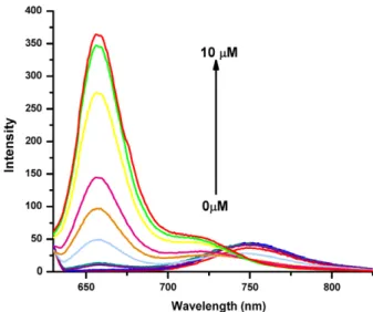
A near IR di-styryl BODIPY-based ratiometric fluorescent chemosensor for Hg(II)
Tam metin
Şekil
Benzer Belgeler
analysis included only 7% of patients within a large cohort with the highest and lowest amount of CT gene expression, we don’t think this is a reason for bias, as the distribution
Keywords: Airborne Ultrasound, Capacitive micromachined ultrasonic transduc- ers, CMUT, transducer array, High Intensity, Beam Steering, MEMS, Unbiased operation, Half frequency
force to protect the ethnic Greek minority in Albania if its existence were threatened by an influx of Albanians from Kosova.66 The then government spokesman,
In this section we describe the improvements we have made on the DST attack which reduce the precomputation time, key search time, space, and plaintext complexities of the attack..
Yapılan analizler sonucunda anne ve babanın birlikte çalıştığı ailelerde tatil satın alma karar sürecinde eşlerin etkisinin ortak olduğu, eşlerden sadece
Tüketicilerin SMS reklamlarına yönelik tutum- ları hakkındaki yukarıda incelenen tüm bu çalışmalara göre; izin, mobil servis sağlayıcı kontrolü, mobil reklamlara
Sosyal Medyanın bu kadar yoğun olarak kullanıldığı bu çağda da pazarlamanın yeni bir şekli olan ve sosyal medyada da etkin olarak kullanılan yeni bir pazarlama
Monodehidroaskorbat redüktaz enzim aktivitesinde gözlenen değişimler Kadmiyum stresi altındaki buğday yapraklarında molibden uygulamalarıyla monodehidroaskorbat redüktaz
