Review
Retinal proteins as model systems for membrane protein folding☆
Oznur Tastan
a,1, Arpana Dutta
b,1, Paula Booth
c, Judith Klein-Seetharaman
d,⁎
a
Department of Computer Engineering, Bilkent University, Ankara, Turkey
b
Department of Physiology and Biophysics, Case Western Reserve University, Cleveland, USA
cSchool of Biochemistry, University of Bristol, UK d
Division of Metabolic and Vascular Health, Warwick Medical School, University of Warwick, Coventry, UK
a b s t r a c t
a r t i c l e i n f o
Article history: Received 30 July 2013
Received in revised form 19 November 2013 Accepted 28 November 2013
Available online 12 December 2013 Keywords:
Rhodopsin Bacteriorhodopsin Membrane protein folding Denatured states
Experimental folding studies of membrane proteins are more challenging than water-soluble proteins because of the higher hydrophobicity content of membrane embedded sequences and the need to provide a hydrophobic milieu for the transmembrane regions. Thefirst challenge is their denaturation: due to the thermodynamic insta-bility of polar groups in the membrane, secondary structures in membrane proteins are more difficult to disrupt than in soluble proteins. The second challenge is to refold from the denatured states. Successful refolding of mem-brane proteins has almost always been from very subtly denatured states. Therefore, it can be useful to analyze membrane protein folding using computational methods, and we will provide results obtained with simulated unfolding of membrane protein structures using the Floppy Inclusions and Rigid Substructure Topography (FIRST) method. Computational methods have the advantage that they allow a direct comparison between di-verse membrane proteins. We will review here both, experimental and FIRST studies of the retinal binding pro-teins bacteriorhodopsin and mammalian rhodopsin, and discuss the extension of thefindings to deriving hypotheses on the mechanisms of folding of membrane proteins in general. This article is part of a Special Issue entitled: Retinal Proteins—You can teach an old dog new tricks.
© 2014 Elsevier B.V. All rights reserved.
1. Introduction
Membrane proteins (MPs) constitute typically more than 20% of genomes, yet compared to soluble proteins, the principles by which they assume their three-dimensional structures are largely unknown. This is predominantly due to the fact that it is experimentally difficult to study MPs, especially under denaturing conditions, the“bread and butter” of protein folding studies. Among MPs, two retinal proteins have model system status for MP folding studies, mammalian rhodopsin (MR) and bacteriorhodopsin (BR). Retinal proteins are excellent models with which to perform such experiments because the retinal chromo-phore acts as a natural reporter of thefinal folded state. Experimental ef-forts to denature and renature these two retinal proteins are therefore reviewed here, together with a comparison of experimental and com-putational studies aimed at understanding the molecular properties of denatured states and pathways of folding. Their possible significance for understanding the principles of folding of other MPs is also discussed. BR is a light-driven proton pump in the purple membrane of Halobacteria salinaria. It consists of seven transmembrane (TM)
α-helices that are connected by short loops and a retinal chromophore covalently bound to a lysine residue. Ground-breaking early work by the late H. Gobind Khorana and his co-workers established BR as a folding paradigm by fully denaturing and refolding BR in vitro[1]. Their subsequent seminal studies of its fragments led to the predomi-nant MP folding theory proposed by Popot and Engelman: the two-stage hypothesis[2,3], see below.
MR is the dim light photoreceptor and a prototypical G protein coupled receptor. It also consists of seven TM helices connected by cytoplasmic and extracellular loops. Mutations in MR have been linked to the retinal degenerative disease, Retinitis Pigmentosa[4]. This is an inherited disorder that causes night blindness and leads to pro-gressive loss of vision in later life due to a gradual reduction of rod and cone photoreceptor cells. Although rare, it is the main inherited ret-inal degeneration disease, with about 1 in 4000 people affected world-wide[5]. According to the human gene mutation database, more than 150 MR mutations are known to cause this disease[6]. Khorana and co-workers have shown that most of these mutations lead to misfolding and/or instability of MR as a result of which the receptor protein is retained in the endoplasmic reticulum and is incapable of binding its chromophore 11-cis retinal[7–9]. Thus, absence of correctly folded MR and presence of misfolded MR in the rod outer segments contribute to the major causes of death of rod cells in autosomal dominant cases[10]. An understanding of folding mechanisms of MR may help design effective strategies to combat Retinitis Pigmentosa by providing deeper insights into the underlying causes of misfolding. In order to
☆ This article is part of a Special Issue entitled: Retinal Proteins—You can teach an old dog new tricks.
⁎ Corresponding author at: Biomedicine and Systems Biology, Division of Metabolic and Vascular Health, Warwick Medical School, Rm. B037, Gibbet Hill, University of Warwick, Coventry CV4 7AL, UK. Tel.: +44 02476 573 806; fax: +44 02476 574 637.
E-mail address:[email protected](J. Klein-Seetharaman).
1Jointfirst co-authors.
0005-2728/$– see front matter © 2014 Elsevier B.V. All rights reserved.
http://dx.doi.org/10.1016/j.bbabio.2013.11.021
Contents lists available atScienceDirect
Biochimica et Biophysica Acta
begin to understand the folding mechanism of MR, denaturation and stability studies in the cell, in vitro and in silico have been carried out (reviewed in[11–19]).
2. Chemical denaturation studies of membrane proteins
Critical to the understanding of protein folding is the reverse pro-cess, denaturation from the native, folded states. This is because chem-ical denaturation studies can provide a useful measure of protein stability, which is intricately linked to folding. The method has been ex-tensively used for water-soluble proteins, for which a denaturation curve is generated typically by the addition of urea. Re-folding from the urea-denatured state then gives a renaturation curve. In the simplest case the denaturation and renaturation curves overlap and the reaction is a two-state equilibrium between the unfolded and folded states. In this case, the equilibrium constant and free energy of unfolding can be deter-mined and plotted against denaturant concentration to reveal a linear free energy relationship that enables extrapolation to a free energy value in the absence of a denaturant[20]. This latter free energy is taken as the intrinsic, thermodynamic stability of the protein. The chem-ical denaturation approach has been successfully applied toα-helical MPs, however it has proved decidedly challenging to refold such proteins and demonstrate equilibrium refolding (see reviews[21–23]). Further-more, there are a number of complications arising from the membrane mimetic used in the experiments[24]. Following thefirst application of this thermodynamic approach to Escherichia coli diacylglycerol kinase, the method has been successfully applied to BR. In both cases, linear free energy relationships have been observed against the concentration of the denaturant, sodium dodecyl sulfate (SDS)[25,26]. The resulting free energy of BR in the absence of a denaturant provides a useful refer-ence measure of the protein's inherent stability. Moreover, it also pro-vides the basis for obtaining information on the strength of interhelical hydrogen bonds and the folding transition state usingϕ value analysis
[27–29].ϕ analysis involves introducing a mutation into the protein and determining the change this mutation induces in the free energy of folding and the activation energy for the reaction. Thus the change in free energy of the transition state can be compared to that in the folded state, which in turn allows transition state structure to be inferred. BR was the focus of thefirst ϕ value study of the folding transition state for a MP[27]. Interestingly, helix formation in the transition state corre-lates with sequence position, with the second helix being largely formed in the transition state while thefinal C-terminal helix is pre-dominantly unstructured[28]. This polarized transition state structure also correlates with the order of TM insertion into the cell membrane, showing that in vitro measurements, correspond well to the cellular sit-uation[24].
In the cell, the folding of MPs may be more complex due to the pres-ence of chaperone functions. For example, the Drosophila protein NinaA is a known chaperone for rhodopsin, but its role in folding of rhodopsin is not clear[30]. More generally, MPs pass through and are partially retained by the translocon, a channel in the endoplasmic reticulum (ER) membrane that allows entry of proteins into the ER for posttranslational modifications. Although it is still not clear whether MPs begin to fold in the translocon and if they are partially or completely folded before entering into the ER membrane, studies using an in vitro transcription– translation system to investigate the role of the translocon in the mecha-nism of MP folding indicate that the translocon takes on the role of a chaperone during MP folding[31,32]. It appears that TM helices 1–4 translocate to the membrane sequentially after each TM helix is synthe-sized, TM helices 5–7 are retained in the translocon until opsin synthesis is complete. It was shown that retention of the TM helix depends on the properties of the helix since replacement of TM7 with TM3 resulted in spontaneous exit of TM3 after its synthesis. This raises the question, why is it necessary for some of the TM segments to be retained in the translocon until opsin synthesis is complete? It is tempting to speculate that long-range interactions may be required for proper folding to occur
in vivo. Furthermore, additional chaperone proteins participate in MP folding in vivo[33].
3. Denaturation and refolding of bacteriorhodopsin
The chemically denatured state is the closest possible equivalent to a nascent polypeptide chain in vivo that can be achieved in vitro. Finding conditions under which a protein can be denatured allows subsequent in depth analysis of the molecular characteristics of the unfolded states, including identification of residual structure (e.g.[34]). Ideally, the denatured conformational ensemble is devoid of secondary and tertiary structure— as much as possible since a true random coil may not exist[35]. However, the random coil ideal is achieved for MPs even less than for soluble proteins. Due to the presence of membranes and membrane mimetics, denaturation is exceedingly difficult for MPs. In fact, BR is one of only two MPs that have been fully denatured to date (the other MP being CopA).
Refolding from the denatured states is the subsequent logical step in the study of folding mechanisms, and has been achieved successfully for many soluble proteins. In contrast, very few MPs can be refolded, even from only partially denatured states. Again, BR is one of the few exceptions. After using trifluoroacetic acid (TFA) as a denaturant, BR was subsequently successfully refolded into phospholipids via an initial transfer into SDS micelles[1]. The possibility of refolding BR in vitro from denatured states established BR as a model system for studying the thermodynamics and kinetics of folding of helical MPs[36], as outlined above. However, these studies involve refolding BR from a par-tially SDS denatured state which retains about half of the native helical structure[26]. Additional kinetic studies of refolding BR from an SDS-denatured state into lipid/detergent micelles have enabled detection of folding intermediates in its folding pathway[11,37–41].
4. Denaturation and efforts to refold rhodopsin
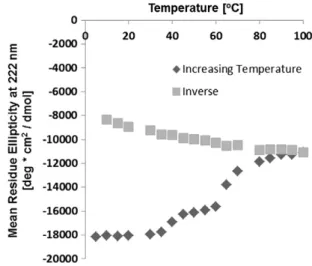
Identification of conditions under which denatured states of MR can be experimentally studied has proven more difficult than for BR. Unlike BR, it has not been possible so far to fully denature MR, and even partially denatured states have not been amenable to refolding. An extreme case demonstrating irreversibility is shown for illustrative purposes inFig. 1, where MR is denatured by high temperatures[42]. While increasing the temperature progressively shows the denaturation of the protein as evi-denced by the change in ellipticity at 222 nm, the subsequent reverse lowering of the temperature does not result in a refolding curve that traces the denaturation curve. This indicates that the protein is not
Fig. 1. Irreversibility of unfolding of MR as evidenced by thermal melting curves. Second-ary structure is measured by circular dichroism. Closed symbols are for the heating cycle (5°
C–100°
C). Open symbols are for the subsequent cooling cycle (100°
C–5°
C). A concen-tration of 2.5μM of MR in 0.05% dodecyl maltoside micelles was used.
refolding, typically due to the irreversible aggregation of the protein. The presence of aggregates of MR at high temperatures has been shown in[18].
Early studies used the denaturants guanidine hydrochloride (GuHCl) and urea to understand the stability of MR in native mem-branes[43,44]. The total decrease in mean residue ellipticity (MRE) at 222 nm, which reports on the amount of helicity, was ~50% for GuHCl. MR in its native environment was shown to refold. In another report, GuHCl denaturation of MR in 2% digitonin was carried out[43]. No changes in secondary structure on the addition of urea and GuHCl were reported, and the focus was therefore on retinal binding and hy-drolysis of the Schiff base. The effect of urea on absorbance at 500 nm of MR showed that even at 8 M concentration, urea has no effect on rho-dopsin[45]. It was reported that opsin, the apo form of MR, is more un-stable towards urea denaturation than MR since urea does begin to denature opsin at the low concentration of 1 M[43]. When compared to GuHCl, urea denatures opsin at lower concentration than GuHCl, with the latter inducing denaturation at 3.5 M[43]. Recently, denatur-ation of opsin in phospholipid bicelles with urea was reported[46]. It was shown that denaturation of opsin in phospholipid/detergent mixed micelles of DMPC/CHAPS with 4 M urea leads to an irreversible unfolding, corresponding to a decrease of 50% in helical content of opsin. A recent survey of different denaturing conditions, including urea, GuHCl, detergents and combinations of different denaturants showed that only high concentrations of SDS and combinations of SDS and urea are suitable for studying denaturation of MR in detergent micelles with-out aggregation[18]. These studies have opened the door to in-depth characterization of MR denatured states[17]. However, refolding from any of the conditions investigated has not been possible even with exten-sive variation of different lipid and detergent systems (unpublished results).
5. Comparison of rhodopsin and bacteriorhodopsin using FIRST The experimental conditions used to (de)stabilize and study BR and MR are different, making a direct comparison difficult. To complement such experimental studies, computational approaches that only require the input of a crystal structure have been applied[16,47,48]. Although crystal structures are static data, it was shown repeatedly that computa-tional methods can predict protein folding nuclei from native state structures[49]. A common approach is normal mode analysis where each amino acid is represented as a bead, and beads are connected by springs based on a cut-off distance. The movement of the beads relative to each other can be described by a matrix version of Newton's second law of motion, which when solved produces a protein's intrinsic mo-tions, called normal modes (with the respective frequencies). Although normal modes describe the motions accessible to a protein, they are themselves static properties that are directly computed from crystal structure coordinates. However, the low frequency/large amplitude motions define the softest directions (that are easiest to excite) on the energy landscape, therefore conformational transitions favorably hap-pen in accordance with those easily accessible directions. Static struc-ture defines the relevant reaction coordinates to monitor a kinetic event and stores sufficient information to find where the softest direc-tions are. Of course, charting those direcdirec-tions, by itself, does not suffice to generate the exact time evolution trajectory of a system. A predicted pathway then does not refer to the complete time trajectory, but to the most likely path to be taken.
The algorithm we deploy, the so-called Floppy Inclusions and Rigid Substructure Topography (FIRST) method[50], is an all atom analysis of the rigidity andflexibility of protein structures. In FIRST, the protein is modeled as a constraint network where each atom is a node and the nodes are subject to constraints defined by the covalent bonds, hy-drogen bonds, hydrophobic tethers and salt bridges[50]. Once the con-straint network is constructed, FIRST calculates for every bond whether it is part of a rigid cluster or part of an underconstrained region. Through
the dilution of hydrogen bond and salt bridge contacts and analyzing the constraint network after each breakage of a hydrogen bond or salt bridge (in the order of their strength), an unfolding pathway is obtain-ed. The applicability of FIRST shows that protein unfolding is inherently similar to the melting of structural glasses, upon which the method is based. The melting of glasses is a second order, continuous transition, thus describing a dynamic process. In its application to proteins, the method was specifically developed to track loss of structural stability as a measure of unfolding and to identify a“folding core” as the rigid cluster that resists dissolution the longest. In an extensive comparison of experimentally determined folding cores in 29 diverse proteins, it was shown that FIRST predicted folding cores provide statistically sig-nificant enhancements over random correlation[49]. We therefore set out to investigate if a folding core can be observed in FIRST simulated unfolding of BR[48]and/or MR[47].
Most structures when analyzed with FIRST start with one large clus-ter, interrupted only byflexible regions within loops, visible here also for BR (Fig. 2A) and MR (Fig. 2B). The vertical axis refers to the hydrogen bond energy and the horizontal axis shows the residues along the pro-tein sequence. Each line in the dilution plot indicates which residues are rigid andflexible at the corresponding hydrogen bond energy cutoff. Thin black lines represent residues with aflexible backbone; whereas each colored block identifies the rigid clusters a residue belongs to with different colors signifying different rigid clusters. The red cluster indicates the largest rigid cluster. Lines are shown only when there is a change in the backbone rigid clusters. For orientation purposes, the positions of the TM helices are outlined by thick black lines in the dilu-tion plots. The simulated unfolding of BR (Fig. 2A) is dominated by inter-actions within individual TM helices which can be seen by the relatively rapid fragmentation of the largest rigid cluster (red) into distinct rigid clusters corresponding to each helix. By contrast, in the denaturation of MR, the largest rigid cluster is observed to contain segments from multiple helices and loops for most of the dilution (Fig. 2B). This persis-tent rigidity results from the interconnectivity of structural elements in MR and portrays a nonlocal cooperativity as opposed to the individual-ity of helices observed in BR denaturation.
In the dilution plot, the folding core intuitively corresponds to the most stable residues that resist denaturation the longest. Thus in previ-ous studies, it was found that a reasonable definition for the folding core is the lowest line in the dilution plot where at least three consecutive res-idues are mutually rigid with at least three consecutive resres-idues of anoth-er secondary structural element[50]. A residue is considered rigid if at least two of the backbone atoms are present in the rigid cluster. The lines of the folding cores of MR and BR according to the above definition are marked by arrows, coinciding with−4.7 and −2.3 kcal/mol in hydrogen bond energy cutoff, respectively. The rigid clusters at the fold-ing core line are mapped onto the BR and MR structures shown in
Fig. 2. In BR (Fig. 2A), we see the coincidence of the folding units with the positions of the TM helices. In MR (Fig. 2B), at the folding core energy cutoff the largest rigid cluster (red) constitutes a core of interconnected residues at the interface between the TM and extracellular domains, lin-ing the retinal bindlin-ing pocket towards the extracellular side. This foldlin-ing core is characterized by long-range interactions involving amino acids close in space but distant in sequence comprising positions from both extracellular loop and TM regions.
Thus, the comparison of BR and MR using FIRST yields very different conclusions on the folding of these two structurally related proteins. In the case of BR (Fig. 2A), early during the simulated denaturation process, individual clusters break off, that correlate with the positions of the TM helices. This observation is consistent with the vast amount of experimental studies that demonstrate helices to be the major folding units in BR, the hallmark of the 2-stage hypothesis[2,3]. In this model, secondary structure elements formfirst, followed by tertiary interac-tions to form the helical bundle. By contrast, when FIRST is applied to MR (Fig. 2B), the initial largest cluster gradually becomes smaller until a region remains with residues at the interface between the TM and
extracellular domain, the folding core. Two additional small clusters remain at the intracellular side. Neither of these clusters correlates with the positions of any helices, indicating that the loop regions in MR may be much more important to the folding of MR than to that of BR. This emphasizes the role of tertiary interactions at the early stages during folding, which has led to the proposal of a long-range interac-tions model for MP folding[16]. It is these long-range interactions that may contribute to the difficulty in experimentally refolding MR as opposed to BR.
6. Comparison of rhodopsin and bacteriorhodopsin folding in experiments
One major difference between BR and MR is the presence of a disul-fide bond between the bottom of TM helix III and the second extracellu-lar loop in MR (Fig. 2B), while there is no disulfide bond in BR and related structures. This disulfide bond is known to be critical for stability of MR and a wrong disulfide bond leads to misfolding of MR[51]. How-ever, the presence or absence of this disulfide bond can be ruled out as a determinant for the differences between MR and BR structures in FIRST simulated unfolding, because a control calculation in which the disulfide bond was removed from the structure prior to application of FIRST[47]
yielded the same result. Thus, while the disulfide bond once formed contributes to additional stability of the folded protein, it is important to distinguish a folding core from a stability core, that may form during later stages in the folding process. The folding core proposed here refers
to a core of structural stability that resists denaturation and is assumed to formfirst in the reverse process. This hypothesis of course requires experimental validation.
While direct experimental proof for the presence of a folding core comprising loop and helix elements in MR is still missing, there is some preliminary evidence supporting the notion of a folding core. Using single-molecule dynamic force spectroscopy, a core of rigid struc-tural segments was observed in MR but not BR[19]. In this study, breakpoints within helices were detected, suggesting that not all helices act as independent single folding units. Furthermore, segments contain-ing loop and TM portions were observed, suggestcontain-ing the involvement of loop residues in assisting secondary structure stabilization. There has also been a forced unfolding study of proteorhodopsin displaying characteristics similar to BR[52]. The second line of evidence is studies from our lab in collaboration with Wayne Hubbell (Arpana Dutta, Christian Altenbach, Sheryll Mangahas, Wayne L. Hubbell and Judith Klein-Seetharaman, unpublished results), in which we attached EPR spin labels in proximity to the folding core and found some restriction of the EPR labels in conformational space in the unfolded states, consis-tent with the idea of a folding core.
Indirect evidence supporting the observation that folding of MR is more complex than that of BR comes from extensive early studies on the role of loops in BR and MR folding through split receptor experi-ments (in detail reviewed in[53]). In these experiments, cuts are intro-duced in a loop connecting two helices and the resulting fragments are isolated and then reconstituted. In the case of BR, all possible
Fig. 2. FIRST unfolding of bacteriorhodopsin and mammalian rhodopsin. Simulated thermal denaturation plot and folding core clusters for A. BR (PDB ID code: 1c3w) and B. MR (PDB ID code: 1l9h). The vertical axis refers to the hydrogen bond energy and the horizontal axis shows the residues along the protein sequence. Each line in the dilution plot indicates which residues are rigid andflexible at the corresponding hydrogen bond energy cutoff. Lines are shown only when there is a change in the backbone rigid clusters. Thin black lines represent residues with aflexible backbone; whereas each colored block identifies the rigid clusters that a residue belongs to with different colors signifying different rigid clusters. Red color represents the largest rigid cluster. Arrows indicate the positions of the predicted folding cores, as defined by the last line in which three residues from independent secondary structure elements are part of the same cluster. The clusters at the folding core energy lines are mapped on the 3D structures of BR (A, right) and MR (B, right). The MR folding core is at the interface between the extracellular and TM domain and it includes the critical disulfide bond. BR does not reveal such a core, and the rigid clusters correspond to helical regions.
combinations of cuts have been made and no loop was found to be es-sential for the formation of a three-dimensional structure able to bind retinal. However, reconstituted fragments did display lower stability, and loop mutations, substitutions and deletions do cause changes in BR stability. Similar studies were also carried out for MR (see[53]for references). However, in contrast to BR, it is not possible to purify frag-ments of MR and reconstitute them later to yield a chromophore bind-ing three-dimensional structure. Instead, split genes were expressed in cells and were characterized subsequent to in vivo reconstitution. In this setup, it is possible to introduce cuts, but only when placed in the loop connecting helices 4 and 5 and the loop connecting helices 5 and 6 in the cytoplasmic domain. No cut in extracellular loops allow formation of a folded structure, consistent with the predicted folding core located at the interface between the extracellular and TM domains.
7. Simulated unfolding of non-retinal binding proteins
Given the structural similarity of seven TM helices and retinal spe-cies covalently bound to the protein, it is remarkable tofind the above described differences in simulated unfolding of BR versus MR. This poses the question how relevant thesefindings are for the folding of other MPs. To address this question, we applied FIRST to all helical MPs with known structure. The list of proteins was retrieved from the PDBTM[54]. Theoretical models and duplicate chains were removed and only proteins with resolution better than 3 Å were chosen. This re-sulted in 237 protein chains. These were clustered using a 35% sequence identity cutoff with BlastClust[55]. Clustering resulted in 52 protein chain groups. When MR and BR related proteins (i.e. MR, BR, sensory rhodopsins I & II, halorhodopsin) were also removed, 47 chain groups remained.
Visual inspection of the dilution plots indicated that some proteins behaved like BR, and some like MR, while some showed characteristics of both, with individual helices breaking off early but a core involving residues from other helices and loops remaining until late. Quantification of the folding core energies exhibited a spectrum of values (Fig. 3). MR and BR are at the two ends of the spectrum (indicated by arrows in
Fig. 3). The dilution of long-range interactions[48]also showed a range of values (data not shown).
Finally, we developed a new way to quantify the dilution plots, shown inFig. 4A. In order to be able to assess to what extent a helix unfolds independently of the other helices, we defined a met-ric,ΔHi= Hu− Hs.For a helix, Hsis defined as the energy level in
the denaturation process where the helix (80% of its residues) becomes independent of other helices. Hu, on the other hand,
marks the energy level where the helix unfolds completely. We
define that a helix unfolds independently if it satisfies one of the two conditions: i) ifΔHsN = − 3 kcal/mol and ΔHiN = −1 kcal/
mol ii) it satisfies ΔHs > –3 kcal/mol and ΔHi b = –2 kcal/mol. The definitions were set such that the average number of helices that fold independently was high for BR-like structures and low for MR-like structures. Based on the above definition, 85% of heli-ces in each of the resolved BR-like structures and only 17% of heliheli-ces in each of the MR-like structures were estimated to unfold independently. Based on the folding behavior of the helices we labeled each of the struc-tures as“BR-like”, “MR-like” or “Mixed”. Therefore, if at least 70% of the helices in a protein are unfolding independently, we classify the unfolding process as BR-like. If less than 30% of the helices in a protein were inde-pendently unfolding, we labeled it as MR-like, otherwise we labeled the structure as mixed. In this way, having labeled each structure within each protein chain group, the group then assigned an overall label based on the labels of the structures within the group. This resulted in five different groups of MP structures labeled as: MR-like, BR-like, mixed, mixed-BR and mixed-MR. MR-like, BR-like and Mixed means all structures in this group are MR-like, BR-like or Mixed, respectively. BR-mixed means the group has structures both BR and Mixed in it and MR-mixed means the group has structures both MR and Mixed in it. The numbers of protein chain groups within each of thesefive major groups is shown inFig. 4B. Approximately half of the MP protein chain groups (25/47) are assigned BR-like, while only 4 are MR-like. The remainder (18/47) shows characteristics of both. A comparison be-tween this classification and the folding core energy is shown in
Fig. 4C. MP groups are ranked by folding core energy (analogous to
Fig. 3) and their class label is shown (the color coding used corresponds toFig. 4B). As one can see all but two groups with less negative values are classified as BR, while the mixed and MR-like groups also have lower folding core energies. This analysis indicates that most helical MPs contain helices as independent folding units, but frequently, there are some regions within the protein structure spanning multiple helices and including loop regions that show high cooperativity in unfolding. This suggests that the contribution of loops to areas of greatest stability is also a common theme among the folding of MPs.
As with all studies that take published crystal structures as input, our analysis may reflect crystallization bias. It is very well possible that BR-like behavior promotes ease of crystallization. While only a diversi fica-tion of crystallized MP structures will help address this quesfica-tion, at this stage we can at least rule out resolution as contributing factor. When we ranked the different MP structure groups according to pre-dicted folding core energy, there was no correlation with the associated resolution of the structures (Supplementary Fig. S1). We also investigat-ed qualitatively if the groups may be correlatinvestigat-ed with membrane origin (bacterial gram negative inner membrane, bacterial gram positive plasma membrane, endoplasmic reticulum membrane, mitochondrial inner membrane, thylakoid membrane, archaebacterial membrane and eukaryotic plasma membrane (Supplementary Fig. S2). We found no correlation. Finally, we began investigating protein functional differences between protein groups (e.g. channels vs. transporters vs. enzymes) and found no correlation (data not shown). However, a rigor-ous hypothesis testing, e.g. using Gene Ontology functional terms, should be carried out to investigate this question further.
8. Extent of denaturation and refolding of non-retinal binding membrane proteins
Testing the predictions made by FIRST experimentally will require studies with denatured states of MPs. There are a number of proteins beyond the model systems BR and MR, for which conditions under which their denatured states can be studied have been established. Bacterial helical MPs for which refolding has been possible include: KcsA, from a trifluoroethanol (TFE)-denatured state[56]; DAGK from SDS[57], urea and GuHCl[58], DsbB from SDS denatured state[59], EmrE from a SDS + urea denatured state[60], CopA from a GuHCl
Fig. 3. FIRST predicted folding core energies of membrane proteins with known structure. Folding core energies were extracted from FIRST simulated unfolding plots as shown in Fig. 3. The averages and standard deviations for all members in the 52 groups of clustered MPs (see text) are plotted in order of decreasing folding core energy. The MR and BR groups are indicated by arrows.
denatured state[61]and LHCII complex in higher plants from a SDS-denatured state[62,63]. In all cases except CopA, refolding was accom-plished only from a partially unfolded, near native state. GuHCl denatur-ation of CopA that led to a substantial decrease in MRE at 222 nm by about 75% could be refolded into the native state[61]. The only eukary-otic helical MP that has been purified in folded form, denatured and refolded so far is the Human Peripheral Myelin Protein 22 (PMP22), a four-helix bundle, whose misfolding has been implicated in Charcot-Marie-Tooth disease. PMP22 displays poor stability even in the absence of denaturants and folding studies were done both in the absence and presence of glycerol which acts as a stabilizing osmolyte. PMP22 was de-natured in mixed micelles of dodecylphosphocholine and n-lauroyl sarcosine and renaturation was achieved by dilution of n-lauroyl sarcosine[64]. A number of other eukaryotic MPs have also been refolded from denatured states, although the details of the denatured states were not a focus of these studies: G protein coupled receptors have been expressed extensively in E. coli and these studies are becom-ing increasbecom-ingly successful[65,66].
Refolding studies on LHCII, a helical MP, provides a good example of MP folding study where a structural insight into folding mechanisms of MP was obtained. EPR spectroscopy was used to investigate the de-tails of refolding LHCII from a 50% lithium dodecyl sulfate denatured state[63]. In this report, distance measurements between two pairs of residues derivatized with EPR labels were carried out during equilibri-um and kinetic unfolding and refolding studies. One pair was placed on either end of a TM helix and the other pair was placed in the luminal side of two different TM helices. Refolding kinetics showed that forma-tion of tertiary contacts between the TM helices occurs after formaforma-tion of TM helices. Thesefindings are in accordance with the two-stage model of folding[2,3]. However, TM helix formation during LHCII refolding was observed to extend beyond thefirst stage of folding there-by not completely following the two-stage model.
9. Discussion and future studies
Analysis of protein folding is greatly facilitated when a protein can be reversibly unfolded, i.e. its native structure can be disrupted and
subsequently restored. A microscopically reversible, dynamic equilibri-um folding reaction allows a thermodynamic analysis of the data, al-though strictly this requires the equilibrium to be solvent independent, which is generally not the case. Thermodynamic studies help to link thefindings of the refolding experiments and denatured states to the biological situation. There are few reports of successful re-covery of fully folded, functional MPs from denatured states. In part this reflects the small number of research laboratories that are studying MP folding and endeavoring to establish defined, controlled refolding con-ditions. However, there are also inherent experimental difficulties compounded by a lack of knowledge of the nature of the denatured state as well as the key structural elements and interactions that are re-quired for correct folding.α-Helical MPs pose considerable challenges, many of which arise from their inherent hydrophobicity and the prob-lems involved in trying to re-create their natural, complex membrane en-vironment. Moreover, some of these proteins are large withflexible structures, making the task of satisfying the requirements for folding of dynamic membrane-embedded, aqueous-exposed and lipid headgroup contacting regions even more difficult. There are four main problems that hinder the re-folding ofα-helical MPs in vitro. Firstly, the precise na-ture of the denana-tured state is unknown. Hence, the residual strucna-ture, protein–denaturant interactions and ensemble of conformations are unknown as well as whether low-order aggregation states are pres-ent. This makes it difficult to identify existing unfavorable interac-tions that could hinder folding through formation of aggregates or incorrectly folded states. A second problem facing folding studies is the lack of information on key structural interactions that are re-quired for correct folding. Thus, it is challenging to optimize the cor-rect conditions for the folding of certain structural elements. It does appear that critical core and/or helical structure aids correct folding
[36]. A third problem is the solvent. Frequently, detergents or mix-tures of detergents and different lipids are used for practicality. These solvents are not accurate mimics of the native environment since a membrane is composed of many different lipids, with asym-metric monolayer composition and lateral heterogeneity together with the presence of other MPs. Moreover, there are differing sol-vent environments at either side of the membrane. The types of
Fig. 4. Independent helix analysis of FIRST simulated unfolding results. A. Definition of parameters quantified from the simulated unfolding plot: Hsis the energy level of a helix before it
starts to unfold; Huis the energy level when a helix becomes independent of other helices;ΔHiis the difference between Huand Hs. In each case, a helix is defined by 80% of all residues in
the helix. B. Helix-based classification to identify BR-like and MR-like behavior in non-retinal membrane proteins. We classified a helix as an “helix that unfolds independently of other helices” if it satisfies ΔHsN = −3 kcal/mol and ΔHiN = −1 kcal/mol or it satisfies ΔHsb −3 kcal/mol and ΔHi N = −2 kcal/mol. Then if at least 70% of the helices in a protein are
unfolding independently, we classify the unfolding process of that protein as BR-like. If less than 30% of the helices in a protein were independently unfolding, we labeled it as MR-like, otherwise we labeled the structure as mixed. In each group, the majority vote was taken. When there was a tie, both labels were assigned. This gives rise tofive groups, MR-like, BR-like, mixed, BR-mixed and MR-mixed. Each group is color coded. C. Relationship between folding core energies and helix-based classification. The 47 groups of MPs (which exclude all retinal protein structures, including MR, BR, sensory rhodopsin, halorhodopsin) were classified according to their predicted folding cores (Fig. 3) and color coded according to their clas-sification from Fig. 4B. As one can see, the less negative folding core values are BR-like folders, while the MR and mixed groups have more negative folding core values.
environments used in experiments in vitro only partially reproduce the native characteristics. Furthermore, it is likely that different sol-vent properties are necessary to optimize different stages of folding
[67]. A fourth issue for MP re-folding in vitro, is tweaking conditions to attain a fully functional state. While regaining the native secondary structure can be proved possible it is often challenging to completely recover the tertiary structure and native ligand binding or function. Thisfinal stage could require further optimization of solvent condi-tions. All four problems are compounded by the lack of experimental methods to probe protein and solvent structures and interactions during the folding process.
Future studies could aim at a molecular description of the denatured states. Since experimental conditions under which MR can be dena-tured without aggregation are now known[18], it will be possible to characterize unfolded states analogous to what has been done with soluble proteins[34]. This could directly test experimentally the predict-ed folding core. Future improvements in computational methods to study MP folding will need to include lipids. The FIRST method was applied without taking the membrane into account, which is clearly a limitation. For example, hydrogen bonds are ranked based on the struc-tures as input only, without considering the presence of the membrane. While the strength of side chain hydrogen bonds in helical MPs appears to be similar to that of soluble proteins[68], backbone hydrogen bonds in membranes will be stronger, and this may affect the ranking of the bonds considered during the dilution step in FIRST. Other methods should also be explored. While molecular dynamics simulations have been applied extensively to lipid systems, none have tackled MP unfolded states. Recent developments in coarse grained modeling of lipids and mem-branes using the MARTINI forcefield[69] may provide alternative avenues for future modeling of MP folding in the presence of lipids. Acknowledgements
We would like to thank Professor Charles Sanders of Vanderbilt University for asking his insightful question“Why can't rhodopsin be refolded?” at the Membrane Protein Folding Meeting of the Biophysi-cal Society in Seoul, South Korea on May 19–22, 2013, inspiring the theme of this article. Thanks also to Fernanda Balem for the preparation ofFig. 2. Research by the authors reviewed in this article has been funded in part by NSF CAREER grant CC044917 and NSF EAGER grant IIS-1144281, National Institutes of Health Grant NLM108730, a DAAD-Helmholtz Fel-lowship and a Royal Society Wolfson Merit Award, Leverhulme Research Fellowship as well as BBSRC grants (BB/G002037/1 and BB/F013183/1) to PJB.
Appendix A. Supplementary data
Supplementary data to this article can be found online athttp://dx. doi.org/10.1016/j.bbabio.2013.11.021.
References
[1] K.S. Huang, et al., Refolding of an integral membrane protein. Denaturation, renatur-ation, and reconstitution of intact bacteriorhodopsin and two proteolytic fragments, J. Biol. Chem. 256 (8) (1981) 3802–3809.
[2] J.L. Popot, D.M. Engelman, Membrane protein folding and oligomerization: the two-stage model, Biochemistry 29 (17) (1990) 4031–4037.
[3] J.L. Popot, S.E. Gerchman, D.M. Engelman, Refolding of bacteriorhodopsin in lipid bilayers. A thermodynamically controlled two-stage process, J. Mol. Biol. 198 (4) (1987) 655–676.
[4] Retnet,http://www.sph.uth.tmc.edu/Retnet/disease.htm#03.202d.
[5] D.T. Hartong, E.L. Berson, T.P. Dryja, Retinitis pigmentosa, Lancet 368 (9549) (2006) 1795–1809.
[6] M.P. Krebs, et al., Molecular mechanisms of rhodopsin retinitis pigmentosa and the efficacy of pharmacological rescue, J. Mol. Biol. 395 (5) (2010) 1063–1078.
[7] C.H. Sung, C.M. Davenport, J. Nathans, Rhodopsin mutations responsible for autoso-mal dominant retinitis pigmentosa. Clustering of functional classes along the poly-peptide chain, J. Biol. Chem. 268 (35) (1993) 26645–26649.
[8] C.H. Sung, et al., Rhodopsin mutations in autosomal dominant retinitis pigmentosa, Proc. Natl. Acad. Sci. U. S. A. 88 (15) (1991) 6481–6485.
[9] S. Kaushal, H.G. Khorana, Structure and function in rhodopsin. 7. Point mutations associated with autosomal dominant retinitis pigmentosa, Biochemistry 33 (20) (1994) 6121–6128.
[10]H.F. Mendes, et al., Mechanisms of cell death in rhodopsin retinitis pigmentosa: implications for therapy, Trends Mol. Med. 11 (4) (2005) 177–185.
[11]P.J. Booth, S. High, Polytopic membrane protein folding and assembly in vitro and in vivo, Mol. Membr. Biol. 21 (2004) 163–170.
[12]J.P. Chapple, et al., Unfolding retinal dystrophies: a role for molecular chaperones, Trends Mol. Med. 7 (2001) 414–421.
[13] C. McKibbin, et al., Opsin stability and folding: modulation by phospholipid bicelles, J. Mol. Biol. 374 (2007) 1319–1332.
[14] C. McKibbin, et al., Opsin stability and folding: the role of Cys 185 and abnormal disul-fide bond formation in the intradiscal domain, J. Mol. Biol. 374 (2007) 1309–1318.
[15] R.S. Saliba, et al., The cellular fate of mutant rhodopsin: quality control, degradation and aggresome formation, J. Cell Sci. 115 (2002) 2907–2918.
[16] J. Klein-Seetharaman, Dual role of interactions between membranous and soluble por-tions of helical membrane receptors for folding and signaling, Trends Pharmacol. Sci. 26 (4) (2005) 183–189.
[17] A. Dutta, et al., Characterization of membrane protein non-native states. 2. The SDS-unfolded states of rhodopsin, Biochemistry 49 (30) (2010) 6329–6340.
[18] A. Dutta, et al., Characterization of membrane protein non-native states. 1. Extent of unfolding and aggregation of rhodopsin in the presence of chemical denaturants, Biochemistry 49 (30) (2010) 6317–6328.
[19]K.T. Sapra, et al., Mechanical properties of bovine rhodopsin and bacteriorhodopsin: possible roles in folding and function, Langmuir 24 (4) (2008) 1330–1337.
[20] A. Fersht, Enzyme Structure and Mechanism, 2nd ed. W.H. Freeman & Co., New York, 1985.
[21]P.J. Booth, P. Curnow, Folding scene investigation: membrane proteins, Curr. Opin. Cell Biol. 19 (2009) 8–13.
[22]P.J. Booth, et al., In vitro studies of membrane protein folding, Crit. Rev. Biochem. Mol. Biol. 36 (2001) 501–603.
[23]H. Hong, et al., Methods for measuring the thermodynamic stability of membrane proteins, Methods Enzymol. 455 (2009) 213–236.
[24] P.J. Booth, A successful change of circumstance: a transition state for membrane pro-tein folding, Curr. Opin. Struct. Biol. 22 (2012) 469–475.
[25] S. Faham, et al., Side-chain contributions to membrane protein structure and stabil-ity, J. Mol. Biol. 335 (2004) 297–305.
[26] P. Curnow, P.J. Booth, Combined kinetic and thermodynamic analysis of alpha-helical membrane protein unfolding, Proc. Natl. Acad. Sci. U. S. A. 104 (2007) 18970–18975.
[27]P. Curnow, P.J. Booth, The transition state for integral membrane protein folding, Proc. Natl. Acad. Sci. U. S. A. 106 (2009) 773–778.
[28] P. Curnow, et al., Stable folding core in the folding transition state of an alpha-helical integral membrane protein, Proc. Natl. Acad. Sci. U. S. A. 108 (34) (2011) 14133–14138.
[29]N.H. Joh, et al., Modest stabilization by most hydrogen-bonded side-chain interac-tions in membrane proteins, Nature 453 (7199) (2008) 1266–1270.
[30]R. Webel, et al., Role of asparagine-linked oligosaccharides in rhodopsin maturation and association with its molecular chaperone, NinaA, J. Biol. Chem. 275 (32) (2000) 24752–24759.
[31] N. Ismail, et al., Specific transmembrane segments are selectively delayed at the ER translocon during opsin biogenesis, Biochem. J. 411 (3) (2008) 495–506.
[32] S.L. Meacock, et al., Different transmembrane domains associate with distinct endoplasmic reticulum components during membrane integration of a polytopic protein, Mol. Biol. Cell 13 (12) (2002) 4114–4129.
[33] D. Athanasiou, M. Aguila, D. Bevilacqua, S.S. Novoselov, D.A. Parfitt, M.E. Cheetham, The cell stress machinery and retinal degeneration, FEBS Lett. 587 (2013) 2008–2017.
[34]J. Klein-Seetharaman, et al., Long-range interactions within a nonnative protein, Science 295 (5560) (2002) 1719–1722.
[35] R.L. Baldwin, B.H. Zimm, Are denatured proteins ever random coils? Proc. Natl. Acad. Sci. U. S. A. 97 (23) (2000) 12391–12392.
[36] P.J. Booth, P. Curnow, Membrane proteins shape up: understanding in vitro folding, Curr. Opin. Struct. Biol. 16 (4) (2006) 480–488.
[37] P.J. Booth, A. Farooq, Intermediates in the assembly of bacteriorhodopsin investigated by time-resolved absorption spectroscopy, Eur. J. Biochem. 246 (3) (1997) 674–680.
[38] P.J. Booth, A. Farooq, S.L. Flitsch, Retinal binding during folding and assembly of the membrane protein bacteriorhodopsin, Biochemistry 35 (18) (1996) 5902–5909.
[39] P.J. Booth, et al., Intermediates in the folding of the membrane protein bacteriorho-dopsin, Nat. Struct. Biol. 2 (2) (1995) 139–143.
[40] M.L. Riley, et al., Slow alpha helix formation during folding of a membrane protein, Biochemistry 36 (1) (1997) 192–196.
[41] S.J. Allen, et al., Folding kinetics of an alpha helical membrane protein in phospholip-id bilayer vesicles, J. Mol. Biol. 342 (4) (2004) 1279–1291.
[42] F. Balem, N. Yanamala, J. Klein-Seetharaman, Additive effects of chlorin e6 and metal ion binding on the thermal stability of rhodopsin in vitro, Photochem. Photobiol. 85 (2) (2009) 471–478.
[43]R. Hubbard, Absorption spectrum of rhodopsin: 500 nm absorption band, Nature 221 (5179) (1969) 432–435.
[44]H. Shichi, Conformational aspects of phodopsin associated with disc membranes, Exp. Eye Res. 17 (6) (1973) 533–543.
[45]E.W. Abrahamson, S.E. Ostroy, The photochemical and macromolecular aspects of vision, Prog. Biophys. Mol. Biol. 17 (1967) 179–215.
[46]C. McKibbin, et al., Urea unfolding of opsin in phospholipid bicelles, Photochem. Photobiol. 85 (2) (2009) 494–500.
[47]A.J. Rader, et al., Identification of core amino acids stabilizing rhodopsin, Proc. Natl. Acad. Sci. U. S. A. 101 (19) (2004) 7246–7251.
[48] O. Tastan, et al., Comparison of stability predictions and simulated unfolding of rho-dopsin structures, Photochem. Photobiol. 83 (2) (2007) 351–362.
[49] A.J. Rader, I. Bahar, Folding core predictions from network models of proteins, Poly-mer 45 (2004) 659–668.
[50]D.J. Jacobs, et al., Proteinflexibility predictions using graph theory, Proteins 44 (2) (2001) 150–165.
[51]J. Hwa, J. Klein-Seetharaman, H.G. Khorana, Structure and function in rhodopsin: mass spectrometric identification of the abnormal intradiscal disulfide bond in misfolded retinitis pigmentosa mutants, Proc. Natl. Acad. Sci. U. S. A. 98 (9) (2001) 4872–4876.
[52]Klyszejko, et al., Folding and assembly of proteorhodopsin, J. Mol. Biol. 376 (2008) 35–41.
[53]O. Tastan, J. Klein-Seetharaman, H. Meirovitch, The effect of loops on the structural organization of alpha-helical membrane proteins, Biophys. J. 96 (6) (2009) 2299–2312.
[54] G.E. Tusnady, Z. Dosztanyi, I. Simon, PDB_TM: selection and membrane localization of transmembrane proteins in the protein data bank, Nucleic Acids Res. 33 (2005) D275–D278(Database issue).
[55] S.F. Altschul, et al., Basic local alignment search tool, J. Mol. Biol. 215 (3) (1990) 403–410.
[56] F.N. Barrera, et al., Unfolding and refolding in vitro of a tetrameric, alpha-helical membrane protein: the prokaryotic potassium channel KcsA, Biochemistry 44 (43) (2005) 14344–14352.
[57]F.W. Lau, J.U. Bowie, A method for assessing the stability of a membrane protein, Biochemistry 36 (19) (1997) 5884–5892.
[58] J.K. Nagy, W.L. Lonzer, C.R. Sanders, Kinetic study of folding and misfolding of diac-ylglycerol kinase in model membranes, Biochemistry 40 (30) (2001) 8971–8980.
[59] D.E. Otzen, Folding of DsbB in mixed micelles: a kinetic analysis of the stability of a bacterial membrane protein, J. Mol. Biol. 330 (4) (2003) 641–649.
[60]D. Miller, et al., In vitro unfolding and refolding of the small multidrug transporter EmrE, J. Mol. Biol. 393 (4) (2009) 815–832.
[61] E.A. Roman, J.M. Arguello, F.L. Gonzalez Flecha, Reversible unfolding of a thermophilic membrane protein in phospholipid/detergent mixed micelles, J. Mol. Biol. 397 (2) (2010) 550–559.
[62] F.G. Plumley, G.W. Schmidt, Reconstitution of chlorophyll a/b light-harvesting com-plexes: xanthophyll-dependent assembly and energy transfer, Proc. Natl. Acad. Sci. U. S. A. 84 (1) (1987) 146–150.
[63] C. Dockter, et al., Refolding of the integral membrane protein light-harvesting com-plex II monitored by pulse EPR, Proc. Natl. Acad. Sci. U. S. A. 106 (44) (2009) 18485–18490.
[64]J.P. Schlebach, et al., Reversible folding of human peripheral myelin protein 22, a tetraspan membrane protein, Biochemistry 52 (19) (2013) 3229–3241.
[65] T. Dahmane, et al., Amphipol-assisted in vitro folding of G protein-coupled receptors, Biochemistry 48 (27) (2009) 6516–6521.
[66] K. Michalke, et al., Mammalian G protein-coupled receptor expression in Escherichia coli: II. Refolding and biophysical characterization of mouse canna-binoid receptor 1 and human parathyroid hormone receptor 1, Anal. Biochem. 401 (1) (2010) 74–80.
[67] P.J. Booth, Sane in the membrane: designing systems to modulate membrane pro-teins, Curr. Opin. Struct. Biol. 15 (2005) 435–440.
[68]J.U. Bowie, Membrane protein folding: how important are hydrogen bonds? Curr. Opin. Struct. Biol. 21 (1) (2011) 42–49.
[69] S.J. Marrink, et al., The MARTINI forcefield: coarse grained model for biomolecular simulations, J. Phys. Chem. B 111 (27) (2007) 7812–7824.