Vol.:(0123456789)
SN Applied Sciences (2019) 1:947 | https://doi.org/10.1007/s42452-019-0997-z Research Article
Synthesis and characterization of hydrothermally synthesized
ɑ‑Fe
2O
3nanostructures with controlled morphology
Cansu Noberi1 · Cengiz Kaya2,3
© Springer Nature Switzerland AG 2019 Abstract
Antimicrobial ɑ-Fe2O3 nanoparticles were synthesized using hydrothermal method (HS) at 150 °C for 12 h. FeCl3 and urea were used to have ɑ-Fe2O3 nanostructures with the help of appropriate amount of surfactants to control the mor-phology. Stable colloidal suspensions were prepared using synthesized nanoparticles and then 3-D metallic filters were coated using electrophoretic deposition under definite voltage and time. It was shown that the obtained particles were effective to eliminate S. Aureus Bacterias.
Keywords ɑ-Fe2O3 · Nanoparticles · Coating · EPD · Antibacterial
1 Introduction
ɑ-Fe2O3 nanoparticles have become critical materials in recent years for various applications and therefore it is widely used due to its unique characteristics, such as high corrosion resistant and antimicrobial behavior [1, 2]. ɑ-Fe2O3 nanoparticles are also used as a photocatalyst n-type semiconductor because its bandgap (nearly 2.1 eV) permits considerable amount of the solar energy [3, 4]. It also shows desirable photocatalytic efficiency and stability in aqueous solutions [4, 5]. Synthesis method and the type of surfactant material used during synthesis play highly important role in order to have oriented morphology with desired properties. For instance, Pu et al. [2] synthesized pure ɑ-Fe2O3 nanoparticles with different morphology using different amount of CTAB as a surfactant material. Spray and Choi [4] used anodic deposition method to syn-thesize highly transparent and uniform films composed of ɑ-Fe2O3 particles.
In the present work, ɑ-Fe2O3 nanoparticles were obtained at 150 °C for 12 h using HS in an attempt to
control the morphology using TWEEN80 as a surfactant. It is also shown that ɑ-Fe2O3 nanoparticles are effective to eliminate S. Aureus Bacterias.
2 Experimental section
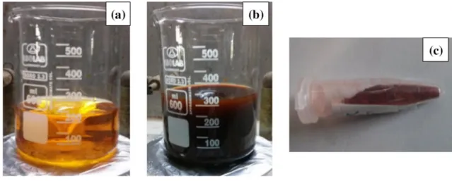
ɑ-Fe2O3 nanoparticles were synthesized hydrothermally at 150 °C for 12 h. FeCl3 and urea were used as starting precursors where both deionized water and butanol were solvents. FeCl3 and urea were solved into the mixture of deionized water and butanol. TWEEN 80 was added as a surfactant material. The obtained solution was put into a Teflon-lined autoclave and sealed. Hydrothermal synthesis has taken place at 150 °C for 12 h. After the synthesis, the color of the obtained solution was changed into red from yellow whilst the pH of the solution was also change to 8.5 from acidic pH range. The solution was washed with dis-tilled water and the red precipitate was dried at 70 °C for 24 h. Dried nanoparticles were calcined at 350 °C for 1 h.
Received: 14 May 2019 / Accepted: 26 July 2019 / Published online: 31 July 2019
* Cansu Noberi, [email protected]; Cengiz Kaya, [email protected] | 1Department of Mechatronics Engineering, Faculty of Engineering and Architecture, Istanbul Gelisim University, Istanbul, Turkey. 2Materials Science and Nanoengineering, Faculty of Natural Sciences and Engineering, Sabancı University, Istanbul, Turkey. 3Nanotechnology Research and Application Centre (SUNUM), Sabancı University, Istanbul, Turkey.
Vol:.(1234567890)
Research Article SN Applied Sciences (2019) 1:947 | https://doi.org/10.1007/s42452-019-0997-z Using hydrothermally synthesized ɑ-Fe2O3
nanoparti-cles stable colloidal suspensions (0.5 wt%) were prepared where the 2-Proponal used as a solvent. TWEEN80 was used as a surfactant material once again in order to pre-vent the flocculation. The prepared stable colloidal sus-pensions were used for the electrophoretic deposition (EPD) process. 3-D nickel based filter materials were coated using EPD using different coating times from 3 to 5 min with a fixed voltage of 30 V DC.
3 Results and discussion
The photos of the obtained solutions before HS, after HS and the final product after drying are shown in Fig. 1a–c, respectively, clearly indicating that change in the color is taking place proofing that the transition is actually occurred.
Figure 2 indicates both SEM and TEM micrographs of the synthesized ɑ-Fe2O3 nanoparticles. Figure 2a reveals that, ɑ-Fe2O3 nanoparticles were clearly synthesized in near spherical morphology while the average particle size is determined to be about 90 nm, as shown in Fig. 2b. However, some degree of aggregation caused by the cal-cination process is also seen in Fig. 2a, b. In order to under-stand the shape and size of the nanoparticles in detail TEM observations are also conducted as shown in Figs. 2c, d which indicate that the shape of ɑ-Fe2O3 nanoparticles have spherical morphology with an average particle size of 90 nm. However, there are also some particles with big-ger diameter as high as 150 nm, as shown in Fig. 2d.
The crystallinity, phase and the purity of the sample was determined using XRD analysis (Fig. 3). XRD patterns exhibited that intense peaks belong to the ɑ-Fe2O3 nano-particles according to the standards (JCPDS card no of ɑ-Fe2O3: 33-0664). Only one peak at around 2θ = 56° could
be attributed to the γ- Fe2O3 phase, as marked in graphic (JCPDS card no of γ-Fe2O3: 39-1346). No other character-istic peaks were observed for impurities.
Figure 4 indicates the antimicrobial activity of hydro-thermally synthesized and calcined ɑ-Fe2O3 nanoparti-cles. It can be seen that as the concentration of nanopar-ticles were increased, number of bacteria colonies were decreased. The most efficient concentration was seen in 1000 µg/ml of ɑ-Fe2O3. That concentration was eliminated almost more than half of the control colony.
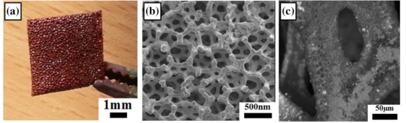
Figure 5a shows the photo of nickel based metallic filter coated using EPD under 30 V for 3 min. Figure 5b shows secondary electron image of the coated filter whereas Fig. 5c indicates back-scattered image of the filter. Macro image of the coated filter (Fig. 5a) shows that the coating process was occurred successfully. SE and BSE micrographs of the filter’s also proof that the success of the EPD. As seen in Fig. 5c the inner parts of the filter coated, too.
4 Conclusion
ɑ-Fe2O3 nanoparticles with an average particle size of 90 nm were synthesized at 150 °C for 12 h by hydrothermal treatment. TWEEN80 was used as a surfactant material in order to have spherical nanoparticles. The antimicrobial effect was determined against to S. Aureus Bacterias and it was shown that ɑ-Fe2O3 nanoparticles have remarkable antimicrobial efficiency in high concentrations. Stable colloidal suspensions were obtained whereas 2-propanol used as a suspension media and Ni-based filters were coated using an applied voltage of 30 V by EPD.
Vol.:(0123456789) SN Applied Sciences (2019) 1:947 | https://doi.org/10.1007/s42452-019-0997-z Research Article
Fig. 2 SEM micrographs (a, b) and TEM micrographs (c, d) of hydrothermally synthesized ɑ-Fe2O3 nanoparticles
Fig. 3 XRD patterns of ɑ-Fe2O3 nanoparticles
Fig. 4 Antimicrobial effect of calcined ɑ-Fe2O3 nanoparticles against to S. Aureus Bacterias
Vol:.(1234567890)
Research Article SN Applied Sciences (2019) 1:947 | https://doi.org/10.1007/s42452-019-0997-z
Acknowledgements Authors would like to special thanks to Prof.
Dr. Adil Allahverdiyev, Dr. Melahat Bağırova and Dr. Emrah Şefik Abamor from Yildiz Technical University, Bioengineering Depart-ment for helping with the antibacterial tests. This work was funded by TUBITAK (The Scientific and Technological Research Council of Turkey) under the contract number 109R007. Financial support from The Office of Scientific Research Fund of Yildiz Technical University is also acknowledged.
Compliance with ethical standards
Conflict of interest The authors declare that they have no conflict of
interest.
References
1. Liu L, Kou HZ, Mo W et al (2006) Surfactant-assisted synthesis of ɑ-Fe2O3 nanotubes and nanorods with shape-dependent magnetic properties. J Phys Chem B 110:15218–15223
2. Pu Z, Cao M, Yang J et al (2006) Controlled synthesis and growth mechanism of hematite nanorhombohedra, nanorods and nanocubes. Nanotechnology 17:799–804
3. Chirita M, Grozescu I (2009) Fe2O3-nanoparticles, physical prop-erties and their photochemical and photoelectrochemical appli-cations. Chem Bull 54(68):1–8
4. Choi RL, Spray KS (2009) Photoactivity of transparent nanocrys-talline Fe2O3 electrodes prepared via anodic electrodeposition. Chem Mater 21:3701–3709
5. Subramaniasiva B, Ntaraj D, Mangalaraj D et al (2009) Highly mesaoporous ɑ-Fe2O3 nanostructures: preparation, charac-terization and improved photocatalytic performance towards Rhodamine B (RhB). J Phys D Appl Phys 43:015501–015509
Publisher’s Note Springer Nature remains neutral with regard to
jurisdictional claims in published maps and institutional affiliations.
Fig. 5 Images of the coated filter using EPD process; macro images (a), secondary electron (b) and back-scattered electron (c) images of the