RESEARCH ARTICLE
An evaluation of the apical seal in oval-shaped root canals prepared with either self-adjusting files or protaper files
Dilek Helvacioglu Yigit, DDS, PhD,aHasan Orucoglu,DDS, PhD,bTevfik Yavuz, DDS, PhD,cCavit Isik Yavuz, DDS, PhD,dAyca Yilmaz, DDS, PhD,eYusuf Nuri KABA,fSamet OZDENf
a
Department of Endodontics, Faculty of Dentistry, Kocaeli University, Kocaeli, Turkey. b
Department of Endodontics, Faculty of Dentistry, Abant Izzet Baysal University, Bolu, Turkey. cDepartment of Prostodontics, Faculty of Dentistry, Selcuk University, Konya, Turkey.
dDepartment of Public Health, Faculty of Medicine, Kocaeli University, Kocaeli, Turkey. eDepartment of Endodontics, Faculty of Dentistry, Istanbul University, Istanbul, Turkey. f Dental Student, Faculty of Dentistry, Kocaeli University, Kocaeli, Turkey.
Received: 24 June 2013 Accepted: 13 October 2013
ABSTRACT
Objectives: This study aimed to evaluate the apical seal obtained with lateral compaction of gutta-percha in
oval-shaped root canals prepared with either self-adjusting files or ProTaper Universal rotary files.
Materials and Methods: Twelve pairs of extracted mandibular premolars with oval-shaped root canals were
randomly divided into two groups. The first group was biomechanically prepared with ProTaper Universal files, while the self-adjusting file system was used in the second group. The roots were better filled using cold lateral compaction of gutta-percha. Apical microleakage was measured with the computerized fluid filtration method. The results were analyzed statistically using the Mann-Whitney U test.
Results: All of the roots in both groups showed leakage. Group 1 demonstrated significantly less microleakage
(p<0.05).
Conclusions: Instrumentation of oval-shaped canals using a self-adjusting file system with cold lateral
compaction of gutta-percha demonstrated significantly greater apical microleakage when compared to ProTaper Universal. The apical leakage with different filling techniques, combined with self-adjusting files, should be evaluated in further studies.
Keywords: ProTaper Universal, self-adjusting file, oval-shaped canals, apical microleakage.
---INTRODUCTION
Effective biomechanical preparation and hermetically filled root canals are considered fundamental requirements for successful endodontic treatment. The aim of root canal preparation is to remove infected pulp tissue remnants and micro-organisms, as well as to shape the root
---Ayça YILMAZ,
Mithat Pasa Mah. Bahcekoy Cad. Cesmeler Vadisi Sitesi B2 34075 Kemerburgaz/Eyup/Istanbul/Turkey Tel: +905325163400
Fax: +902123623004
e-mail: [email protected]
canal system to facilitate irrigation and the placement of filling material.1 Preparing a consistent and uniform canal shape enhances hermetic obturation of root canal fillings, which in turn provide the apical seal.2 The goal of three-dimensional filling is to provide an impermeable, fluid-tight seal within the entire root canal system, to prevent oral and apical microleakage.3
Oval-shaped canals are relatively common configurations with a prevalence of about 25% in the apical third of teeth. Mandibular incisors and single canal premolars demonstrated long-oval canals
with a prevalence greater than 50 % at 5 mm from the apex.4 (Wu 2000). A micro-CT analyses of teeth have shown the frequent presence of oval canals along the entire root with a higher buccal and lingual wall thickness than the mesial and distal, including in the last few apical millimeters of the root canal.5Grande et al.5also found that the premolar root canals became more oval toward the coronal aspect.
Many different nickel-titanium (NiTi) rotary instruments are currently used to prepare root canals. NiTi files with good centering ability within the root canal lumen remove less dentin and produce rounder canals.6 NiTi rotary files provide consistent and uniform canal preparation in the apical region7, improving the apical seal.8 However, it has been demonstrated that these files perform poorly in oval-shaped canals.9Moreover, there have been only a few studies regarding whether use of NiTi rotary files has any effect on the apical seal of oval canals.10 The self-adjusting file (SAF) system (ReDent-Nova, Ra’anana, Israel) was recently introduced, with the intention of keeping the natural shape of the root canal. The vibrating metal lattice of the file adapts itself to the canal walls, even in oval-shaped or C-shaped canals. This system is also designed to maintain continuous flow of irrigants while removing a uniform layer of dentin from canal walls.11
The quality of root fillings has been evaluated using such leakage models as fluid filtration and dye leakage.12In fact, it has been demonstrated that all root canal fillings leak13 and that complete fluid-tight seal of canals are uncommon. Oval-shaped canals represent a challenge, both in biomechanical preparation and in the filling phase of root canal treatment.14,15 Such canals require adequate cleaning and shaping procedures, as well as effective filling, to achieve hermetic sealing of the root canal system. The purpose of this study was to evaluate the apical seal obtained with lateral compaction of
gutta-percha in oval-shaped root canals prepared with either SAF or ProTaper Universal (Dentsply Maillefer, Ballaigues, Switzerland).
MATERIALS AND METHODS Selection and Preparation of Teeth
A total of 105 extracted mandibular single-root premolar teeth with no large decay or previous root canal filling were examined for the selection of canals with oval cross-sections. The buccolingual and mesiodistal diameters were measured at 6 mm coronal from the apex with a digital caliper (Mitutoyo Corporation, Japan). Teeth presenting with apical curvature and two canals were excluded from the study. A total of 52 teeth with buccolingual/mesiodistal diameter ratios of ≥2 were selected as oval-shaped canals. Four teeth formed positive and negative control groups. Radiographs from the remaining 48 teeth were evaluated to match pairs on the basis of similar root canal morphology. A total of 24 teeth (12 pairs) were chosen as the study group and were randomly placed in one of two experimental groups: Group 1: ProTaper Universal; and Group 2: SAF). Working length and apical patency were determined with #10 K-File (Mani Inc., Tochigi-Ken, Japan) inserted into the root canal until its tip was visible at the apex. Tooth length was standardized to 10 mm by cutting off the crowns of teeth for permeability test.
Biomechanical Preparation:
ProTaper Universal
The first group of 12 teeth was prepared with ProTaper Universal rotary instruments (Dentsply Maillefer). A ProTaper Universal file was operated at 300 rpm with 2 N/cm of torque, using a pre-programmed motor (WaveOne,
Dentsply Maillefer, Ballaigues, Switzerland). The root canals were instrumented following the manufacturer’s instructions up to an F3 file. One set of instruments was used for each root canal.
The canals were irrigated with 4 ml of 2.5 % NaOCl between each instrument, resulting in a total volume of 20 ml of NaOCl. Irrigation was performed with a syringe, and a 27-gauge needle was placed at 2 mm from the working length. The smear layer was removed using 3 ml of 17 % EDTA for 2 mins, followed by 3 ml of distilled water. The negative control group was prepared in the same manner as the first group, but the positive control group was left uninstrumented.
Biomechanical Preparation: SAF
Second group of twelve teeth was prepared using the SAF system. A glide path was established using hand files up to a size of #20 K-File at the working length. The SAF was operated using handpiece head (RDT3, ReDent-Nova, Ra’anana, Israel) for 4 minutes at 5000 rpm and amplitude of 0.4 mm. One instrument was used for each root canal. The irrigation hub on the SAF file was connected to a special irrigation pump (VATEA Irrigation device, ReDent-Nova, Ra’anana, Israel), which provided a total of 20 ml of NaOCl irrigation at a flow rate of 5 ml/min throughout the instrumentation. Three milliliters of 17 % EDTA for 2 mins were used, followed by 3 ml of distilled water.
Root Canal Filling
After completion of the biomechanical instrumentation procedure, the canals were dried with paper points (Dentsply De Trey, Konstanz, Germany). Adequate master gutta-percha cones with 0.02 tapers were coated with AHPlus sealer (Dentsply DeTrey, Konstanz, Germany) and inserted to working length. The root canals were then filled with cold, laterally condensed gutta-percha and AHPlus sealer. Size 25, 20 and 15 accessory gutta-percha points were condensed using stainless steel finger spreaders until the filling was completed. Coronal access was sealed with temporary filling material (Cavit G, 3M Espe, Seefeld, Germany). The filled roots
were stored in 100% humidity at 37°C for 1 week to allow the sealer to set. The negative and positive control groups were filled with lateral compaction of gutta-percha with AHPlus sealer. The negative control group was completely coated with three layers of nail polish, while an area of 1 mm around the apex was left uncoated in the positive control group.
Fluid Filtration Test
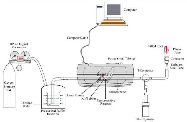
Apical microleakage was measured using a computerized fluid filtration meter, as described by Orucoglu et al.16The roots were inserted into the plastic tube from the apical side and were connected to 18-gauge stainless steel tubes. A cyanoacrylate adhesive (Zapit, Dental Venture of America Inc., Anaheim Hills, CA, USA) was applied circumferentially between the roots and plastic tubes. The computerized fluid filtration meter with laser system used in this study had a 25-µl micropipette (Microcaps, Fisher Scientific, Philadelphia, PA, USA) mounted horizontally.16 O2 from a pressure tank of
120 kPa (1.2 atm) was applied on the apical side. The pressure was kept constant throughout the experiment by means of a digital air pressure regulator added to the pressure tank. A 25-µl micropipette (Microcaps, Fisher Scientific, Pittsburgh, PA, USA) was connected to the pressure reservoir with polyethylene tubing (Microcaps, Fisher Scientific, Pittsburgh, PA, USA). All of the pipettes and syringes and the plastic tubes on the apical side of the sample were filled with distilled water. The water was sucked back with the microsyringe to approximately 2 mm. In this manner, an air bubble was created in the micropipette, and this bubble was adjusted to a suitable position within the syringe. The computerized fluid filtration meter was based on the basic light refraction at the starting and ending positions of air bubble movement inside the micropipette (Figure 1). Through one side of the micropipette inside the device,
an infrared light was passed. Two light-sensitive photodiodes were arranged on the opposite side of the micropipette to detect any movement of the air bubble inside micropipette. All of the operations were controlled with PC-compatible software (Fluid Filtration’03, Konya, Turkey). A 5-min pressurization preload of the system was completed before readings were taken. Measurements of fluid movement for each sample were automatically obtained at 2 minutes during the 8 minutes of testing using PC-compatible software. The software converted the minute linear movements of the bubble into nanoliter movements at a rate of one measurement. This information was then input into the PC-compatible software. The leakage quantity is expressed in units of µl.cm H2O.min-1, and the means were calculated.
Statistical Analysis
The results were analyzed statistically using the Mann-Whitney U test between
the two experimental groups, using SPSS for Windows (SPSS Inc., Chicago, IL, USA), version 15.0. The significance level used was p<0.05.
RESULTS
Table 1 shows the mean leakage scores and standard deviations for the two experimental groups. All of the roots in both groups showed leakage. The teeth in Group 1 (ProTaper Universal) exhibited a mean leakage of 0.000446 μl.cm H2
O-1.min-1.10-4 1.2 atm. The teeth in Group 2 (SAF) showed a mean leakage of 0.000635 μl.cm H2O-1.min-1.10-4 1.2 atm with only
one measuring 0.00102 μl.cm H2
O-1.min-1.10-4 1.2 atm. In applying the Mann-Whitney U test, Group 1 demonstrated significantly less microleakage (p < 0.05). The positive controls showed excessive amounts of leakage (0.0026 μl.cm H2
O-1.min-1.10-4 1.2 atm), while the negative controls exhibited no leakage at 1.2 atm.
Table 1. Apical microleakage values of groups (in μl.cm H2O-1.min-1.10-4 1.2 atm).
Group n Mean SD Range
1 12 0.000446 0.000081 0.000448-0.001020
2 12 0.000635 0.000168 0.000249-0.000556
SD: Standard deviation
DISCUSSION
Three-dimensional filling aims to provide an impermeable, fluid-tight seal over the entire root canal system, preventing coronal and apical microleakage.3 Oval-shaped root canals tend to have buccal and lingual recesses, which can present a challenge to the clinician at the filling stage. In the present study, the apical seal obtained in oval-shaped root canals using SAF were compared with those using ProTaper Universal. ProTaper Universal was chosen because its effectiveness in root canals has been well investigated in the literature.17-20 The SAF system was recently introduced, and it has been claimed to affect a larger percentage of the root canal wall than rotary NiTi files do.21
Several methods have been proposed to establish the sealing ability of procedures or materials in root canal filling. Fluid filtration testing is the most widely accepted leakage-measurement method compared to other methods, including linear dye leakage and electrochemical methods. With the fluid filtration technique, the specimens are well preserved after each assay, allowing for analysis at different study periods. This method was proved advantageous, with sensitive and reproducible results.22 The computerized fluid filtration meter used in this study had the primary advantages of computer controlled and digital air pressure arrangement, compared to conventional meters. Moreover, the movement of a small air bubble can be
observed with computer-controlled laser diodes.23
NiTi rotary instruments might enlarge the root canal sufficiently to achieve a certain thickness of master cone. However, the instruments are less effective when dealing with canals with oval cross-sections. These instruments create round preparations, leaving untouched buccal and/or lingual extensions in oval-shaped canals.19 In contrast, most root canal systems have round cross-sections in the apical parts, even if the middle and coronal parts of the roots present oval canal shapes.4 Previous studies have reported that oval-shaped root canals taper to rounder shapes apically because the long diameters of oval canals decrease in apical areas.25,26 In one study, no significant difference was observed in the percentages of unprepared root canal walls using SAF or rotary instruments in the apical third of the canal.21 Versiani et al21 reported that the size of the SAF preparation in the apical third of the canal was equivalent to those prepared with #40 rotary file with a 0,02 taper. Taha et al24compared the canal shapes created in oval-shaped root canals by three preparation techniques, and they stated that the use of rotary NiTi files in the apical third should be considered the preferred technique for oval-shaped canals. In the present study, specimens prepared with ProTaper Universal yielded better results with regard to apical seal. This result might be attributable to the apical cross-sectional shape of the root canals.
Dentinal debris can become wedged in isthmus areas and recesses when root canals are prepared with rotary instruments.27 Incomplete removal of this debris can result in root canal blockage28, negatively affecting the quality of filling.27,29 The untreated parts of the root canal system can serve as potential areas for leakage. The remaining debris can also influence the short-term results of apical leakage tests.
We assumed that the SAF system could reach all of the areas of the root canals superiorly; however, this assumption did not positively affect the sealing ability in the present study. These conflicting results might have been due mainly to the filling technique. Lateral compaction, being the most popular technique, was chosen as the filling method in the present study. However, uninstrumented recesses might not be completely filled by cold lateral condensation in oval-shaped canals.26 Because it might have only limited ability to fill entirely the empty recesses cleaned of debris with the SAF, cold lateral condensation might not be achieved properly. De-Deus et al17 evaluated the filling ability of a thermoplasticized carrier-based technique in canals prepared with the SAF system. They reported that the SAF system improved the filling quality of oval-shaped canals. The SAF system, in combination warm gutta-percha techniques, should be investigated in further studies to obtain more comparable results.
In conclusion, instrumentation of oval-shaped canals using the SAF system, with cold lateral compaction of gutta-percha, demonstrated significantly higher apical microleakage compared to ProTaper Universal. Homogenous preparation of the root canal walls could not provide complete apical seal in oval canals. The apical microleakage with different filling techniques, in combination with SAF, may be evaluated in future studies.
ACKNOWLEDGEMENT
This research was supported by Kocaeli University Research Fund (Grant No: BAP-2011-66-HDP).
REFERENCES
1. Schilder H. Cleaning and shaping the root canal. Dent Clin North Am 1974;18:269-296.
2. Dow Pr, Ingle JI. Isotope determination of root canal failure. Oral Surgery, Oral Medicine, Oral Pathology 1955;8:1100-1108
3. Delivanis PD, Mattison GD, Mendel RW. The survivability of F43 strain of Streptococcus sanguis in root canals filled with gutta-percha and Procosol cement. J Endod 1983;9:407-410.
4. Wu MK, R'oris A, Barkis D, Wesselink PR.Prevalence and extent of long oval canals in the apical third. Oral Surg Oral Med Oral Pathol Oral Radiol Endod 2000;89:739-743.
5. Grande NM, Plotino G, Pecci R, Bedini R, Pameijer CH, Somma F. Micro-computerized tomographic analysis of radicular and canal morphology of premolars with long oval canals. Oral Surg Oral Med Oral Pathol Oral Radiol Endod 2008;106:70-76.
6. Bergmans L, Van Cleynenbreugel J, Wevers M, Lambrechts P. Mechanical root canal preparation with NiTi rotary instruments: rationale, performance and safety. Status report for the American Journal of Dentistry. Am J Dent. 2001;14:324-333.
7. Glosson CR, Haller RH, Dove SB, DelRio CE. A comparison of root canal preparations using ni-ti hand, ni-ti engine-driven, and k-flex endodontic instruments. J Endod 1995; 21:146-151.
8. Weine FS, Kelly RF, Lio PJ. The effect of preparation procedures on
original canal shape and on apical foramen shape. J Endod 1975;1:255-262.
9. Metzger Z, Teperovich E, Cohen R, Zary R, Paqué F, Hülsmann M. The self-adjusting file (SAF). Part 3: removal of debris and smear layer-A scanning electron microscope study. J Endod 2010;36:697-702.
10. Von Fraunhofer JA, Fagundes DK, McDonald NJ, Dumsha TC. The effect of root canal preparation on microleakage within endodontically treated teeth: an in vitro study. Int Endod J 2000;33:355-360.
11. Metzger Z, Teperovich E, Zary R, Cohen R, Hof R. The self-adjusting file (SAF). Part 1: respecting the root canal anatomy--a new concept of endodontic files and its implementation. J Endod 2010;36:679-690.
12. De-Deus G, Leal F, Soares J, Luna AS, Murad C, Fidel S, Fidel RA. Dye extraction results on bacterial leakproof root fillings. J Endod 2008;34:1093-1095.
13. Marshall FJ, Massler J. The sealing of pulpless teeth evaluated with radioisotopes. Journal of Dental Medicine 1961;16:172-184.
14. Hilaly Eid GE, Wanees Amin SA. Changes in diameter, cross-sectional area, and extent of canal-wall touching on using 3 instrumentation techniques in long-oval canals. Oral Surg Oral Med Oral Pathol Oral Radiol Endod. 2011;112:688-695
15. Ruckman JE, Whitten B, Sedgley CM, Svec T. Comparison of the Self-Adjusting file with Rotary and Hand Instrumentation in Long-oval-shaped Root Canals. J Endod 2013;39:92-95.
16. Oruçoğlu H, Sengun A, Yilmaz N. Apical leakage of resin based root canal sealers with a new computerized fluid filtration meter. J Endod 2005;31:886-890.
17. De-Deus G, Barino B, Marins J, Magalhães K, Thuanne E, Kfir A. Self-adjusting file cleaning-shaping-irrigation system optimizes the filling of oval-shaped canals with thermoplasticized gutta-percha. J Endod 2012;38:846-849.
18. Elayouti A, Chu AL, Kimionis I, Klein C, Weiger R, Löst C. Efficacy of rotary instruments with greater taper in preparing oval root canals. Int Endod J 2008;41:1088-1092.
19. Metzger Z, Zary R, Cohen R, Teperovich E, Paqué F.The quality of root canal preparation and root canal obturation in canals treated with rotary versus self-adjusting files: a three-dimensional micro-computed tomographic study. J Endod 2010;36:1569-1573.
20. Paqué F, Balmer M, Attin T, Peters OA. Preparation of oval-shaped root canals in mandibular molars using nickel-titanium rotary instruments: a micro-computed tomography study. J Endod 2010;36:703-707.
21. Versiani MA, Pécora JD, de Sousa-Neto MD. Flat-oval root canal preparation with self-adjusting file instrument: a micro-computed tomography study. J Endod 2011;37:1002-1007.
22. Kardon BP, Kuttler S, Hardigan P, Dorn SO.An in vitro evaluation of the sealing ability of a new root-canal-obturation system. J Endod 2003;29:658-661.
23. Gençoglu N, Oruçoglu H, Helvacıoḡlu D. Apical leakage of different gutta-percha techniques: thermafil, js quick-fill, soft core, microseal, system B and lateral condensation with a computerized fluid filtration meter. Eur J Dent 2007;1:97-103.
24. Taha NA, Ozawa T, Messer HH. Comparison of three techniques for preparing oval-shaped root canals. J Endod 2010;36:532-535.
25. Baisden MK, Kulild JC, Weller RN.Root canal configuration of the mandibular first premolar. J Endod 1992;18:505-508.
26. Wu MK, Wesselink PR.A primary observation on the preparation and obturation of oval canals. Int Endod J 2001; 34:137-141.
27. Paqué F, Ganahl D, Peters OA.Effects of root canal preparation on apical geometry assessed by micro-computed tomography. J Endod 2009; 35:1056-1059.
28. al-Omari MA, Dummer PM. Canal blockage and debris extrusion with eight preparation techniques. J Endod 1995 21:154-158
29. Wu MK, de Schwartz FB, van der Sluis LW, Wesselink PR. The quality of root fillings remaining in mandibular incisors after root-end cavity preparation. Int Endod J 2001;34:613-619.