Introduction
Glandular odontogenic cyst (GOC) is an uncommon jaw cyst that arises from odontogenic origin and was first described by Gardner et al. as a definite pathology in 1988.[1] In 1992, the World Health Organization (WHO) classified odontogenic tumors histologically and described GOC as “a cyst arising in the tooth‑bearing areas of the jaws and characterized by an epithelial lining with cuboidal or columnar cells both at the surface and lining crypts or cyst‑like spaces within the thickness of the epithelium.”[2] The prevalence of GOC varies from 0.012% to 1.3% of all jaw cyts with a mean of 0.17%.[3,4] Its clinical importance arises from two properties, “high recurrence rate”[3] and “aggressive growth pattern.”[4] Multicytic lesions treated by conservative surgery had a recurrence rate of 55% with an average duration of 4.9 years.[3,4] Padayachee and Van Wyk in 1987 reported two cases of botryoid odontogenic cyst (BOC) but with glandular element, so they proposed a term of sialo‑odontogenic cyst. Later, in the following years, more evidence supported its odontogenic origin rather than sialogenic origin depending on lack or minimal marker expression besides several cases of hybrid lesion composed of GOC and other odontogenic lesions.[5‑8]
Clinically, it is most commonly present at the mandibular anterior region and appears usually as asymptomatic slow
Address for correspondence: Dr. Gokhan Gurler, Department of Oral and Maxillofacial Surgery, School of Dentistry, Istanbul Medipol University, Ataturk Bulvari No 27, 34083 Unkapani-Fatih-Istanbul, Turkey. E‑mail: [email protected] Abstract Glandular odontogenic cyst (GOC) is an uncommon and aggressive jaw cyst with a high recurrence rate. It may grow into a large size. Diagnosis of the cyst is challenging since it may be confused with some other jaw cysts and malignancies. Treatment methods vary from conservative surgery to radical bone resection. In this case series, we briefly present five cases of GOC diagnosed and treated at our clinic. Thorough histopathological diagnosis and long‑term follow‑up are necessary in patients with GOC.
Keywords: Glandular odontogenic cyst, histopathology, recurrence, surgical treatment
Glandular Odontogenic Cyst: Case Series
Case Report
Gokhan Gurler, Humam Al-Ghamian, Nihan Aksakalli1, Cagri Delilbasi Department of Oral and Maxillofacial Surgery, School of Dentistry, Istanbul Medipol University, 1Departmentof Tumor Pathology and Oncological Cytology, Istanbul University Institute of Oncology, Istanbul, Turkey
growing swelling. GOC occurs most commonly in the middle age with a slight male predilection.[5,6] Radiographically, it is an intraosseous localized lesion that appears unilocular or multilocular with well‑defined margins with or without root resorption.[7,8] Histologically, it also mimics lateral periodontal cyst (LPC), BOC, radicular cyst and residual cyst with mucous metaplasia, and low‑grade mucoepidermoid carcinoma. Thus, it is really hard to make definitive diagnosis.[3] Treatment methods vary from conservative surgery to aggressive resection.
In this article, we summarized clinical, radiological, and histopathological features of five cases diagnosed as GOC.
Case Report
Five patients (1 female and 4 males) were admitted to our clinic with a complaint of “facial swelling” [Figure 1]. The patients reported that swelling was slow growing, firm, and painless, but they could not exactly remember the time of onset of swelling. They also did not specify any numbness or pain. The age of the patient ranged from 33 to 51 years with a mean of 41.8 years. Patients’ medical histories did not reveal any significant evidence. Extraoral examination showed enlargement of the facial counter with an intact overlying skin. Intraoral examination showed vestibular swelling, but the mucosa appeared healthy. Panoramic radiography and cone‑beam computed tomography revealed either well‑defined How to cite this article: Gurler G, Al-Ghamian H, Aksakalli N, Delilbasi C. Glandular odontogenic cyst: Case series. Contemp Clin Dent 2017;8:653-7.
Access this article online
Website:
www.contempclindent.org
DOI: 10.4103/ccd.ccd_554_17 Quick Response Code:
This is an open access article distributed under the terms of the Creative Commons Attribution‑NonCommercial‑ShareAlike 3.0 License, which allows others to remix, tweak, and build upon the work non‑commercially, as long as the author is credited and the new creations are licensed under the identical terms.
unilocular or multilocular radiolucent lesions in all patients. Lingual cortical perforation close to the base of the mandible was present in two patients (patient #3 and patient #5) [Figures 2 and 3]. Depression of the inferior alveolar canal was observed in patients with mandibular lesions. Root resorption was evident in two patients (patient #1 and patient #3). Histopathological diagnosis of GOC was made for all cases after incisional biopsy under local anesthesia. Marsupialization followed by enucleation and curettage (patient# 1) or only enucleation and curettage were the performed treatment methods (other patients). Intraoperative view of a patient who underwent enucleation is given in Figure 4. The removed lesions were reprocessed for pathological diagnosis which confirmed the initial diagnosis of GOC. Postoperative courses were uneventful, and the patients are still being called for follow‑up. No recurrence was seen in the follow‑up period of 2–4 years. Data of the patients are given in Table 1. Histopathological features are given in Figures 5‑8.
Discussion
GOC is a local aggressive cyst from odontogenic origin with a high recurrence rate. The average age at diagnosis of
the cyst is 51 with a peak ranged from the 5th to 7th decade. GOC has no gender predilection, and the mandible is the most common site for development, especially the anterior region.[9] Maxillary lesions usually localize in the canine region. [10] Radiographically, it appears as a unilocular or multilocular radiolucency surrounded by radiopaque sclerotic margin with or without root resorption.[11]
Histologically, the identification of ciliated cell of surface epithelium and duct‑like spaces filled with mucin pool easily differentiates LPC and BOC from GOC and is highly suggestive of GOC.[12] The identification of superficial cuboidal cells, epithelial whorls, ciliated cells, and intraepithelial microcysts highly suggests GOC in differentiating GOC and low‑grade mucoepidermoid carcinoma.[13]
In 2008, Kaplan et al. introduced specific criteria for histological diagnosis of GOC.[3] Five major criteria should be present for diagnosis, and four minor criteria can support the diagnosis if present.
Figure 4: Intraoperative view after enucleation (Patient #4) Figure 1: Extraoral view of the patient (patient #4)
Figure 3: (a) Panoramic cone-beam computed tomography view showing the lesion located in the base of the left mandible in patient no. 5. (b) Lingual perforation and depression of the inferior alveolar nerve can be seen in the sagittal section (arrow). (c) Six months following enucleation. (d) Follow-up view at 4 years. The patient was rehabilitated with dental implants and fixed prosthesis
d c
b a
Figure 2: (a) Extension of the lesion from the right mandibular incisor to the sigmoid notch in patient no. 3. Note the thinning of the mandibular base (arrows). Root resorption is evident in the second premolar and first molar. An impacted tooth can be noticed within the lesion. (b) Lingual perforation was noted in cone-beam computed tomography sagittal section. (c) 1 year following marsupialization. At this period, residual lesion was curettaged together with impacted tooth removal and extraction of mobile teeth with root resorption. (d) Postoperative 3-year follow-up with no evidence of recurrence
d c
b a
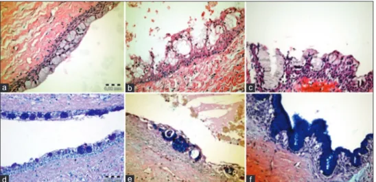
Figure 6: Epithelial lining surface had eosinophilic cuboidal or columnar cells, so-called «hobnail cells», showed papillary appearance into the cyst lumen (arrows) H and E, ×400
Figure 7: Different histopathological areas of glandular odontogenic cysts. The squamous epithelial lining showed numerous mucous cell (a; H and E, ×400) and duct-like structures (b and c; H and E, ×400, ×400, respectively). Periodic acid-Schiff + alcian blue staining highlighted the mucin in the cytoplasm (d-f; ×40, ×200, ×400, respectively) d c b f a e The major criteria include:
• Nonkeratinized squamous epithelial lining with flat connective tissue wall interface
• Various thickness of epithelial lining with or without epithelial sphere or whorls or focal luminal proliferation
Figure 5: Epithelial lining showed plaque-like thickenings into the cyst wall. Epithelial cells assumed a swirled appearance (stars). H and E, ×200
Figure 8: Intraepithelial microcysts (mucin pools). (a-c) H and E, ×40, ×200, ×200, respectively. (d-f) Periodic acid–Schiff stain ×200, ×200, ×200, respectively
d c b f a e
• Cuboidal “hobnail” cells
• Intraepithelial mucous pool with mucous cell, with or without crypts lined by mucous‑producing cells • Microcyst or duct‑like structure in the epithelial layer. The minor criteria include: • Lining epithelium with papillary proliferation • Ciliated cells • Multicystic or multiluminal architecture • Clear or vacuolated cells in basal or spinous layer. Fowler et al. suggested some criteria to distinguish GOC from GOC mimickers. The following criteria should be present and considered individually: microcysts, epithelial sphere, clear cells, and variable thickness of cyst lining. Furthermore, the presence of seven or more microscopic parameters of Kaplan is highly predictive of GOC while the presence of five or less microscopic features is highly predictive of GOC mimickers.[9] Several studies support the aggressive pattern and high recurrence rate of GOC, and this can be explained by multilocular nature of GOC and easy separation of epithelium from underlying connective tissue.[14] Furthermore, the conservative treatment methods may cause high recurrence.[6,10] The recurrence mechanism may be due to the thinness of the cyst wall and the presence of microcysts which make completely removal of the cyst very difficult.[15] The treatment of choice is controversial ranging from enucleation, curettage, or en block resection.[16] Most of the GOC cases were treated conservatively, and a recurrence rate of 30% has been reported.[10] The treatment of choice in our study was conservative treatment as all of them were primary lesion. During follow‑up period of the patients, if any signs of recurrence appear, we will perform for more aggressive surgical treatment.
GOC is a rare jaw cyst of odontogenic origin. It has a potentially aggressive behavior and high recurrence rate, so definite diagnosis is very important and should be carefully determined by correlating the microscopic criteria with
clinical and radiographical findings. Furthermore, long‑term follow‑up is mandatory not to overlook any recurrence. Declaration of patient consent
The authors certify that they have obtained all appropriate patient consent forms. In the form the patient(s) has/have given his/her/their consent for his/her/their images and other clinical information to be reported in the journal. The patients understand that their names and initials will not be published and due efforts will be made to conceal their identity, but anonymity cannot be guaranteed.
Financial support and sponsorship Nil.
Conflicts of interest
There are no conflicts of interest.
References
1. Gardner DG, Kessler HP, Morency R, Schaffner DL. The glandular odontogenic cyst: An apparent entity. J Oral Pathol 1988;17:359‑66.
2. Kramer IR, Pindborg JJ, Shear M. The WHO histological typing of odontogenic tumours. A commentary on the second edition. Cancer 1992;70:2988‑94.
3. Kaplan I, Anavi Y, Hirshberg A. Glandular odontogenic cyst: A challenge in diagnosis and treatment. Oral Dis 2008;14:575‑81. 4. Mervyn Shear PS. Cysts of the Oral and Maxillofacial Regions.
4th ed. Oxford, UK: Blackwell Publishing Ltd; 2007.
5. Magnusson B, Göransson L, Odesjö B, Gröndahl K, Hirsch JM. Glandular odontogenic cyst. Report of seven cases. Dentomaxillofac Radiol 1997;26:26‑31.
6. Gardner DG, Morency R. The glandular odontogenic cyst, a rare lesion that tends to recur. J Can Dent Assoc 1993;59:929‑30. 7. Tran PT, Cunningham CJ, Baughman RA. Glandular odontogenic
cyst. J Endod 2004;30:182‑4.
8. Noffke C, Raubenheimer EJ. The glandular odontogenic cyst: Clinical and radiological features; Review of the literature and report of nine cases. Dentomaxillofac Radiol 2002;31:333‑8. 9. Fowler CB, Brannon RB, Kessler HP, Castle JT, Kahn MA.
Table 1: Data of cases diagnosed as glandular odontogenic cyst Patient Age Sex Location of lesion Clinical symptom Size
(cm) Radiological features Treatment Follow‑up (years)
1 44 Male Posterior mandibular body right Buccal painless swelling 3×1 Unilocular radiolucency with well‑defined margin, #46 roots resorbed Curettage 3 2 39 Female Posterior maxilla
right Labial painless swelling 2×1 Unilocular, well‑defined expansion, no root resorption Enucleation 2 3 51 Male Posterior mandibular body and ramus Buccal and lingual painless swelling, tooth mobility 6×2 Multilocular, well‑defined expansion, #45,46 roots resorbed, 48 impacted and displaced lingual cortex perforation Marsupialization + curettage 3 4 33 Male Anterior maxilla Labial painless swelling 4×2 Unilocular, well‑defined
expansion, no root resorption Enucleation 2 5 42 Male Posterior
Glandular odontogenic cyst: Analysis of 46 cases with special emphasis on microscopic criteria for diagnosis. Head Neck Pathol 2011;5:364‑75.
10. Hussain K, Edmondson HD, Browne RM. Glandular odontogenic cysts. Diagnosis and treatment. Oral Surg Oral Med Oral Pathol Oral Radiol Endod 1995;79:593‑602.
11. Manor R, Anavi Y, Kaplan I, Calderon S. Radiological features of glandular odontogenic cyst. Dentomaxillofac Radiol 2003;32:73‑9.
12. Krishnamurthy A, Sherlin HJ, Ramalingam K, Natesan A, Premkumar P, Ramani P, et al. Glandular odontogenic cyst: Report of two cases and review of literature. Head Neck Pathol 2009;3:153‑8.
13. Chavez JA, Richter KJ. Glandular odontogenic cyst of the mandible. J Oral Maxillofac Surg 1999;57:461‑4.
14. Shen J, Fan M, Chen X, Wang S, Wang L, Li Y, et al. Glandular odontogenic cyst in China: Report of 12 cases and immunohistochemical study. J Oral Pathol Med 2006;35:175‑82.
15. Kaplan I, Anavi Y, Manor R, Sulkes J, Calderon S. The use of molecular markers as an aid in the diagnosis of glandular odontogenic cyst. Oral Oncol 2005;41:895‑902.
16. Economopoulou P, Patrikiou A. Glandular odontogenic cyst of the maxilla: Report of case. J Oral Maxillofac Surg 1995;53:834‑7.