Abstract
The first aim of this study is to determine the changes of cardiac biomarkers and coagulation profiles in parvoviral enteritis and present the importance of these parametres for prognosis of the disease. The second aim is to determine the presence of myocarditis in the enteritis form of the disease via cardiac biomarkers. Twenty seven dogs with parvoviral enteritis (experiment group) and 6 healthy dogs (control group) which were aged between 1.5 and 6 months, weighted between 5 - 15 kg were used as a material of this study. Anorexia, fever, depression, lethargy, vomitting and haemorrhagic diarrhea were determined in dogs with parvoviral enteritis. Parvovirus infection in dogs were verified via feces parvovirus antigen test. Blood samples were collected from all dogs and electrocardiographies (ECG’s) were performed. Standard treatment is applied for dogs with parvoviral enteritis. Twenty three of these dogs were treated successfully; however four of them died. Mild to intermediate acute myocarditis were determined in the histopathological examination of the dead dogs. Plasma protrombin (PT), actived parsiel tromboplastin time (aPTT), antitrombin III (AT-III), fibrinogen and D-dimer concentrations and serum creatin kinase-MB (CK-MB), cardiac troponin I (cTnI) and brain natriuretic peptid (BNP) concentrations were measured. Increase in levels of plasma PT (P<0.001) and aPTT (P<0.001), fibrinogen (P<0.001) and D-dimer (P<0.05), and decrease in level of III AT-III (P<0.05) were detected in dogs with parvoviral enteritis. Increased level of serum CK-MB (P<0.05) and BNP (P<0.001) were also determined. No important change detected in serum cTnI levels. As results, in dogs with parvoviral enterit there were increase in PT, APTT, fibrinogen and D-dimer levels, on the other hand there was a decrease on AT III level resulting in DIC. In addition to this an increase was observed on plasma serum CK-MB and BNP levels. Considering the increase on CK-MB and BNP levels a long with results of histopathological dead dogs, it should be taken into account that acute miocarditis also occures simultaneous with hemoragic parvoviral enterit.
Keywords: Dog, Cardiac biomarkers, Parvoviral Enteritis, Coagulation profile
Parvoviral Enteritli Köpeklerde Pıhtılaşma Profilleri ve
Kalp Biyomarkır Düzeyleri
Özet
Bu çalışmanın birinci amacı, köpeklerin parvoviral enteritinde kalp biyomarkırları ve pıhtılaşma profilindeki değişimleri belirlemek, hastalığın tanı ve prognozunda bu parametrelerin önemini ortaya koymaktır. İkinci amacı ise enterit formunda miyokart hasarı gelişip gelişmediğini kalp biyomarkırları ile belirlemektir. Bu çalışmanın materyalini yaşları 1.5 ile 6 ay, canlı ağırlıkları 5-15 kg arasında değişen 27 parvoviral enteritli (deney grubu) ve 6 sağlıklı köpek (kontrol grubu) oluşturdu. Parvoviral enteritli köpeklerde anoreksi, ateş, depresyon, latherji, kusma ve kanlı diyare belirlendi. Köpeklerin parvoviral enfeksiyonu dışkı parvovirus antijeni testi ile doğrulandı. Bütün köpeklerden kan örnekleri alındı ve kalp EKG traseleri çekildi. Parvoviral enteritli köpeklere standart tedavi uygulandı. Bu köpeklerin 23’ü iyileşti, 4’ü ise öldü. Ölen hayvanların histopatolojik muaynesinde hafif veya orta derecede akut miyokardit saptandı. Bütün köpeklerin plazma protrombin zamanı (PT), active edilmiş parsiyel tromboplastin zamanı (aPPT), antitrombin III (AT-III), fibrinojen ve D-dimer düzeyleri ve serum kretin kinaz-MB (CK-MB), beyin natriüretik peptit (BNP) ve kardiak troponin I (kTnI) düzeyleri ölçüldü. Parvoviral enteritli köpeklerde plazma PT (P<0.001) ve APTT (P<0.001) süreleri ile fibrinojen (P<0.001) ve D-dimer (P<0.05) düzeylerinde artış, AT-III (P<0.05) düzeyinde ise azalma belirlendi. Serum CK–MB (P<0.05) ve BNP (P<0.001) düzeylerinde artış, kTnI düzeyinde ise önemli bir farklılık saptanmadı. Sonuç olarak köpeklerin parvoviral enteritinde PT, APPT, fibrinojen ve D-dimer düzeylerinde artış, AT-III düzeyinde azalma ve buna ilişkin DİK meydana gelmektedir. Ayrıca parvoviral enteritli köpeklerde plazma CK-MB ve BNP düzeyindeki artış tespit edildi. Gerek CK-MB ve BNP düzeyindeki artış gerekse ölen hayvanların histopatolojik sonucuna göre köpeklerde hemorajik enterit formuyla birlikte akut miyokardit formununda gelişebileceği göz önünde bulundurulmalıdır.
Anahtar sözcükler: Köpek, Kalp biomarkırları, Parvoviral enterit, Pıhtılaşma profili
Levels of Cardiac Biomarkers and Coagulation Profiles in
Dogs with Parvoviral Enteritis
[1]Cenk ER
1
Mahmut OK
1[1] 1
This study was funded by Selcuk University Coordinates of Scientific Research Projects (Project No:11102015)E Selçuk Üniversitesi, Veteriner Fakültesi, İç Hastalıklar Anabilim Dalı, TR-42031 Selçuklu, Konya - TÜRKİYE
İletişim (Correspondence)
+90 535 7224314
[email protected]INTRODUCTION
Canine parvovirus infection is an acute, high contagious and mortal viral disease of dogs. Although the disease may develop in dogs within all age ranges, it may cause severe and mortal infections in dogs that are younger than 12 months. Infection has two clinical forms as acute hemorrhagic enteritis and myocarditis. Acute enteritis form is mostly seen in puppies up to 12 months of age. Typical symptoms include depression, vomiting and small bowel hemorrhagic diarrhea. Myocarditis form can develop from infection in utero or in puppies less than 8 weeks old. In this form, the infected puppies may be found dead without any symptoms within 24 h [1-3]. Virus affinites
predominantly cript epitels in the small intestine, pre-cursor cells of bone marrow, lymphoid cells and cardiac muscle cells due to fast reproduction speed of the virus [4].
Disseminated intravascular coagulation is a severe problem that threats lives of both people and animals. In this syndrome, disseminated intravascular micro trombosises shape and cause the circulation disorders, thus multiple organ failure develops and leads to death [5,6]. Protrombin
time, activated parsiyel tromboplastin time, D-dimer, fibrinogen levels, activity of Anti-trombin III and trombosit count should be considered regarding DIC [7]. It is pointed
that, important changes can be seen in CPV infections due to gastrointestinal hemorhagia and bone marrow depression [2]. On the other hand it is argued that the
reason of hemorrhagic enteritis which occures in CPV may be due to endotoxemia caused by coliform bacteries and increased level of cytokines [8].
Acute cardiomyopathies are rooted from bacterial, viral, parasitical, metabolic disorders, toxication and deficiency of vitamins and elements [9]. One of the important reason
of the acute cardiomyopathies is parvoviral infections. It is noticed that cardiac enzymes and neurohormons are the most confidental methods for the diagnosis of myocardial damage [10-15]. Troponins (cTnI, cTnT), creatin kinase-MB
(CK-MB) and brain natriuretic peptid (BNP) are the leading cardiac biomarkers which are used in the diagnosis of myocard damages [10-12]. Vartner et Ingwall [16] indicated
that the first symptoms is the increase in the level of serum CK-MB when the cardiac muscle is damaged. Yılmaz et Şentürk [17] noticed that significiant increase in the level
of serum CK-MB of dogs with parvoviral enteritis. On the other hand, Burgener et al.[18] determined that cTnI level
increases significantly in the first 24 to 48 h in the case of acute miocardial damage in dogs. The cTnI level returns to it’s normal level after 48 h. Another caridac biomarker used to diagnose ventriculer damage is B type natriuretic peptid [19-21]. It is noticed that especially BNP can be used to
evaluate the response to therapy and prognosis of disease in patients with congestive heart failure[22]. Macdonald et
al.[23] noticed that BNP level signicantly increase in dogs
with cardiac failure, Donker et al.[24] also reports that BNP
level increases within several days in dogs through the
artifically generated atrio-ventricular block.
The first aim of this study is to determine the changes in some selected cardiac biomarkers such as BNP, CK-MB and cTnI and coagulation profiles in parvoviral enteritis and present the importance of these parametres for prognosis of the disease. The second aim is to determine the presence of myocarditis in the enteritis form of the disease via cardiac biomarkers.
MATERIAL and METHODS
Animals and Clinical Examination
Authorization to conduct this study has been taken from S.U. Faculty of Veterinary Medicine Animal Ethics Comittee (2011/10). The materials of the study consist of twenty seven dogs with parvoviral enteritis (experiment group) and 6 healthy dogs (control group) which were aged between 1.5 and 6 months, weighted between 5 - 15 kg, were brought into University of Selcuk, Faculty of Veterinary Medicine, Department of Internal Medicine. History of diseases of dogs were found to be 1 to 2 days. First, routin clinical examinations performed for all dogs. Feaces samples were collected from clinically parvovirus suspected dogs (anorexia, depression, lethargy, vomiting and hemorhhagic diarrhea) by using rectal swab. Feaces samples were evaluated by the quick parvovirus antigen detection test (CPV Ag test, rapidy test kits, Vettek Medical İstanbul/Türkiye). The dogs with positive results were incorporated into the study. Twenty two of these dogs had not been vaccinated and 5 others had been. Electrocardiogram examination is performed to all healthy and parvoviral enteritis infected dogs to determine any ritmic problems in the heart. Standard therapy is applied to dogs with parvoviral enteritis.
Colection of Blood Samples
Blood samples with and without anti-coagulant (Na-citrat) collected from dogs with parvoviral enteritis and healthy dogs; plasma and serums were separated. Samples, which would be used in the evaluation of coagulation profiles and cardiac biomarkers, were kept in -80°C deep freeze until the measurement is completed.
Measurement of Coagulation Profile
Protrombin time (Kat no: OUHP-49) and APPT (Kat no:OQGS-29), AT III activity (Kat no: OWWR-15), fibrinogen level (Kat no: OWZG-15) and D-dimer (Kat no: OPBP-03) levels were measured by coagulomethric method in Sysmex CA 1500 device (Siemens, A-7799, Germany).
Measurement of Cardiac Biomarkers
CK-MB (ADVIA Centaur for human, Kat no: RF420) and cTnI (ADVIA Centaur for human, Kat no: RF421C) levels were measured by ELISA method in Dimension Xpand
Plus device (Siemens, 2004080651, Germany), BNP (ADVIA Centaur for human, Kat no: 02816634) level was measured by ELISA method in Immunassay Systems device (Siemens, TRL 93450905, Germany) [25,26]. Measurable sensitivity and
test interval of CK-MB enzym level is 0.18 ng/mL and 300 ng/mL, measurable sensitivity and test interval of cTnI enzym level is 0.006 pg/mL and 50 ng/mL, and for BNP, it is 1 pg/mL and 5.000 pg/ml.
Histopathological Examination
Necropsy performed for all dead dogs. Tissue samples collected from ilio-secal valvule, some areas of small intestines where bleeding detected, cardiac tissue and other organ tissues such liver and spleen to control if any other pathological findings appear.
Statistical Analysis
Unpaired student test was used to determine the differences between groups. SPSS 19.0 for Windows® was used to perform the test. The test procedure is designed according to Akgül [27].
RESULTS
Clinical Results
Anorexia (27 case), lethargy (27 case), depression (27 case), fever (10 case >39.5°C), vomiting (23 case) and hemorrhagic diarrhea (21 case) were seen in dogs which formed the experiment group of the study. Twenty three of these dogs responsed well to the therapy and four of the dogs did not. No anormal condition was seen in ECG examination of all healthy and 23 of the sick dogs. However, sinus tachicardia was diagnosed in 3 of 4 dead dogs.
Coagulation Profile Results
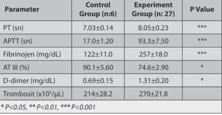
Statistically, significant increase in PT (P<0.001) and APTT (P<0.001) times, fibrinogen (P<0.001) and D-dimer (P<0.05) levels, and significant decrease in AT-III (P<0.05) activity were observed in dogs with parvoviral enteritis compared with healthy dogs. No significant change was seen in trombosit count (Table 1).
Cardiac Biomarker Results
Statistically, significant increase in CK-MB (P<0.05) and BNP (P<0.001) levels were measured in dogs with parvoviral enteritis compared to healthy dogs. No significant change was detected in cardiac troponin I level (Table 2).
Histopathological Findings
Villous atrophy in small bowels, mono nucleer cell infiltration in lamina propria and mild fibrosis, degeneration in intestinal cript epitels and mild or intermediate acute miocarditis were detected during the histopathological examination of the dead dogs.
DISCUSSION
Anorexia, depression, lethargy, fever, tachicardia and tachipnea, vomiting, diarrhea (that can change to mucoid or hemorrhagic) are the common symptoms among dogs with parvoviral enteritis. Severe hypovolemia, due to fluid and electrolit loss caused by vomiting and diarrhea, leads to death if not treated [17,28-31]. Anorexia, depression,
lethargy, fever, tachicardia and tachipnea, vomiting, diarrhea and hemorrhagic diarrhea were diagnosed in this study as well. The diagnosis was finalized after the parvovirus antigen test was resulted positive. Twenty three of these dogs responded well to the therapy and four of them could not survive. Villous atrophy in small bowels, mono nuclear cell infiltration in lamina propria and mild fibrosis, degeneration in intestinal cript epitels and mild or intermediate acute miocarditis were detected during the histopathological examination of the dead dogs.
Disseminated intravascular coagualtion may develop due to viremia, septisemi, parazitic infection, severe tissue damage, toxication, intravascular hemolizis, autoantibody, hepatitis, pancreatitis and neoplasma [7,32-34]. Protrombin
time, activated parsiyel tromboplastin time, D-dimer, fibrinogen levels, activity of Anti-trombin III and trombosit count should be considered in DIC [7]. It is pointed that,
important changes due to coagulopathie can be seen in CPV infections due to gastrointestinal hemorrhagia and bone marrow depression [2]. At the same time, it is argued
that the reason of hemorrhagic enteritis which occures
Table 1. Mean values and statistical importance of coagulation parametres
in healthy and infected dogs
Tablo 1. Sağlıklı ve enfekte köpeklerde pıhtılaşma parametrelerinin
ortalama değeri ve istatistiksel önemi
Parameter Group (n:6)Control Group (n: 27)Experiment P Value
PT (sn) 7.03±0.14 8.05±0.23 *** APTT (sn) 17.0±1.20 93.3±7.50 *** Fibrinojen (mg/dL) 122±11.0 257±18.0 *** AT III (%) 90.1±5.60 74.6±2.90 * D-dimer (mg/dL) 0.69±0.15 1.31±0.20 * Trombosit (x103/µL) 214±28.2 270±21.8 * P<0.05, ** P<0.01, *** P<0.001
Table 2. Mean values and statistical importance of cardiac biomarkers in
healthy and infected dogs
Tablo 2. Sağlıklı ve enfekte köpeklerde kalp biyomarkırlarının ortalama
değeri ve istatistiksel önemi
Parameter Group (n:6)Control Group (n: 27)Experiment P Value
CK–MB (ng/mL) 2.20±1.00 8.51±1.25 *
cTnI (ng/mL) 0.06±0.02 0.10±0.03
BNP (pg/ mL) 1.40±0.05 30.1±2.68 ***
in CPV may be due to endotoxemia caused by coliform bacteries and increased level of sitokins [8]. Otto et al.[35]
noticed that DIC may occure in dogs with parvoviral enteritis. For an accurate diagnosis, prolonged PT and APTT, decrease in AT-III activity, trombosit count and fibrinogen level, increase in FDP or D-dimer level must be detected. At least three of these changes must be detected for a suspicious DIC case [36]. Feldman et al.[37]
indicated that prolong in PT and APTT, increase in FDP level, decrease in AT-III activity and trombosit count, develop in dogs with DIC. In recent years D-dimer is used instead of FDP and it is proved that D-dimer is more sensitive and spesific incompared to FDP [7,38]. On the
other hand, Carr et al.[39] noticed that increased level of
D-dimer does not always indicate the presence of DIC. Laforcade et al.[40] noticed that they had found prolong in
PT and APTT, increase in D-dimer, decrease in AT-III level and no changes in trombosit count. Esmon [41] and Wada [42]
noticed that fibrinogen level may increase proportional to inflammation if there is no high consumption.
Prolonged PT and APTT, decreased AT-III and increase in D-dimer level may show the development of DIC in the dogs with parvoviral enteritis in this study. Although prolonged PT and APTT, increased D-dimer level and decreased AT-III activity were detected as other authors also noticed [7,36,43]; however, compared to other studies,
in this study there were no decrease in fibrinogen and trombosit levels. On the other hand, Otto et al.[35] noticed
that they found increase in fibrinogen level and no change in D-dimer level and trombosit count. In this study, detection of prolonged PT and APTT, decreased AT-III and increased D-dimer level shows the coagulation activation (PT↑, APTT↑, AT-III↓) which is the first step of DIC and fibrinolitic activation (D-dimer↑) which is the last step of DIC. To conclude the findings coincides with the findings of Otto et al.[35] and Laforcade et al.[40].
Some authors [35,41,42]report that fibrinogen level may
increase in the first period of DIC due to inflamatory response; then, the level decreases due to fibrinolisis; some other authors [7,43] report significant decrease in
fibrinogen level. In this study, increase in fibrinogen level was observed. Increase in fibrinogen level may be due to the increase of acute phase proteins in spite of DIC. Our findings on fibrinogen verifies the studies of most of the other mentioned authors [35,41,42].
The most important method to determine the myo-cardial damage is to measure cardiac biomarkers [10-15].
Troponins, CK-MB and BNP are the most common used biomarkers in the diagnosis of acute myocardial damage [10-12,15,44]. CK-MB is a first degree trusted and
spesific enzyme in acute cardiac tissue damage [45].
Smithline et al.[46] indicate that serum CK-MB level starts
to increase within 4 h following the myocardial damage, and reaches it’s peak level in approximately within the 12th h then, decrease to normal level in 24 to 72 h. Troponin
I is considered a reliable serum biomarker for myocardial ischemia and necrosis in human and animals. After acute myocardial injury, cTnI is released from the cytoplasmic pool, resulting in increased blood concentrations within 2 h, with a peak after 12-24 h. Persistently increased cTnI blood level suggest irreversible and active on going damage to cardiomyocytes [25,47]. On the other hand,
Mair et al.[48] report that cTnI starts to increase in 4 to
6 hours and it may take 7 to 10 days to reach the basal level. Vartner et Ingwall [16] noticed that CK-MB is the
first enzyme that can be detected in serum following to myocardial damage. Yılmaz and Şentürk [17] noticed the
significant increase of CK-MB serum level in dogs with parvoviral enteritis. Burgener et al.[18] showed that cTnI
level significantly increase in 24 to 48 h and then returns to basal level. Bastan et al.[49] found that increased serum
cTn-I levels were consistent with short survival times in dogs with CPV-2. B type natriuretic peptid is also an important indicator of ventricular damages [19-21].
Especially, BNP level may inform the clinician about the prognosis and response to treatment of patient with congestive heart failure [22]. Macdonald et al.[23] report that
BNP level significantly increase in dogs with congestive heart failure. Donker et al.[24] report that BNP level
through artifical atrio ventricular block increase in several days. Haßdenteufel et al.[50] determined that NT-proBNP
represents a useful additional diagnostic parameter in veterinary clinical cardiology to assess the severity of cardiac disease.
In this study, CK–MB (P<0.05) and BNP (P<0.001) levels statistically increased in dogs with parvoviral enteritis compared to the healthy dogs. Although not statistically No significant, cTnI concentration was numerically increased in dogs with parvoviral enteritis compared to the healthy dogs (Table 2). Increase in CK-MB and BNP concentrations may show that mild to intermediate myocarditis might be ocurred in the enteritis form of CPV. On the other hand, possible reason of numerically increased in cTnI level may be that there is no severe damage in myocard. Detection of mild to intermediate myocarditis in histopathological examination of 4 dead dogs, increase in CK-MB (15-28 ng/ ml) and BNP (44-58 pg/ml) levels, and numerically increase in cTnI (0.1 ng/ml) levels of these dogs may be confirm the result. It must be considered that CK-MB level may increase in blood serum rapidly, in mild to intermediate myocarditis thus it may be more diagnostic than cTnI. CK- MB, BNP and cTnI result of this study were appropriated with findings of some authors [12,17,18,22-25,44,46-49].
In conclusion, PT, APTT, fibrinogen and D-dimer levels increase, AT-III level decreases in parvoviral enteritis of dogs due to DIC. In addition, increase in serum CK-MB and BNP levels were detected. It is concluded that acute myocarditis form may develop in the enteritis form of disease according to histopathological results of dead animals and increase in CK-MB and BNP levels.
REFERENCES
1. Truyen U: Canine Parvovirus. 2000, Available from URL: www.ivis.org,
Document No: A0106.0100. cited, Feb 2013.
2. Goddard A, Leisewitz AL: Canine parvovirus. Vet Clin Small Anim,
40, 1041-1053, 2010.
3. Ok M, Şen İ, Birdane FM, Bekteş HG, Turgut K: Diagnostic importence
of ELISA and hemaglutination inhibition tests in canine parvoviral infection of dogs. Indian Vet J, 77 (6): 465-467, 2000.
4. LammCG, Rezabek GB: Parvovirus infection in domestic companion
animals. Vet Clin North Am: Small Anim Pract, 38, 837-850, 2008. DOI: 10.1016/j.cvsm.2008.03.008
5. Bruchim Y, Aroch I, Saragusty J, Waner T: Disseminated intravascular
coagulation. Compendium, 3 (10): 1-16, 2008.
6. Laforcade AM, Rozanski EA, Freeman LM, Li W: Serial evaluation of
protein C and antitrombin in dogs with sepsis. J Vet Intern Med, 22, 26- 30, 2008. DOI: 10.1111/j.1939-1676.2007.0021.x
7. Caldin M, Furlanello T, Lubas G: Validation of an immunoturbidimetric
D-dimer assay in canine citrated plasma. Vet Clin Pathol, 29, 51-54, 2000. DOI: 10.1111/j.1939-165X.2000.tb00398.x
8. Isogai E, Isogai H, Onuma M, Mizukoshi N, Hayashi M, Namioka S:
Escherichia coli associated endotoxemia in dogs with parvovirus infection. Jpn J Vet Sci, 51 (3): 597-606, 1989.
9. Osterziel KJ, Hassfeld S, Geier C: Familial dilated cardiaomyopathy.
Herz, 30, 529-534, 2005. DOI: 10.1007/s00059-005-2732-3
10. Apple FS: Tissue specificity of cardiac troponin I, cardiac troponin T
and creatine kinase-MB. Clin Chim Acta, 284, 151-159, 1999. DOI: 10.1016/ S0009-8981(99)00077-7
11. Robinson DJ, Christenson RH: Creatine kinase and it’s
CK-MB isoenzyme: The conventional marker fort he diagnosis of acute myocardial infarction. J Emerg Med, 17 (1): 95-104, 1999. DOI: 10.1016/ S0736-4679(98)00129-2
12. Saavedra JM, De Oliveira AM, Jöhren O, Tonelli L: Chapter IV: Brain
endothelin and natriuretic peptid receptors. In, Quirion R, Björklund A, Hökfelt T (Eds): Handbook of Chemical Neuroanatomy, Peptide Receptors. Part I. 125-162, Elsevier Scinces BV, Amsterdam, 2000.
13. Duygu H, Türk U, Zoghi M, Nalbantgil S: Plazma B-tipi natriüretik
peptid düzeylerinin kardiyovasküler hastalıklardaki yeri ve önemi. Anadolu Kardiyol Derg, 5, 305-311, 2005.
14. Elmalı E, Karaeren Z, Çağdaş Ö, Akan ÖA: Akut koroner sendrom
şüpheli hastalarda kardiyak troponin T ve troponin I’nın karşılaştırılması.
Turk J Biochem, 30 (3): 212-215, 2005
15. Ok M, Sağkan Öztürk A, Er C: Üç köpekte kalp yetmezliği. Eurasian
J Vet Sci, 26 (1): 57-62, 2010.
16. Vartner DE, Ingwall JS: Effect of moderate pressure overload cardiac
hypertrophy on the distrubition of ceratine kinase isoenzymes. Proc Soc
Exp Biol Med, 175, 5-9, 1984. DOI: 10.3181/00379727-175-1-RC2
17. Yılmaz Z, Şentürk S: Characterisation of lipid profiles in dogs
with parvoviral enterit. J Small Anim Pract, 48, 643–650, 2007. DOI: 10.1111/j.1748-5827.2007.00391.x
18. Burgener IA, Kovacevic A, Mauldin GN, Lombard CW: Cardiac
troponins as indicators of acute myocardial damage in dogs. J Vet
Intern Med, 20, 277-283, 2006. DOI: 10.1111/j.1939-1676.2006.tb02857.x
19. Nakagawa O, OgawaY, Itoh H, Suga S, Komatsu Y, Kishimoto I, Nishino K, Yoshimasa T, Nakao K: Rapid transcriptional activation and
early mRNA turnover of brain natriuretic peptide in cardiocyte hyper-trophy. Evidence for brain natriuretic peptride as an emergency cardiac hormone against ventricular overload. J Clin Invest, 96, 1280-1287, 1995.
20. Dickstein K: Natriuretic peptides in detection of heart failure. The
Lancet, 351, 4-9, 1998.
21. Maisel AS, Krishnaswamy P, Nowak RM, McCord J, Hollander JE, Duc P, Omland T, Storrow AB, Abraham WT, Wu AHB, Clopton P, Steg PG, Westheim A, Knudsen CW, Perez A, Kazanegra R, Herrmann HC, McCullough PA: Rapid measurement of B-type natriuretic peptide in the
emergency diagnosis of heart failure. N Engl J Med, 347, 161-166, 2002. DOI: 10.1056/NEJMoa020233
22. Boswood A: Biomarkers in cardiovascular disease: Beyond natriuretic
peptides. J Vet Cardiol, 11, 23-32, 2009. DOI: 10.1016/j.jvc.2009.01.003
23. Macdonald KA, Kittleson MD, Munro C, Kass P: Brain natriuretic
peptide concentration in dog with heart disease and congestive heart failure. J Vet Intern Med, 17, 172-177, 2003.
24. Donker DW, Maessen JG, Verheyen F, Ramaekers FC, Spatjens RL,Kuijpers H, Ramakers C, Sciffers PMH, Vos MA Crijins HJGM, Volders PGA: Impacat of acute and enduring volume overload on
mechonatransduction and cytoskeletal integrity of canine left ventricular myocardium. Am J Physiol Heart Circ Physiol, 292 (H): 2324-2332, 2007. DOI: 10.1152/ajpheart.00392.2006
25. O’Brien PJ, Smith DE, Knetchel TJ, Marchak MA, Pruimboo-Brees I,Brees DJ, Spart DP, Archer FJ, Butler P, Poter AN: Cardiac troponin I is
a sensitive, specific biomarker of cardiac injury in laboratory animals.
Lab Anim, 40, 153-171, 2008. DOI: 10.1258/002367706776319042
26. DeClue AE, Osterbur K, Bigio A, Sharp CR: Evaluation of serum
NT-pCNP as a diagnostic and prognostic biomarker for sepsis in dogs.
J Vet Intern Med, 25, 453-459, 2011. DOI: 10.1111/j.1939-1676.2011.0713.x
27. Akgül A: Tıbbi Araştırmalarda İstatistiki Analiz Teknikleri “SPSS
Uygulamaları”, 2. Baskı, Emek Ofset Ltd Şti, Ankara, 2003.
28. Bloom ME, Kerr JR: Pathogenesis of parvovirus infections. In, Kerr
JR, Cotmore SF, Bloom ME, Linden RM, Parrish CR (Eds): Parvoviruses. 323-325, Oxford University Press Inc , New York, 2006.
29. Pollock RVH, Coyne MJ: Canine Parvovirus. Vet Clin North Am: Small
Anim Pract, 23, 555-568, 1993. DOI: 10.1016/S0195-5616(93)50305-4
30. Yeşilbağ K, Yılmaz Z, Özkul A, Pratelli A: Aetiological role of viruses
in puppies with diarrhoea. Vet Rec, 161, 169-170, 2007. DOI: 10.1136/ vr.161.5.169
31. Kocatürk M, Martinez S, Eralp O,Tvarijonaviciute A, Ceron J, Yılmaz Z: Prognostic value of serum acute-phase proteins in dogs
with parvoviral enterit. J Small Anim Pract, 51, 478-480, 2010. DOI: 10.1111/ j.1748-5827.2010.00965.x
32. Levi M: Pathogenesis and treatment of DIC. Thromb Res, 1, 54-55,
2005.
33. Mischke R: Acute haemostatic changes in accidentally traumatised
dogs. Vet J, 169, 60-64, 2005. DOI: 10.1016/j.tvjl.2004.01.008
34. Çöl R, Durgun Z: Sepsis, lökositler, sitokinler ve disseminant
intravasküler koagulasyon. Vet Bil Derg, 23, 97-106, 2007.
35. Otto CM, Rieser TM, Brooks MB, Russell MW: Evidence of
hypercoagulability in dogs with parvoviral enterit. JAVMA, 217 (10): 1500-1509, 2000. DOI: 10.2460/javma.2000.217.1500
36. Turgut K: Veteriner Klinik Laboratuvar Teşhis. İkinci Baskı, 885-886,
Bahçıvanlar Basım Sanayi, Konya, 2000.
37. Feldman BF, Madewell BR, O’Neill S: Disseminated intravascular
coagulation: Antithrombin, plasminogen, and coagulation abnormalities in 41 dogs. J Am Vet Med Assoc, 179 (2): 151-154, 1981.
38. Stokol E, Brooks MB, Erb HN, Mauldin GE: D-dimer concentrations
in healthy dogs and dogs with Disseminated intravascular coagulation.
Am J Vet Res, 61, 393-398, 2000. DOI: 10.2460/ajvr.2000.61.393
39. Carr HJ, McKinney M, MacDonagh J: Diagnosis of disseminated
intravascular coagulation: Role of D-dimer. Am J Clin Pathol, 91, 280-287, 1989.
40. Laforcade AM, Freeman LMS, Shaw SP, Brooks MB, Rozanski EA, Rush JE: Hemostatic changes in dogs with naturally occuring sepsis. J Vet
Intern Med, 17, 674–679, 2003. DOI: 10.1111/j.1939-1676.2003.tb02499.x
41. Esmon CT: Inflammation and thrombosis. J Thromb Haemost, 1,
1343-1348, 2003. DOI: 10.1046/j.1538-7836.2003.00261.x
42. Wada H: Disseminated intravascular coagulation. Clin Cbim Acta, 344
(1-2): 13-21, 2004.
43. Prins M, Schellens CJMM, van Leeuwen MW, Rothuizen J, Teske E: Coagulation disorders in dogs with hepatic disease. Vet J, 185, 163-
44. Welsh TM, Kukes GD, Sandweiss LM: Differences of creatine kinase
MB and cardiac troponin I concentrations in normal and diseased human myocardium. Ann Clin Lab Sci, 32 (1): 44-49, 2002.
45. Gillum RF, Fortmann SP, Prineas RJ, Kottke TE: International
diagnostic criteria for acute myocardial infarction and acute stroke. Am
Heart J, 108, 150-158, 1984. DOI: 10.1016/0002-8703(84)90558-1
46. Smithline HA, Thompson M, Moran C, Mader TJ: Can CK-MB and
kTnI be detected in the first 10 min of acute coronary ischemia? Am
Fam Physician, 60 (4): 598-602, 2003.
47. Wells SM, Sleeper M: Cardiac troponins. J Vet Emerg Crit Care. 18,
235-245, 2008. DOI: 10.1111/j.1476-4431.2008.00307.x
48. Mair J, Jartner Dowarzak E, Dienstl A: Early detection of acute
myocardial infarction by measurement of mass concentration of creatine kinase-MB. Am J Cardiol, 68, 1545-1550, 1991. DOI: 10.1016/0002-9149(91)90307-7
49. Bastan İ, Kurtdede A, Sel T, Özen D, Yumuşak N, Timurkan MÖ, Baydın A: Serum cardiac troponin-I dogs with CPV-2 infection. Ankara
Üniv Vet Fak Derg, 60, 251-255, 2013.
50. Haßdenteufel E1, Kresken JG, Henrich E, Hildebrandt N, Schneider C, Stosic A, Schneider M: NT-proBNP as a diagnostic marker in dogs with
dyspnea and in asymptomatic dogs with heart murmur. Tierarztl Prax