Summary
The purpose of this study was to determine the presence of virulence genes, perform typing for the characterization of Arcanobacterium pyogenes field strains, and investigate the correlation between clonal types, virulence genes and occurence of disease. The isolates (n=51) used in this study were isolated from different sources (liver (n=4) of sheep; liver (n=25), lungs (n=5), broncho-alveolar lavage (BAL) fluid (n=3), milk (n=12) and suppurative tissue (n=1) of dairy cows and synovial fluid (n=1) of a calf ). The presence of haemolytic activity in A. pyogenes isolates was determined using rabbit, sheep, cattle, chicken, dog and human erythrocytes. Also, the presence of any cytotoxic effect was investigated by growth in Vero cell cultures. Genomic DNA fingerprinting for clonal analysis was generated by BOX-PCR typing. Conventional PCR was used for the determination of the presence of eight A. pyogenes virulence factor genes, namely, nanH (encoding neuraminidase H), nanP (encoding neuraminidase P), plo (encoding pyolysin-PLO), cbpA (encoding collagen-binding protein A) and fimA, fimC, fimE and fimG (encoding the major fimbrial subunit of four different fimbriae). Furthermore, the correlation between clonal types, virulence factors and the occurence of disease was investigated. The haemolysins of all strains had haemolytic effect on rabbit, sheep, cattle, chicken, dog and human erythrocytes. In addition, all strains were found to be cytotoxic to Vero cells. According to clonal analysis results, the A. pyogenes isolates were determined to belong to 12 different types. While all A. pyogenes strains were positive for the plo gene, the positivity rate was 62% for the nanH gene, 84% for the nanP gene, 58% for the cbp gene, 96% for the fimA gene, 66% for the fimC gene, 42% for the fimE gene and 10% for the fimG gene. It was determined that no correlation existed between the clonal types and virulence factors of A. pyogenes isolates and occurence of disease.
Keywords: Arcanobacterium pyogenes, Clonal typing, Virulence genes, Molecular characterization
Koyun ve Sığırlardan İzole Edilen Arcanobacterium pyogenes
Suşlarının Moleküler Karakterizasyonu
Özet
Bu çalışmanın amacı, Arcanobacterium pyogenes saha izolatlatlarının karakterizasyonu için virulens genlerinin varlığını belirlemek, tiplendirmek ve klonal tipleri, virülens genleri ve hastalık oluşumu arasındaki ilişkiyi incelemektir. Bu çalışmada, farklı örneklerden izole edilen A. pyogenes izolatı (51) kullanıldı: koyun (n=4) ve sığır (n=25) apseli karaciğerlerinden, süt ineklerine ait akciğerler (n=5), bronko-alveolar yıkantılardan (n=3), mastitisli sütlerden (n=12), suppuratif doku örneğinden (n=1) ve buzağı eklem sıvısından (n=1). Bütün A. pyogenes izolatlarında, tavşan, koyun, sığır, tavuk, köpek ve insan ertitrositleri ile hemolitik aktivite varlığı belirlendi. Aynı zamanda, vero hücre kültüründe sitotoksik etkileri incelendi. Klonal analiz için genomik DNA parmak izi BOX-PCR tiplendirme ile yapıldı. Sekiz virulens faktör genlerinin varlığının değerlendirilmesi için klasik PCR kullanıldı: nanH (nöraminidaz H), nanP (nöraminidaz P), plo (pyolysin-PLO), cbpA (kollejen bağlayan protein A) ve fimA, fimC, fimE, fimG (4 farklı fimribanın önemli fimbrial yapılar). Aynı zamanda, klonal tipleri, virulens faktörleri ve hastalık varlığı arasında bir ilişki incelendi. Bütün suşların hemolizinleri, tavşan, koyun, sığır, tavuk, köpek ve insan eritrositlerine hemolitik etkisine sahipti. Ayrıca, bütün suşların vero hücresine sitotoksik olduğu bulundu. Klonal analiz sonuçlarına göre; A. pyogenes izolatlarının 12 farklı tipte olduğu belirlendi. Bütün suşlar plo geni pozitif iken, nanH geni %62, nanP geni %84, cbp geni %58, fimA geni %96, fimC geni %66, fimE geni %42 ve fimG geni %10’du. Klonal tipler, virulens faktörler ve hastalık varlığı için A. pyogenes izolatları arasında bir ilişki bulunmadı.
Anahtar sözcükler: Arcanobacterium pyogenes, Klonal tiplendirme, Virulens gen, Moleküler karakterizasyon
The Molecular Characterization of Arcanobacterium pyogenes
Strains Isolated from Samples of Sheep and Cattle
H. Hüseyin HADİMLİ *
Kürşat KAV *
* Department of Microbiology, Faculty of Veterinary Medicine, Selcuk University, TR-42075 Campus/Konya - TURKEY
Makale Kodu (Article Code): KVFD-2011-4636
İletişim (Correspondence)
+90 332 2233622INTRODUCTION
Arcanobacterium pyogenes is an important opportunistic pathogen of mucosal surfaces in livestock, including cattle, sheep and goats 1, and is also responsible for suppurative infectious disease 2. A. pyogenes causes mastitis, abortion, pyometra, arthritis, and orchitis in livestock and foot abscesses in poultry, and can be recovered as either pure or mixed cultures 3-5. It is also a secondary pathogen frequently isolated from liver and kidney abscesses in cattle with or without Fusobacterium necrophorum 6-8. As it has also been isolated from the rumen of cattle and stomach of pigs, it is considered that A. pyogenes could be a common bacterium of the digestive system in animals 2. In addition to animals, it has been reported that A.pyogenes has also been isolated from cases of arthritis and subcutaneous abscesses in humans dealing with animals 9,10.
A. pyogenes expresses several known and putative virulence factors, including pyolysin (plo), neuraminidase (nanH and nanP) and collagen-binding protein (cbpA), which may contribute to its pathogenicity 11-16. Pyolysin (PLO), a haemolytic exotoxin expressed by A. pyogenes, is a member of the thiol-activated cytolysin family of bacterial toxins. These toxins can play a primary role in the pathogenesis of infections caused by various Gram-positive pathogens. In this context, it has been demonstrated that PLO acts as the most important virulence determinant in A. pyogenes infections 15,17. PLO is produced by all strains of A. pyogenes 17. The collagen-binding protein (cbpA) is required by the infectious agent to adhere to collagen-rich tissue 15,16. In addition, fimbriae can be involved in the adhesion of the agent to host cells 1.
In our previous study 18, we isolated, biochemically identified and confirmed with PCR the pylosin gene in A. pyogenes strains. Furthermore, the antibiotic susceptibility of A. pyogenes isolates was determined. Silva et al.19 described the genomic characterization of A. pyogenes isolates based on the screening of eight known and putative virulence factors using conventional PCR and BOX-PCR typing. The present study was aimed at establishing a research model similar to that described by Silva et al.19.
The purpose of this study was to determine the virulence genes, describe the genomic characterization of Arcanobacterium pyogenes field strains by typing, and investigate the correlation between clonal types, virulence genes and persistence of disease.
MATERIAL and METHODS
Bacterial Isolates
The isolates (n=51) used in this study were recovered from different sources (liver of sheep; liver, lungs, broncho-alveolar lavage fluid, milk and suppurative tissue of dairy
cows; and synovial fluid of a calf ). Twenty-nine of the isolates were obtained from liver abscesses of slaughtered feed-lot cattle (n=25) and sheep (n=4) at a slaughterhouse in Konya province. Twelve strains were isolated from the milk of dairy cows with clinical mastitis. The lung (n=5) and broncho-alveolar lavage fluid (n=3) isolates were recovered from dairy cows suffering from respiratory problems. One strain was isolated from the suppurative tissue of a dead dairy cow. One other isolate was recovered from the synovial fluid of a calf.
Determination of the Presence of Haemolysin and Cytotoxic Effect
Each isolate was grown in brain-heart infusion medium at 37ºC for 48 h and the supernatants were harvested by centrifugation at 6.000 rpm. In order to determine the presence of haemolytic activity, rabbit, sheep, cattle, chicken, dog and human erythrocytes were added to each supernatant and the samples were incubated at 37°C for 18-20 h. Furthermore, the supernatant of each isolate was added to Vero cell cultures, which were observed for a period of 48-72 h for the development of cytopathological effect.
Genomic DNA Isolation
Genomic DNA was extracted from 48 h-old-cultures of A. pyogenesusing a commercial DNA purification kit (Wizard®, Promega, USA). Theprocedure was performed according to the manufacturer’s instructions. Firstly, isolates of A. pyogenes were grown at 37ºC for 48 h and harvested by centrifugation at 12.000g for 3 min. After washed 3 times in PBS, the pelleted bacterial cells were first treatedwith 60 µl of lysozyme (10 mg ml-1;Merck, Germany) for 1 h at 37°C. The preparations were analysed on 0.7% agarose gel and the quantity and quality ofDNA were determined spectrophotometrically using a ND 2000 spectrophotometer (Nanodrop, Germany). The amount of DNA for each isolate was adjusted to the required concentration for the detection of virulence factor genes.
Clonal Analysis
Genomic DNA fingerprinting was generated by PCR using the BOX-A1R (5’-CTACGGCAAGGCGACGCTGACG-3’) primer 19. Amplification reactions occurred in a Thermal Cycler (Eppendorf, Germany) in a 25 µl volume of reaction mixture containing a primer concentration of 2 mM, 100 ng of genomic DNA and 1 U of DNA Taq polymerase (Fermentas, EU). An initial denaturation step (95°C, 2 min) was followed by 34 cycles of denaturation (95°C, 1 min), annealing (53°C, 1 min) and extension (72°C, 5 min), with a single final extension cycle at 72°C for 10 min 19. A PCR mixture without DNA was used as a negative control.
PCR products (5 µl) were electrophoresed in 1.5% agarose gel at 90 V for 3 h and stained with ethidium bromide. Band sizes were determined by comparison with
a standard DNA ladder (Fermentas, EU). DNA banding profiles were imaged using Infinity Capture Version 12.4 for Windows (Vilbert Lourmat, France). Intra-species relation- ships on the basis of the similarity of DNA band profiles were calculated using the Bio1D++ program (Vilbert Lourmat, France), according to the NEI and LI (1979) similarity coefficient, i.e. a=2nxy/(nx + ny) where nx and ny are the number of bands in lane “x” and “y”, respectively, and nxy the number of shared bands between the two lanes. Dendrograms were produced from the similarity values in the matrix using the UPGMA (unweighted pair group match average) algorithm.
Screening of Genes Encoding Known and Putative Virulence Factors
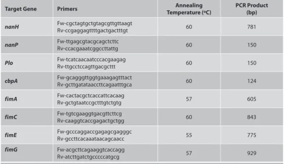
The PCR protocols were performed as described by Silva et al.19. Conventional PCR was used for the determination of the presence of eight A. pyogenes virulence factor genes: nanH (encoding neuraminidase H), nanP (encoding neuraminidase P), plo (encoding pyolysin-PLO), cbpA (encoding CbpA) (Jost and Billington 1) and four fimbrial genes, fimA, fimC, fimE and fimG (encoding the major fimbrial subunit of four different fimbriae) (Table 1). Also, plo gene in our previous study (Hadimli et al.18) were determined by used different specific primers.
PCR reactions were carried out in a 25 µl reaction mixture containing 25 pmol of each primer, 0.1 mM of each deoxynucleotide triphosphate (Fermentas), 1xPCR buffer, 2 mM MgCl2, 100 ng of genomic DNA and 1 U of Taq DNA polymerase (Fermentas). Except for the annealing temperatures, the thermal cycling conditions were identical for all sets of primers: 3 min at 94ºC followed by 35 cycles of denaturation (94ºC for 1 min), annealing (1 min) and extension (72ºC for 3 min) and a final step at 72ºC
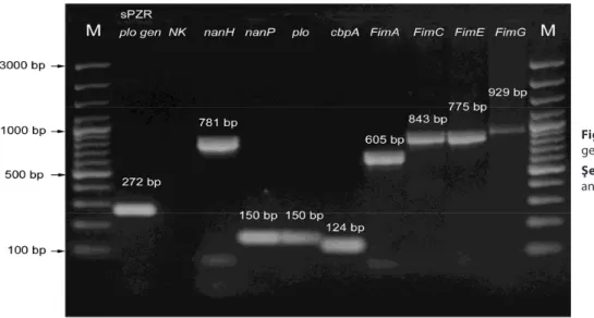
for 7 min. The annealing temperature was set between 55ºC and 60ºC. Positive controls for each virulens gene were used (Fig. 1). Also, A PCR mixture without DNA was used as a negative control.
The identity of PCR products was initially confirmed by DNA sequencing. Amplification products were separated by electrophoresis in 2.5% or 1% agarose gel (plo, cbpA, nanH, nanP, fimA, fimC, fimE and fimG), stained with ethidium bromide and the bands were visualized using Infinity-Capture Version 12.4 (Vilbert Lourmat, France). The result was considered to be positive if the amplification product was of the expected molecular size.
RESULTS
Haemolysins of all A. pyogenes strains had haemolytic effect on rabbit, sheep, cattle, chicken, dog and human erythrocytes. Furthermore, all isolates produced β- haemolysis on blood agar base containing sheep blood. In addition, all strains were found to be cytotoxic to Vero cell cultures in vitro.
Virulence Factor Genes
The results obtained for clonal types and virulence factor genes, as well as the number of isolates, and sources of samples are shown in Table 2. While all A. pyogenes strains were positive for the plo gene, the positivity rate was 62% for the nanH gene, 84% for the nanP gene, 58% for the cbp gene, 96% for the fimA gene, 66% for the fimC gene, 42% for the fimE gene, and 10% for the fimG gene.
Clonal Type Analysis
It was determined that, the A. pyogenes strains were of
Table 1. PCR primers used to amplify eight A. pyogenes virulence factor genes 19
Tablo 1. A. pyogenes’in 8 virülens faktör genini çoğaltmak için kullanılan PZR primerleri 19
Target Gene Primers Temperature (ºC)Annealing PCR Product (bp)
nanH Fw-cgctagtgctgtagcgttgttaagtRv-ccgaggagttttgactgactttgt 60 781
nanP Fw-ttgagcgtacgcagctcttcRv-ccacgaaatcggccttattg 60 150
Plo Fw-tcatcaacaatcccacgaagagRv-ttgcctccagttgacgcttt 60 150
cbpA Fw-gcagggttggtgaaagagtttactRv-gcttgatataaccttcagaatttgca 60 124
fimA Fw-cactacgctcaccattcacaagRv-gctgtaatccgctttgtctgtg 57 605
fimC Fw-tgtcgaaggtgacgttcttcgRv-caaggtcaccgagactgctgg 60 843
fimE Fw-gcccaggaccgagagcgagggcRv-gccttcacaaataacagcaacc 55 775
fimG Fw-acgcttcagaaggtcaccagg
12 clonal types, according to the source, case or sample they were recovered from. Of the 25 strains isolated from the liver of cattle, 6 belonged to type VIII, 5 to type VI, 4 to each of the types IV and VII, 3 to type XI and 1 to each of the types I, IX and X. It was determined that, of the 12 strains isolated from the milk of dairy cows with clinical mastitis, 2 belonged to each of the types II, VI and XII, and 1 to each of the types I, III, V, VII, VIII and XI. Of the isolates recovered from the lung samples of dairy cows, 3 were of the type II and 1 of each of the types IV and VI. On the other hand, of the strains isolated from the liver of sheep, 2 were classified as type XI and 1 as each of the types I and IV. Of the isolates recovered from broncho-alveolar lavage fluid (BAL), 2 belonged to type II and 1 to type I, whereas the two isolates recovered from the synovial fluid of a calf and from suppurative tissue were type VII.
DISCUSSION
Arcanobacterium pyogenes is a ubiquitous opportunistic pathogen, which causes suppurative diseases in animals of economic value, including cattle and sheep, and in other animal species. A. pyogenes exists commensally in the upper respiratory tract and uro-genital system of animals 1-8.
It has been reported that, synergistic and antagonistic haemolytic properties can be used as additional criteria for the identification of bacteria belonging to the genus Arcanobacterium 20. In the present study, it was observed that, haemolysins of all A. pyogenes strains had a haemolytic effect on rabbit, sheep, cattle, chicken, dog and human erythrocytes. Furthermore, all isolates produced β-haemolysis on blood agar containing sheep blood.
Table 2. Correlation between clonal types, virulence factor genes and sources of samples Tablo 2. Klonal tipler, virülens factor genleri ve örneklerin kaynakları
BOX-PCR
Type İsolatesNo of Source
Virulens Gene Factors
plo* nanH nanP cbpA fimA fimC fimE fimG
I 4 BAL, CM, CL, SL 4/4 4/4 ¾ 4/4 4/4 3/4 2/4 0/4 II 7 BAL(2), CM(2), L(3) 7/7 5/7 4/7 6/7 6/7 5/7 5/7 0/7 III 1 CM 1/1 0/1 1/1 0/1 1/1 0/1 0/1 1/1 IV 6 CL (4), SL, L 6/6 6/6 5/6 6/6 6/6 6/6 6/6 0/6 V 1 CM 1/1 0/1 1/1 0/1 1/1 0/1 0/1 0/1 VI 8 CL (5), CM (2), L 8/8 5/8 7/8 5/8 7/8 6/8 3/8 0/8 VII 7 CL(4), CFJ, CM, ST 7/7 1/7 6/7 3/7 7/7 5/7 2/7 1/7 VIII 7 CL (6), CM 7/7 3/7 6/7 3/7 7/7 4/7 1/7 1/7 IX 1 CL 1/1 0/1 1/1 0/1 1/1 1/1 1/1 0/1 X 1 CL 1/1 0/1 1/1 1/1 1/1 1/1 0/1 0/1 XI 6 CL (3), CM, SL (2) 6/6 6/6 6/6 1/6 6/6 2/6 1/6 2/6 XII 2 CM (2) 2/2 2/2 2/2 1/2 2/2 1/2 1/2 1/2 Total 51 51/51 32/51 43/51 30/51 49/51 34/51 22/51 6/51
BAL: Broncho-alveolar lavage, CM: Cow’s milk, CL: Cattle’s liver, SL: Sheep liver, L: Cow’s Lung, CFJ: Calf fluid of joint, ST: Suppurative tissue,
* plo gen was deteArmined using two different primers and all isolates given same result
Fig 1. PCR analysis of virulence factor genes
Şekil 1. Virulens faktör genlerinin PZR analizleri
The similar studies were reported that molecular epidemiological analyses of isolates were done from the same flock or cases of infection 21, 22. In some cases, while it showed that isolates seems to be originate by a single clone, sometimes the heterogeneity determined among strains 22. While some study 23, were firstly given to date of properties of phenotypic and genotypic in the isolates from infections of animals, in other study 24, molecular studies are undertaken to improve the diagnosis of the newly described bacteria.
Silva et al.19 genotypically characterized the A. pyogenes isolates they recovered from dairy cows with either normal puerperium or clinical metritis, and identified the clonal
types of the isolates, which they had recovered from post-partum dairy cows belonging to the same herd. While some clonal types were found to be strictly associated with the development of clinical metritis, other types were identified from strains recovered from dairy cows with normal puerperium and clinical metritis. It was stated by these researchers that the presence of the virulence factor genes was not correlated with the capability of the agent in mastitis and other infections, and that A. pyogenes clonal types may not be a determinant factor in the development of the disease 19,25. In the present study, the molecular identification of ovine and bovine A. pyogenes strains recovered from different cases of infection was assessed.
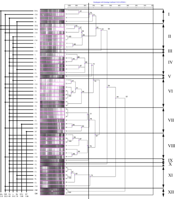
Fig 2. Dendogram of BOX-PCR from A. pyogenes 51 isolates. The degree of correlaction showed on scala
In the present study, it was determined using BOX-PCR that, the A. pyogenes isolates were of 12 clonal types, whilst Silva et al.19 referred to 10 herd-specific clonal types (5 clonal types in each of the herds A and B). While Silva et al.19 reported the rate of maximum similarity between the isolates of the two herds as 83.5%; in the present study, the maximum similarity rate of the isolates was determined to be 84.8%. In both studies, the maximum similarity rates of the isolates were rather high. While Silva et al.19 reported that 82% of the isolates belonged to 5 clonal types, in the present study, 80.4% of the isolates belonged to 6 different clonal types (types II, IV, VI, VII, VIII and XI).
Silva et al.19 pointed out to a vast heterogeneity among A. pyogenes isolates colonizing the uterus of dairy cows with normal puerperium and clinical metritis. Similarly, in the present study, it was determined that, based on the sample/case/source the isolates were recovered from, the A. pyogenes isolates displayed a high rate of heterogeneity for clonal types. The clonal type with greatest heterogeneity was determined as type VII (4 bovine livers, 1 calf synovial fluid, milk and suppurative tissue isolates); whilst, the clonal type with greatest homogeneity was type VIII (6 liver isolates and 1 milk isolate).
The number of clonal types was identified as 8 in isolates recovered from the liver of cattle slaughtered at a slaughterhouse, 9 in isolates recovered from the milk of dairy cows with mastitis, 3 in isolates recovered from the liver of sheep, and 2 in isolates recovered from the broncho-alveolar lavage fluid of dairy cows. The isolation of certain clonal types from different cases of infection and the identification of different clonal types from A. pyogenes isolates in the same case suggest that the pathogenicity of A. pyogenes should be further investigated in detail.
All of the clonal types identified were determined in isolates that were recovered from different sources and that are capable of inducing various infections. While the clonal types III, V and XII were similar strains isolated from only bovine mastitis, types IX and X were strains isolated from only bovine liver. On the other hand, some of the clonal types (I, II, IV, VI, VII, VIII, XI) were associated with strains isolated from different sources (broncho-alveolar lavage fluid, cow’s milk, bovine liver, ovine liver, bovine lung, calf synovial fluid, suppurative tissue). The isolation of these clonal types from different infections and sources suggests that their potential to cause infection could be greater.
The genome of A. pyogenes contains a variety of known and putative virulence factors. The pyolysin is well known as a major virulence factor of this bacteria 1,9,26 and it is generally present in all A. pyogenes isolates 8,17,20. In the present study, 2 different primers referred to by different researchers 8,19 were used for the plo gene. In result, all A. pyogenes isolates were found to be positive for the plo gene (band widths of 272 bp and 150 bp).
While Silva et al.19 reported that all investigated A. pyogenes strains carried nanH and nanP, according to some studies 1,25, some of A. pyogenes isolates were found to be positive for nanH (100% and 87%) and nanP (64.2% and 75%). In present study, positivity rates were determined as 62.7% and 84.3% for the nanH and nanP, respectively. The predominance of the nanP gene in bovine isolates has been indicated not to be surprising 1,19. It has been stated that the NanH and nanP genes may contain different host substances and that the nanP gene may encode tropism for adhesion of the agent to host tissues 12.
In many of studies 1,19,25,27 reported that fimA gene seems to be involved in adhesion processes and this gene was found to be high positive (98%, 94%, 100% and 90.9%) rates. The results of the present study corresponded to the findings of others 1,19,25,27.
While Esmay et al.14 reported the presence of the cbpA gene (encoding adhesion to the HeLA and 3T6 cells) in only 48% of the strains isolated from cases of different infections, in the present study, it was determined that 58.8% of the isolates were positive for the cbpA gene. While the rates of positivity determined in the present study were lower than those reported by Silva et al.19, it was observed that these results were close to those reported by Esmay et al.14.
The difference between the results obtained in this study and other researchers 1,14,16,19,25,27 is considered to have arisen from the isolates studied by the above mentioned researchers all being of uterine origin, and the isolates investigated in the present study having been recovered from different species, tissues and infections.
Silva et al.19 reported that in A. pyogenes strains; clonal types, virulence gene profiles and pathogenicity (development of disease) were correlated with each other. However, no such correlation was determined in the present study. The underlying reason of the difference in the results of the two studies may be Silva et al.19 having studied only on uterine samples obtained from two herds, and the strains investigated in the present study having been recovered from a wider range of sources.
The determination of the presence of virulence factors in the bacterial genome may not be adequate to show that the bacterium is capable of inducing disease. A. pyogenes isolates may possess their virulence factor genes differently under specific conditions. Host-intrinsic factors, synergic action between other bacteria and A. pyogenes, and differential gene expressions of virulence factor genes may have a more relevant effect on disease development 19. The variety of factors expressed by A. pyogenes may explain how this microorganism is able to colonize many different host tissues and cause such diverse disease processes 1,11,12,13,16.
isolated from different tissues and infections of sheep and cattle were identified in the present study. While all of the A. pyogenes isolates were positive for the plo gene, the positivity rates for other virulence factors differed among the isolates. Nonetheless, no correlation was determined between the virulence genes or clonal types and cases of infection.
REFERENCE
1. Jost BH, Billington SJ: Arcanobacterium pyogenes: Molecular patho-genesis of an animal opportunist. Antonie van Leeuwenhoek, 88, 87-102, 2005.
2. Nagaraja TG, Chengappa MM: Liver abscesses in feedlot cattle: A review. J Anim Sci, 76, 287-298, 1998.
3. Quinn AK, Vermont JJ, Twiss DP: Arcanobacterium pyogenes mastitis in a 18-month-old heifer. New Zealand Vet J, 50, 167-168, 2002.
4. Gouletsou PG, Ethenakis GC, Tzora A, Cripps PJ, Saratsis P: Isolation of Arcanobacterium pyogenes from the scrotal skin and the prepuce of healthy rams or from rams with testicular abnormalities. Small Rumin Res, 63, 177-182, 2006.
5. Miller ANA, Williams EJ, Sibley K, Herath S, Lane EA, Fishwick J, Nash DM, Rycroft AN, Dobson H, Bryant CE, Sheldon IM: The effects of Arcanobacterium pyogenes on endometrial function in vitro, and on uterine and ovarian function in vivo. Theriogenol, 68, 972-980, 2007. 6. Narayanan S, Nagaraja TG, Staats J, Chengappa MM, Oberst RD: Biochemical and biological characterizations and ribotyping of
Actinomyces pyogenes and Actinomyces pyogenes-like organisms from
liver abscesses in cattle. Vet Microbiol, 61, 289-303, 1998.
7. Nagaraja TG, Beharka AB, Chengappa MM, Carroll LH, Raun AP, Laudert SB, Parrott JC: Bacterial flora of liver abscesses in feedlot cattle fed tylosin or no tylosin. J Anim Sci, 77, 973-978, 1999.
8. Ertaş HB, Kılıç A, Özbey G, Muz A: Isolation of Arcanobacterium
(Actynomyces) pyogenes from abcessed kidney and identification by PCR. Turk J Vet Anim Sci, 29, 455-459, 2005.
9. Dias CAG, Cauduro PF, Mezzari A, Cantarelli V: Actynomyces pyogenes isolated from a subcutaneous abcess in a dairy farmer. Clin Microbiol Lett, 18, 38-40, 1996.
10. Lynch M, O’Leary J, Murnaghan D, Cryan B: Actinomyces pyogenes septic arthritis in a diabetic farmer. J Infection, 37, 71-73, 1998.
11. Jost BH, Songer JG, Billington SJ: An Arcanobacterium (Actinomyces)
pyogenes mutant deficient in production of the pore-forming cytolysin
pyolysin has reduced virulence. Infect Immun, 67, 1723-1728, 1999. 12. Jost BH, Songer JG, Billington SJ: Cloning, expression and characterization of a neuraminidase gene from Arcanobacterium
pyogenes. Infect Immun, 69, 4430-4437, 2001.
13. Jost BH, Songer JG, Billington SJ: Identification of a second
Arcanobacterium pyogenes neuraminidase and involvement of
neuraminidase activity in host cell adhesion. Infect Immun, 70, 1106-1112, 2002.
14. Esmay PA, Billington SJ, Link MA, Songer JG, Jost BH: The
Arcanobacterium pyogenes collagen-binding protein, CbpA, promotes
adhesion to host cells. Infect Immun, 71, 4368-4374, 2003.
15. Rudnick ST, Jost BH, Songer JG, Billington SJ: The gene encoding pyolosin, the pore-forming toxin of Arcanobacterium pyogenes, resides within a genomic islet flanked by essential genes. FEMS Microbiol. Letters, 225, 241-247, 2003.
16. Pietrocola G, Valtulina V, Rindi S, Jost BH, Speziale P: Functional and structural properties of CbpA, a collagen binding protein from
Arcanobacterium pyogenes. Microbiology 153, 3380-3389, 2007.
17. Billington SJ, Songer JG, Jost BH: Molecular characterization of the pore-forming toxin, pyolosin, a major virulence determinant of
Arcanobacterium pyogenes. Vet Microbiol, 82, 261-274, 2001.
18. Hadimli HH, Erganiş O, Kav K, Sayın Z: Isolation of Arcanobacterium
pyogenes from samples of sheep and cattle and identification by
Polimerase Chain Reaction. Kafkas Univ Vet Fak Derg, 16 (4): 611-616, 2010. 19. Silva E, Gaiva M, Leita S, Jost BH, Carneiro C, Vilela CL, Lopes da Costa L, Mateus L: Genomic characterization of Arcanobacterium
pyogenes isolates recovered from the uterus of dairy cows with normal
puerperium or clinical metritis, Vet Microbiol, 25, 111-118, 2008.
20. Ülbegi-Mohyla H, Hassan AA, Kanbar T, Alber J, Lämmler C, Prenger-Berninghoff E, Weib R, Siebert U, Zschöck M: Synergistic and antagonistic hemolytic activities of bacteria of genus Arcanobacterium and CAMP-like hemolysis of Arcanobacterium phocae and Arcanobacterium
haemolyticum with Psychrobacter phenylpyruvicus. Res Vet Sci, 87 186-188,
2009.
21. Kapur V, Sischo WM, Greer RS, Whittam TS, Musser JM: Molecular population genetic analysis of Staphylococcus aureus recovered from cows. J Clin Microbiol, 33 (Suppl. 2), 376-380, 1995.
22. Fitzgerald JR, Meaney WJ, Hartigan PJ, Smyth CJ, Kapur V: Fine-structure molecular epidemiological analysis of Staphylococcus aureus recovered from cows. Epidemiol Infect, 119, 261-269, 1997.
23. Hassan AA, Ülbegi-Mohyla H, Kanbar T, Alber J, Lämmler C, Abdulmawjood A, Zschöck M, Weiss R: Phenotypic and genotypic characterization of Arcanobacterium hamolyticum isolates from infections of horses. J Clin Microbiol, 47, 124-128, 2009.
24. Hassan AA, Mohyla H, Kanbar T, Alber J, Lämmler C, Abdulmawjood A, Speck S, Zschöck M, Weiss R: Molecular identification of Arcanobacterium bialowiezense and Arcanobacterium bonazi based on 16S-23S rRNA intergenic spacer region sequences. Vet Microbiol, 130, 410-414, 2008.
25. Hijazin M, Ülbegi-Mohyla H, Alber J, Lämmler C, Hassan AA, Abdulmawjood A, Prenger-Berninghoff E, Weib R, Zschöck M: Molecular identification and further characterization of Arcanobacterium
pyogenes isolated from bovine mastitis and from various other origins. J Dairy Sci, 94, 1813-1819, 2011.
26. Ding H, Lammler C: Purification and further characterization of a haemolysin of Actinomyces pyogenes. Zentralbl Veterinarmed, B 43, 179-188, 1996.
27. Santos TMA, Caixeta LS, Machado VS, Rauf AK, Gilbert RO, Bicalho RC: Antimicrobial resistance and presence of virulence factor genes in
Arcanobacterium pyogenes isolated from the uterus of postpartum dairy