R E S E A R C H A R T I C L E
Open Access
Uncovering sperm metabolome to discover
biomarkers for bull fertility
E. B. Menezes
1†, A. L. C. Velho
1,2†, F. Santos
1,2†, T. Dinh
1, A. Kaya
3, E. Topper
4, A. A. Moura
2and E. Memili
1*Abstract
Background: Subfertility decreases the efficiency of the cattle industry because artificial insemination employs spermatozoa from a single bull to inseminate thousands of cows. Variation in bull fertility has been demonstrated even among those animals exhibiting normal sperm numbers, motility, and morphology. Despite advances in research, molecular and cellular mechanisms underlying the causes of low fertility in some bulls have not been fully elucidated. In this study, we investigated the metabolic profile of bull spermatozoa using non-targeted metabolomics. Statistical analysis and bioinformatic tools were employed to evaluate the metabolic profiles high and low fertility groups. Metabolic pathways associated with the sperm metabolome were also reported.
Results: A total of 22 distinct metabolites were detected in spermatozoa from bulls with high fertility (HF) or low fertility (LF) phenotype. The major metabolite classes of bovine sperm were organic acids/derivatives and fatty acids/conjugates. We demonstrated that the abundance ratios of five sperm metabolites were statistically different between HF and LF groups including gamma-aminobutyric acid (GABA), carbamate, benzoic acid, lactic acid, and palmitic acid. Metabolites with different abundances in HF and LF bulls had also VIP scores of greater than 1.5 and AUC- ROC curves of more than 80%. In addition, four metabolic pathways associated with differential metabolites namely alanine, aspartate and glutamate metabolism, β-alanine metabolism, glycolysis or gluconeogenesis, and pyruvate metabolism were also explored.
Conclusions: This is the first study aimed at ascertaining the metabolome of spermatozoa from bulls with different fertility phenotype using gas chromatography-mass spectrometry. We identified five metabolites in the two groups of sires and such molecules can be used, in the future, as key indicators of bull fertility. Keywords: Metabolomics, Bovine, Spermatozoa, Gas chromatography, Mass spectrometry
Background
Fertility is the key to success for sustainability and eco-nomics of the livestock system in both beef and dairy cattle industries [1]. In cattle breeding, artificial insemin-ation (AI) is the most common assisted reproductive technology, and it employs ejaculates from genetically superior sires to inseminate a large number of cows [2]. However, only around 50% of such inseminations result in successful pregnancies, leading to considerable eco-nomic losses [3, 4]. Some studies reported that a signifi-cant percentage of reproductive failure (i.e., low numbers of pregnant females) is caused by the low fertilizing
capacity of the male gamete [5,6]. The accurate evaluation of bull fertility is currently attempted through routine ana-lysis of semen, but such conventional anaana-lysis is unable to determine a priori the full potential and actual fertility of the sires [4, 7, 8]. In addition, conventional analysis of semen has limited use for identification and prediction of sub-fertile animals [9]. The molecular events linked to sperm physiology are important because they serve as the foundation for identification of indicators of the fertilizing capacity of sires, improving the outcomes of AI [10].
The complex nature of events involved in fertilization is affected by the fluctuating concentrations of
macro-molecules found in the spermatozoon itself [11, 12],
seminal plasma [13], and by the microenvironment of fe-male reproductive tract [14]. At ejaculation, spermatozoa become coated with seminal plasma proteins, and even though the seminal fluid is diluted into the female
© The Author(s). 2019 Open Access This article is distributed under the terms of the Creative Commons Attribution 4.0 International License (http://creativecommons.org/licenses/by/4.0/), which permits unrestricted use, distribution, and reproduction in any medium, provided you give appropriate credit to the original author(s) and the source, provide a link to the Creative Commons license, and indicate if changes were made. The Creative Commons Public Domain Dedication waiver (http://creativecommons.org/publicdomain/zero/1.0/) applies to the data made available in this article, unless otherwise stated.
* Correspondence:[email protected]
†E. B. Menezes, A. L. C. Velho and F. Santos contributed equally to this work. 1Department of Animal and Dairy Sciences, Mississippi State University, 4025
Wise Center, Mississippi State, MS 39762, USA
Full list of author information is available at the end of the article Menezeset al. BMC Genomics (2019) 20:714
reproductive tract, the effects of those molecules are likely to be maintained as they quickly adhere to the sperm surface. Several studies have reported that sem-inal plasma proteins improve the fertilizing capacity of sperm [15–19]. Furthermore, recent reports provide evi-dence that the expression of microRNAs in bull sperm-atozoa is associated with fertility outcomes [20,21], and proteomics approaches have also been used for the dis-covery of potential fertility biomarkers. Metabolomics is vitally important because low molecular weight com-pounds may provide a clear picture of the regulatory pathways within spermatozoa [22, 23]. Also, metabolites are linked to physiological events through a cascade of biochemical complex networks [24,25] and also contrib-ute to the definition of the phenotype of an individual
[25]. Growing evidence suggests that spermatozoa
metabolize a wide spectrum of exogenous substrates that directly or indirectly regulate the signaling pathways in-volved in sperm motility, hyperactivation, capacitation, acrosome reaction, and sperm-oocyte fusion [26].
In recent years, studies using approaches in metabolo-mics have revealed potential fertility biomarkers in the seminal plasma of humans [27, 28], stallions [12], and bulls [29]. In a recent publication, we reported the iden-tification of 63 compounds in bull seminal plasma by using gas chromatography-mass spectroscopy (GC-MS). Fructose was the most abundant metabolites in the bo-vine seminal fluid, followed by citric acid, lactic acid, urea, and phosphoric acid [30]. A wide range of metabo-lites has also been detected in spermatozoa from humans [31–33], bulls [34], boars [35] and goats [36], bringing evidence that both seminal plasma and sperm metabolites could be meaningfully related to the fertility of males. The present study was conducted to test the hypothesis that metabolites of ejaculated sperm were as-sociated with fertility scores of dairy bulls.
Results
Metabolome profile of bull spermatozoa
Twenty-two metabolites were structurally identified in the bull spermatozoa, regardless of fertility phenotype of the animals. As the full scan spectra of the metabolites showed consistency in all samples, the retention time, target ion, two quantitative ions, and chemical structure of the derivative product of each metabolite were used
for the metabolite identification (Table 1). The NIST
Mass Spectral Search Program (NIST/EPA/NIH Mass Spectral Library, Version 2.0) was also employed to iden-tify all peaks in the chromatograms. An example of a chromatogram of bull sperm metabolites is depicted in
Fig.1. In our study, some eluting compounds in GC-MS
spectra could not be identified because of the limitation of single quadrupole technology and the lack of spectral information in the current databases.
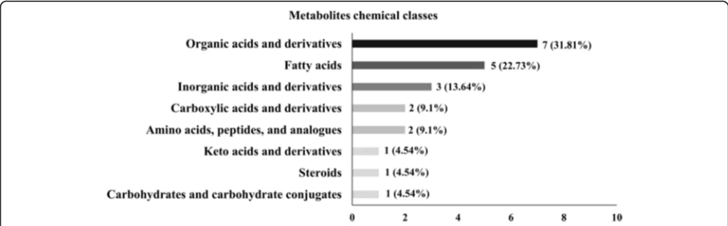
The metabolites identified in samples of bovine sperm-atozoa were categorized into eight major chemical clas-ses, as determined by hierarchical clustering analysis: organic acids/derivatives, fatty acids and conjugates, in-organic acids and derivatives, carboxylic acids and deriv-atives, amino acids, peptides/analogues, keto acids and derivatives, steroids and derivatives, and carbohydrates/ carbohydrate conjugates (Table1).
Organic acids/derivatives were the largest group of metabolites identified in the bull sperm by GC-MS, representing 31.81% of all metabolites in those cell types. Detected organic acids/derivatives were lactic acid (9.7E-04± 1E-03; 3.4E-03/0.016), oxalic acid (0.01 ± 4.3E-04; 9.1E-03/0.013), urea (5.9E-03± 8.2E-04; 1.1E-03/9.1E-03), ben-zoic acid (1.2E-03± 8.6E-05; 9.9E-03/1.9E-03), carbonate (5.7E-04± 1.4E-04; 3.5E-07/0.001) as well as carbamate (0.05 ± 0.01; 0.044/0.057) and (0.14 ± 1E-03; 0.061/0.186). The second largest group of sperm metabolites covered a remarkable diversity of fatty acids (22.73%), including nonanoic acid (4.5E-03± 4E-04; 2E-03/6 E-03), azelaic acid (0.02 ± 2.4E-04; 1E-03/ 3E-03), oleic acid (0.33 ± 0.02; 0.256/ 0.446), oleanitrile (5.9E-03± 5.5E-04; 0.018/0.035), and pal-mitic acid (5.9E-03± 5.5E-04; 3E-03/8E-03). In addition, there were three inorganic compounds, such as phos-phoric acid, (0.2 ± 0.06; 0.065/0.82), borate (0.071 ± 9.3E-03; 0.033/0.12), and phosphine (0.14 ± 8.1E-03; 0.109/ 0.176), making 13.64% of all metabolites detected by GC-MS in the bull spermatozoa. Carboxylic acids and derivatives (9.1%) including acetic acid (8.2E-04± 1E-04; 1.2E-04/0.002) and acetate (6.0E-03± 5.9E-03; 3.1E-03/9.1E-03) were also identified. Amino acids such as gamma-aminobutyric acid (GABA; 7.7E-03± 8.2E-03; 3.6E-03/0.01) and L-serine (6.1E-04± 1E-01; 4.6E-05/1E-03) comprised 9.1% of the bull sperm metabolite profile. Carbohydrates/ carbohydrate conjugates (4.54%) such as glycerol (0.09 ± 4.7E-03; 0.075/0.123) as well as steroids and de-rivatives (4.54%) such as cholesterol (7.1E-03± 9.4E-04;
3.7E-03/1.3E-02) were also detected. Furthermore, a
member of keto acids and derivative class (4.5%), 2-ketobutyric acid (4.5E-04± 1E-04; 1.1E-04/1E-03), was identified in bull spermatozoa (Table 1 and Fig. 2).
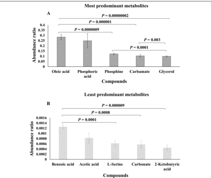
Oleic acid, phosphoric acid, phosphine, carbamate, and glycerol were the most abundant metabolites in bull
spermatozoa (Fig. 3a). In contrast, the least abundant
metabolites were benzoic acid, acetic acid, L-serine,
car-bonate, and 2-ketobutyric acid (Fig. 3b). Based on the
analysis of variance, the abundance ratio of oleic acid in the bull sperm was greater than those of phosphine (P = 0.0000009), carbamate (P = 0.000001), and glycerol (P = 0.00000002) (Fig. 3a). Moreover, the abundance ratio of phosphine was greater than those of glycerol (P = 0.0001), and the abundance ratio of carbamate was higher than those of glycerol (P = 0.003). In addition, the
metabolites had also displayed significant differences. Bull spermatozoa had a greater abundance ratio of ben-zoic acid as compared with L-serine (P = 0.0001),
car-bonate (P = 0.0008), and 2-ketobutyric acid (P =
0.000009), as shown in Fig.3b.
Associations between sperm metabolites and bull fertility
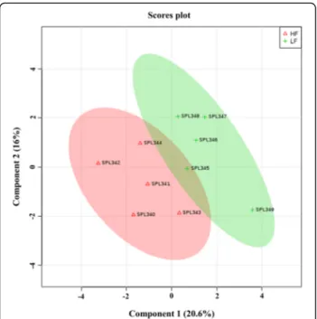
Partial Least Square-Discriminant Analysis (PLS-DA) analysis was assessed to determine the contribution of metabolites for the separation of high fertility (HF) and low fertility (LF) groups. PLS-DA two-dimensional score plots of sperm metabolites demonstrated that HF and LF phenotypes of bulls were separated from each other in two distinct clusters with a small overlap (Fig.4). The
first two components (1 and 2) explained 20.6 and 16.0% of the variance in the data set, respectively. In addition, the first five components explained 75.6% of the total variance. The metabolites that contributed most to the separation of LF and HF groups were GABA, carbamate, benzoic acid, and lactic acid. The performance charac-teristics of this multivariate model were R2= 0.428 and Q2= 0.874, respectively.
The Variable Importance in Projection (VIP) score based on the PLS-DA model represents the potential of
the metabolite as a biomarker (Fig. 5) and those
vari-ables with VIP score greater than 1.5 were considered important towards the classification model. Five metabo-lites had VIP scores > 1.5, including GABA (VIP = 2.01),
Table 1 Metabolites identified in bull spermatozoa by GC-MS
Compounds RT TI QI 1 QI 2 Mean relative abundance
(± SD)
Min/Max values FC t-test P-value Amino acids, peptides, and analogs
GABA 11.40 147 73 174 7.7E-03± 8.2E-04 3.6E-03/0.01 1.61 0.023
L-Serine 13.19 147 73 103 6.1E-04± 1E-04 4.6E-05/1E-03 1.33 0.211
Carbohydrates and carbohydrate conjugates
Glycerol 12.62 147.2 73 205.2 0.09 ± 4.7E-03 0.075/0.123 1.04 0.390
Carboxylic acids and derivatives
Acetic acid 13.29 191 73 146.8 8.2E-04± 1E-04 1.2E-04/0.002 0.68 0.211
Acetate 10.10 147 73 155 6.0E-03± 5.9E-03 3.1E-03/9.1E-03 0.81 0.142
Fatty acids and conjugates
Nonanoic acid (C9:0) 13.94 117 73 202 4.5E-03± 4E-04 2E-03/6E-03 1.01 0.246
Azelaic acid (C9H16O4) 20.51 317.2 73 201 0.02 ± 2.4E-04 1E-03/ 3E-03 1.09 0.397
Oleic acid (C18:1 n-9) 27.56 131.2 73 144.2 0.33 ± 0.02 0.256/0.446 0.99 0.481
Oleanitrile (C18H33N) 23.96 122.2 69.2 55 5.9E-03± 5.5E-04 0.018/0.035 1.05 0.395
Palmitic acid (C16:0) 23.54 313.4 73 117 5.9E-03± 5.5E-04 3E-03/ 8E-03 0.77 0.040
Inorganic compounds
Phosphoric acid 12.58 299 73 357 0.2 ± 0.06 0.065/0.82 0.73 0.228
Borate 6.59 221 73 157 0.071 ± 9.3E-03 0.033/0.12 0.91 0.366
Phosphine 9.53 116 73 204 0.14 ± 8.1E-03 0.109/0.176 0.93 0.390
Keto acids and derivatives
2-Ketobutyric acid 14.31 202.2 73 111.8 4.5E-04± 1E-04 1.1E-04/1E-03 0.89 0.474
Organic acids and derivatives
Lactic acid 8.20 117 73 147 9.7E-04± 1E-03 3.4E-03/0.016 1.59 0.040
Oxalic acid 8.16 147 73 133 0.01 ± 4.3E-04 9.1E-03/0.013 1.02 0.403
Urea 11.83 147 73 189 5.9E-03± 8.2E-04 1.1E-03/9.1E-03 1.03 0.452
Benzoic acid 11.90 105 77 179 1.2E-03± 8.6E-05 9.9E-03/1.9E-03 1.46 0.034
Carbonate 12.32 191 73 199 5.7E-04± 1.4E-04 3.5E-07/0.001 1.00 0.493
Carbamate 7.651 8.019 147 174 73 73 204 198 0.05 ± 0.01 0.14 ± 1E-03 0.044/0.057 0.061/0.186 1.08 1.47 0.171 0.033 Steroids and steroid derivatives
Cholesterol 34.50 129.2 73 207.2 7.1E-03± 9.4E-04 3.7E-03/1.3E-02 1.03 0.442
RT Retention time, TI Target ion, QI Quantitate ion, SD Standard deviation, Min Minimum, Max Maximum, and FC Fold change. P-value is determined by the t-test to determine the statistical differences in metabolite abundances between HF and LF groups.P-values that are < 0.05 are italicized
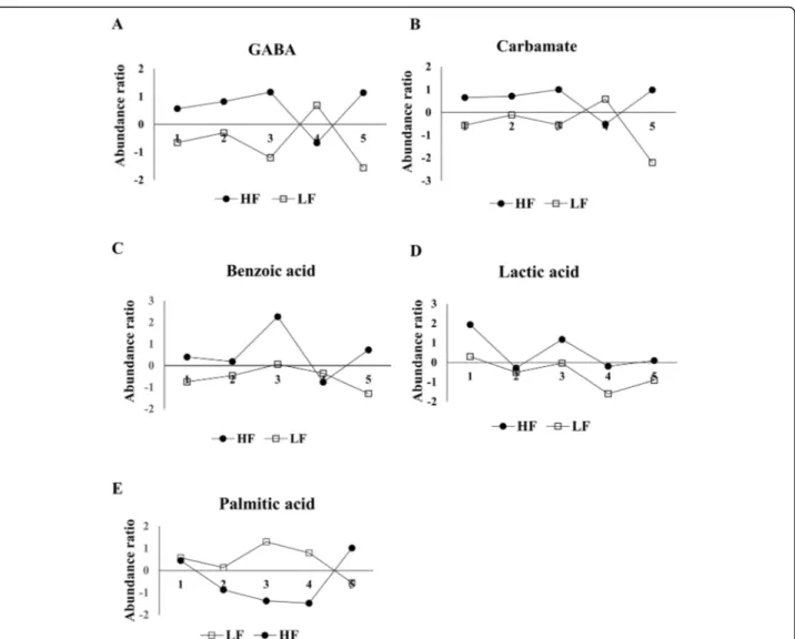
carbamate (VIP = 1.88), benzoic acid (VIP = 1.86), lactic acid (VIP = 1.81), and palmitic acid (VIP = 1.50). Al-though our results also indicate that differences in me-tabolite abundance are not consistent between fertility groups (Fig. 6), the abundance ratios of five sperm me-tabolites were statistically different between LF and HF
groups (Fig. 6): GABA, carbamate, benzoic acid, lactic
acid, and palmitic acid, as determined by univariate stat-istical analysis (Table1).
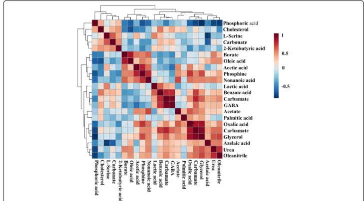
The correlation matrix shows positive (red) and nega-tive (blue) associations between the abundance ratios of
the metabolites in HF and LF bulls (Fig. 7). Thus,
phosphoric acid was inversely associated with oleic
acid (r =− 0.64), phosphine (r = − 0.67), oxalic acid
(r =− 0.61), glycerol (r = − 0.82), urea (r = − 0.73), and oleanitrile (r =− 0.76). Phosphine had a positive asso-ciation with acetic acid (r = 0.50), nonanoic acid (r = 0.73), oxalic acid (r = 0.71), and glycerol (r = 0.64). In addition, carbamate was positively correlated with benzoic acid (r = 0.75) and glycerol abundance was related to that of oxalic acid (r = 0.59), carbamate (r = 0.83), urea (r = 0.62) and oleanitrile (r = 0.66). L-serine had positive correlation with carbonate (r = 0.61) and negatively linked to azelaic acid (r =− 0.78) Fig. 1 Representative chromatogram of metabolites from spermatozoa bulls. Peaks of GABA, carbamate, benzoic acid, lactic acid, and
heptadecanoic acid (internal standard) are indicated
Fig. 2 Number of metabolites per chemical class. Metabolites identified in bull spermatozoa according to their chemical classes, defined as amino acids, peptides/analogs, carbohydrates/carbohydrate conjugates, fatty acids, steroids/steroid derivatives, keto acids and derivatives, organic and inorganic compounds, carboxylic acids and derivatives
in the bovine sperm. L-serine had also a negative
as-sociation with azelaic acid (r =− 0.78) whole GABA
was positively correlated with carbamate (r = 0.94) and benzoic acid (r = 0.74).
Diagnosis evaluation of the biomarkers
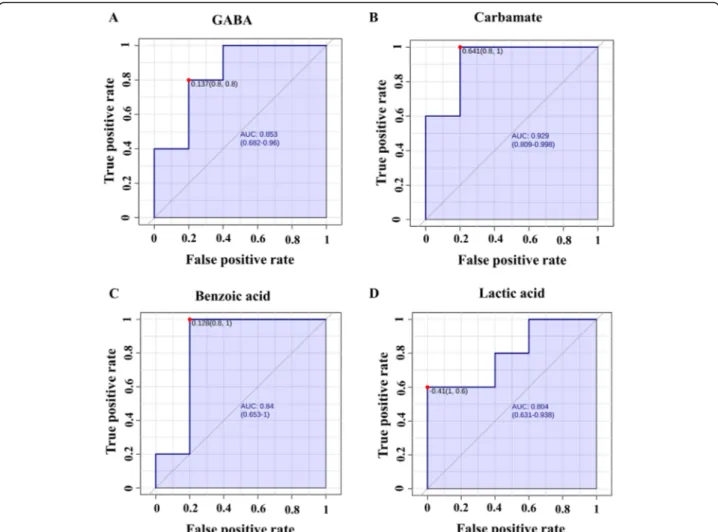
Multivariate ROC analyses were used to assess the sensi-tivity and specificity of the potential biomarkers of bull fertility. By analyzing the data, we demonstrated that all the area under the receiver operating characteristic (ROC) curve (AUC) of the sperm metabolites ranged from 0.52 to 0.92. Metabolites with an AUC > 80% were
carbamate (AUC = 0.92; P = 0.005), GABA (AUC = 0.84;
P = 0.001), benzoic acid (AUC = 0.84; P = 0.006), and lac-tic acid (AUC = 0.80;P = 0.008; Fig.8).
Functional biochemical pathway analysis
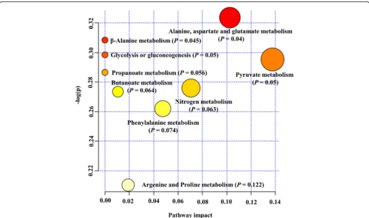
Metabolic pathway analysis was performed to evaluate the most relevant pathways associated with differential metabolites in the sperm of HF and LF bulls. The differ-ential metabolites encompass four biochemical path-ways, which may reveal the metabolic mechanisms within spermatozoa that might affect fertility. The meta-bolic pathways were alanine, aspartate and glutamate metabolism (P = 0.04), β-alanine metabolism (P = 0.045), glycolysis or gluconeogenesis (P = 0.05), and pyruvate metabolism (P = 0.05), as shown in Fig.9.
Discussion
Characterization of the sperm metabolic signatures is a powerful approach that can potentially lead to the Fig. 3 Abundance ratios of the most and least predominant metabolites present in bull spermatozoa. a Abundance ratios of the five most abundant metabolites such as oleic acid, phosphoric acid, phosphine, carbamate, and glycerol. b Abundance ratios of the five least metabolites identified as benzoic acid, acetic acid, L-serine, carbamate, and 2-ketobutyric acid. The abundance ratio of the metabolites was calculated by dividing the abundance of target ions of metabolites by that of target ion of the internal standard. Error bars represent standard error of the mean. P < 0.05 was considered significant
development of biomarkers for male fertility. In the present study, we investigated the metabolic profiles of spermatozoa from bulls with high vs. low fertility status using non-targeted metabolomics as well as statistical and bioinformatics tools. Results presented here are an important foundation to further understand the mecha-nisms by which metabolites of spermatozoa may affect fertility outcomes and to help to predict the fertilizing potential of sires.
Metabolites play key roles in sperm physiology and are related to differences in male fertility phenotypes
[31–33, 37]. Recently, NMR- and GC-MS-based
stud-ies showed that pathways for nucleoside, amino acid, and energy metabolism were disturbed in
astheno-zoospermic men [32], and that metabolites found in
human spermatozoa are associated with semen
pa-rameters [37]. In this study, our analyses by GC-MS
revealed that the majority of metabolites of bovine sperm are organic acids and derivatives, followed by a group of fatty acids and conjugates. Organic acids are produced by the breakdown of amino acids and fatty acids, and the degradation of such metabolites gener-ates energy substrgener-ates for tricarboxylic acid (TCA)
cycle and respiratory chain [38]. The presence of
or-ganic acid suggests that bull spermatozoa have active
energy metabolism [8, 39, 40]. In addition, organic
acids play crucial roles during anabolism by providing
C-atom backbones [38]. Studies have previously
re-ported the presence of organic acids in human spermatozoa [31, 32] and seminal plasma of bulls [30] and humans [27, 41, 42]. Fatty acids, in turn, are in-volved in the structural organization of the sperm membranes, energy metabolism, and signaling mole-cules [12, 43, 44]. The enzymatic machinery for
beta-oxidation is present in human spermatozoa [45, 46],
suggesting that sperm may obtain energy also through the oxidation of fatty acids [47]. Several types of fatty acids have also been reported in seminal plasma of humans and bulls [27, 30, 31].
The most predominant metabolites of the bull sperm-atozoa were oleic acid, phosphoric acid, phosphine, car-bamate, and glycerol; whereas benzoic acid, acetic acid, L-serine, carbonate, and 2 ketobutyric acid were among the least abundant metabolites. Oleic acid (C18:1 n-9) is the most abundant monounsaturated fatty acids of the plasma membrane of ejaculated stallion [48], boar [49],
and ram spermatozoa [50]. Oleic acid is negatively
linked to sperm motility and concentration in humans [51–53]. In addition, high levels of oleic acid have been reported to increase lipid oxidation [53], leading to
dis-orders in sperm membrane metabolism in men [27]. On
the other hand, addition of oleic acid maintains bull sperm viability and lowers the production of reactive oxygen species (ROS) in vitro [54]. The high content of Fig. 4 Partial-Least Squares Discriminant Analysis (PLS-DA) scores
plot of metabolite profiles generated by GC–MS analysis of bull spermatozoa from high (HF) and low fertility (LF) bulls. Plots showed a clear cluster distinction between HF (n = 5) and LF (n = 5) groups. Supervised PLS-DA was obtained with 2 components. The explained variances are shown in parentheses
Fig. 5 Variable importance in projection (VIP) scores of spermatozoa metabolites in high (HF) and low fertility (LF) bulls. The selected metabolites were those with VIP > 1.5. Heat map with red or green boxes on the right indicates high and low abundance ratio, respectively, of the corresponding metabolite in HF and LF bulls. VIP score was based on the PLS-DA model. GABA (VIP = 2.01), carbamate (VIP = 1.88), benzoic acid (VIP = 1.86), lactic acid (VIP = 1.81), and palmitic acid (VIP = 1.5)
oleic acid in bovine sperm suggests its contribution to
reduce ROS production [54] and to generate energy for
sperm hyperactivation [55]. Phosphoric acid was the sec-ond most abundant metabolite in bull spermatozoa. In normal sperm cells, phosphoric acid is produced by the breakdown of ATP in a reaction catalyzed by inorganic
pyrophosphatase (PPA1) [56]. PPA1 catalyzes the
hy-drolysis of one molecule of inorganic pyrophosphate (PPi) to two molecules of phosphoric acid, leading to the release of energy in form of adenosine triphosphate (ATP). The transport of PPi from spermatozoa to the seminal plasma may be regulated by a transmembrane protein, called progressive ankylosis protein (ANKH). In mammals, PPA1 is present in the post-acrosomal sheath of the sperm head and in the distal part of sperm acro-some. The energy produced from the conversion of PPi to phosphoric acid could be used for sperm motility and
for acrosomal function during sperm-zona penetration [57,58]. In addition, inorganic phosphate resulted from the hydrolysis of ATP positively affect both motility and
fertilizing capacity of human sperm [59]. Thus, high
levels of inorganic phosphate in bull spermatozoa may be required to maintain sperm motility status and to achieve normal fertility.
Our GC-MS-based analyses indicated that carbamate is the fourth most abundant metabolite of the bovine spermatozoa with the second highest VIP score. Carba-mate was first reported in seminal plasma from healthy and asthenozoospermic men [27]. However, it is new the description of carbamate in bull spermatozoa as well as its higher abundance in sperm from HF bulls. Endogen-ous carbamate is generated by the interaction of cellular
carbon dioxide (CO2) with an NH2 group of primary
and secondary amines [60, 61] when the concentration
Fig. 6 Abundance ratio of five differential metabolites in spermatozoa from high and low fertility bulls. (a) GABA (P = 0.023), (b) Carbamate (P = 0.033), (c) Benzoic acid (P = 0.035), (d) Lactic acid (P = 0.040), and (e) Palmitic acid (P = 0.040) were significantly different between high (HF) and low (LF) bulls
of CO2increases [62]. The formation of carbamate
influ-ences the function of hemoglobin as well [63]. Although the importance of carbamate in sperm physiology is un-known, we can speculate that the spermatozoon, like other cells, employs several mechanisms to maintain the
cell pH [64]. Therefore, carbamate formation might be
an important mechanism by which spermatozoa regulate their intracellular pH.
The principal inhibitory neurotransmitter in the adult brain, GABA, was found with greater abundance in spermatozoa from HF sires and it had the highest VIP score. The key enzyme in the synthesis of the GABA,
glutamate decarboxylase, and GABAA/GABABreceptors
were previously identified in human spermatozoa [65].
GABA has been also detected in seminal plasma and
spermatozoa from humans [66], as well as in seminal
plasma of bulls [30]. GABA induces capacitation of
spermatozoa from bulls [67,68], rats [69], and rams [70]
and acrosome reaction of bovine sperm [68]. The high
abundance of GABA in the sperm of HF bulls may be explained by the roles described above, and in sperm hy-peractivation [71]. In the present work, abundance ratios of GABA and carbamate were positively associated, and this may be related to the fact that carbamate modulates GABAAreceptor [72]. Moreover, a positive link was also
found for GABA and benzoic acid in bull spermatozoa. A previous in vitro study reported that benzoic acid
increases efflux of glutamate [73] and levels of benzoic acid may regulate sperm function since GABA is formed
by decarboxylation of L-glutamate [74]. The abundance
ratios of benzoic acid were increased in spermatozoa of HF bulls as compared to LH sires. The presence of ben-zoic acid was reported in seminal plasma of bulls [30]
and asthenozoospermic and normozoospermic men [27]
as well as in spermatozoa from asthenozoospermic and
normozoospermic men [32]. A recent study reported a
positive correlation between the abundance ratios of benzoic acid and sperm counts in rats [75], suggesting that benzoic acid plays a role in male fertility.
Lactate is an important energy source for bull, human, stallion, and boar spermatozoa [8, 46]. The production of lactate by the bull sperm occurs mainly through gly-colysis and mitochondrial oxidative phosphorylation (OxPhos) [8, 45, 47, 76]. Multivariate statistical analysis conducted in the present study demonstrated that lac-tate was one of the metabolites contributing to fertility phenotype with the fourth highest VIP associated with HF bulls. Greater lactate abundance in HF bulls suggests that these animals utilize anaerobic glycolysis more effi-ciently than LF sires [8,77]. It is well-known that sperm mitochondria compensate for decreased energy produc-tion by increasing lactate yield under hypoxia. The effi-cient glycolysis is dependent on either endogenous or
exogenous pyruvate, which indirectly feeds the
Fig. 7 Heatmap visualization of Pearson’s correlations among metabolites present in bull spermatozoa. The scale is based on colors from red (positive) to blue (negative) representing associations between the relative abundance of bull sperm metabolites that related to each other in the groups
accelerated glycolysis with nicotinamide adenine
di-nucleotide (NAD+) through the lactate
dehydrogenase-mediated conversion of pyruvate to lactate [8, 76]. The
oxidation of NAD+ in the electron transport chain
gen-erates the ATP molecules by oxidative phosphorylation [8]. Thus, when high energy is required for sperm motil-ity and other events, spermatozoa efficiently metabolize glycolysable substrates to yield ATP [8, 46]. In fact, the inhibition of lactate dehydrogenase blocks sperm capaci-tation in bulls [78], humans [79], mice [80], and goats [81]. Therefore, the level of lactate in sperm could be considered as an early indicator of bull fertility [82].
Palmitic acid (C16:0), another metabolite found in bull spermatozoa, had the fifth highest VIP score associated with LF animals. This is consistent with previous studies showing increased levels of palmitic acid in infertile men
[27, 83, 84] and in asthenozoospermic semen as
com-pared to normozoospermic ones [85]. Another study
re-ports that high palmitic acid in seminal plasma from
asthenozoospermic men indicates a disorder in sperm
membrane metabolism [27].
The importance of lipid metabolism for the produc-tion of energy for spermatozoa has been discussed in
previous studies [43]. Our analytical methods allowed
the detection of considerable amounts of nonanoic acid and azelaic acid in bull spermatozoa. Nonanoic acid (C9: 0), also known as pelargonic, is a 9-carbon chain fatty acid, and it was previously reported in goat epididymal sperm membrane [86] and mouse epididymal fluid [87]. The importance of nonanoic acid for sperm physiology is still unknown, but it is possible that it contributes to
sperm maturation [87]. In addition, OR51E1, a known
receptor of nonanoic acid, was detected in the acrosomal
cap of human spermatozoa [88, 89], and activation of
OR51E1 with nonanoic acid led to the phosphorylation of various protein kinases [90]. Also, OR51E1 level de-creased upon acrosomal exocytosis [91], and such results suggest that nonanoic acid is involved in acrosome Fig. 8 Receiver operating characteristic curves based on PSL-DA analysis models. a GABA (AUC: 0.84; P = 0.001), b Carbamate (AUC: 0.92; P = 0.005), c Benzoic acid (AUC: 0.84; P = 0.006), and d Lactic acid (AUC: 0.80; P = 0.008). AUC: Area under curves
reaction, possibly by triggering protein tyrosine phos-phorylation during sperm capacitation.
The present study reported for the first time the pres-ence of azelaic acid (nonanedioic acid; C9H16O4) in bull spermatozoa. Azelaic acid is a nine-carbon saturated ali-phatic dicarboxylic acid, and it has been reported in tes-tes of rats [92]. This metabolite was also found in mouse [93] and human spermatozoa [31]. Azelaic acid is the end product of linoleic acid peroxidation [94] and acts as a ROS scavenger [95], protecting spermatozoa. More-over, studies mention additional roles for azelaic acid in-cluding inhibition of tyrosinases [96, 97], mitochondrial enzymes [98], anaerobic glycolysis [98], mitochondrial oxidoreductase activity, and DNA synthesis [99]. A study showed evidence that the incubation of mouse sperm in fructose-containing media resulted in a high concentra-tion of azelaic acid in sperm when compared with glucose-containing media [93]. Given that azelaic acid modulates the activity of glycolytic key enzymes [100], we hypothesize that this metabolite is essential for en-ergy metabolism of the sperm cells.
We also evaluated the metabolic pathways of certain molecules and their potential contributions to male fer-tility. There were four significant pathways associated with differential sperm metabolites including alanine,
as-partate and glutamate metabolism, β-alanine
metabol-ism, glycolysis or gluconeogenesis, and pyruvate
metabolism. Alanine, aspartate, and glutamate are linked to amino acid metabolism. As amino acids play key roles in multiple cellular processes, they influence the meta-bolic activity of the spermatozoa [101]. The GABA is in-volved in sperm motility, acrosome reaction, and fertilization in human spermatozoa [102]. Thus, we as-sume that as spermatozoa of HF animals have more GABA their fertility rate increases. Another amino
acid-related pathway identified in bull sperm was β-alanine
metabolism. β-alanine is structurally intermediate
be-tween alpha-amino acid (glycine, glutamate) and GABA neurotransmitters [103], and β-alanine is a ubiquitous amino acid correlated with the TCA cycle. In fact, both TCA intermediates and amino acids have been found as part of the metabolomic profile of bull seminal plasma, as recently described by Velho et al. [30]. The glycolysis consists of a series of biochemical reactions to generate
energy in the form of ATP [77]. Glycolytic metabolite
such as lactate was significantly elevated in spermatozoa from HF bulls as compared to LF animals, suggesting that the maintenance of intracellular energy status is es-sential for sperm function. Considering the pathways an-alyzed in the present study, pyruvate metabolism is crucial for understanding the contributions of OxPhos for the fertilizing capacity of spermatozoa [45]. Bull spermatozoa also rely on OxPhos to maintain sperm functions [77]. A recent study showed that pyruvate is Fig. 9 Pathway analysis of differentially present metabolites in bull as determined by MetaboAnalyst 4.0. Each point represents one metabolic pathway; the size of the dot is in positive correlation with the impaction of the metabolic pathway
the most important source of energy for stallion sperm
[8] and that the impairment of sperm mitochondrial
ultrastructure may affect male fertility [104].
Although the sample size represents a limitation of the present study, the sensitivity of the GC-MS approach, together with the bioinformatic tools, enabled us to construct a metabolomics analytical model of sperm from bulls with different fertility phenotypes. Metabo-lites with different abundances in bulls of high and low fertility (GABA, carbamate, benzoic acid, lactic acid, and palmitic acid) are potential biomarkers of bull fertility.
Conclusions
The metabolomic signatures of bull spermatozoa ad-vance our current understanding of the multifactorial and complex processes related to the physiology of male fertility. The present study uncovered vital pieces of in-formation about sperm metabolites for diagnosing male fertility. In addition, because of the strong similarities in physiology and genetics between cattle and other mam-mals, including humans and endangered mammam-mals, the knowledge generated in the present investigation can be applied to enhance reproductive biotechnology of other species.
Methods
Study design
Metabolomic analysis of bull spermatozoa with two dis-tinct and reliable fertility phenotypic scores was
per-formed by GC-MS. Univariate and multivariate
statistical models were employed to identify key differ-ences between the two groups, HF (n = 5) and LF (n = 5) bulls. Statistical and bioinformatics tools were also used to identify potential biomarkers of bull fertility.
Determination of fertility phenotypes of dairy bulls
In the current study, the field fertility data were collected for the evaluation of fertility scores of mature Holstein bulls (Table2), as previously described by Peddinti et al. [105]. Fertility data were obtained from the Alta Advan-tage Program (Alta Genetics, Inc., Watertown, WI, USA), which periodically updates results from AI in the partnering herds [105]. The conception rates were con-firmed in the field by either ultrasound or veterinary pal-pation. The method used for the calculation of bull fertility was similar to the one employed in previous in-vestigations [17–19,29]. Factors that influenced the
fer-tility of sires such as environmental and herd
management were adjusted using a model previously de-scribed [106, 107]. The average conception of breeding records and conception rates was calculated using the Probit F90 software [108]. The bulls were selected with conception rates of two standard deviations above and below the average conception rates of the population of sires available in Alta Genetics database. When bulls had percent differences in their conception rates above
aver-age, we defined them as “HF”; in contrast, if sires had
percent differences in their conception rates below aver-age, we designated them as“LF” (Table2).
Sperm collection and preparation
Semen samples from 10 mature Holstein bulls with different fertility scores were provided by Alta Genet-ics (Watertown, WI, USA). All animals were raised under the same management conditions and received the same nutrition. Ten ejaculates, one per bull, were collected using an artificial vagina and spermatozoa were separated from seminal plasma by centrifugation (700 g, 4 °C, 10 min). Then, the pellet containing spermatozoa was washed twice (700 g; 4 °C; 15 min.) with cold phosphate-buffered saline (PBS) and further
aliquoted (100μl) into a new 2 ml Cryotube®
(Sigma-Table 2 Fertility scores of mature Holstein bulls. High fertility (HF) bulls were designed from 1 to 5 and bulls 5 to 10 were grouped as low fertility (LF). Fertility score of each bull was expressed as the percent difference of its conception rate from the average conception rate of all bulls. Probit.F90 software was used to estimate bull fertility
Bull # Fertility status Number of breedings Conception rates difference from average (%) Std of difference Conception rates (%)
1 HF 5293 5.42 2.02 45.3 2 HF 825 5.1 1.90 40.4 3 HF 2032 4.8 1.79 40.3 4 HF 2487 3.59 1.34 45.7 5 HF 5751 3.56 1.33 39.8 6 LF 1604 −3.75 −1.40 35.7 7 LF 2276 −4.06 −1.52 37.8 8 LF 967 −4.49 −1.68 34.4 9 LF 5603 −6.76 −2.52 34.6 10 LF 674 −10.61 −3.96 23.3
Aldrich, St Louis, MO, USA). Following the second centrifugation, spermatozoa were snap-frozen in liquid nitrogen and transported to Mississippi State Univer-sity (MSU). At MSU, snap-frozen spermatozoa were
stored at − 80 °C until preparation for GC-MS
ana-lyses. The metabolomic profiles of the seminal plasma obtained from the same bull semen samples have been reported in our previous publication [30].
Chemicals and materials
GC-MS grade chemicals and solvents were all purchased from Sigma Aldrich in the highest purity available. Phosphate-buffered solution (137 mM NaCl, 2.7 mM
KCl, 8 mM Na2HPO4, and 2 mM KH2PO4; pH 7.4) was
purchased from Thermo Fisher Scientific (Waltham, MA, USA). Amber glass vials (2 ml) with 300μl fixed in-sert were obtained from Agilent Technologies Inc. (Santa Clara, CA, USA).
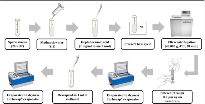
Metabolite extraction
Sperm metabolites were isolated from bull spermatozoa as previously described by Paiva et al. [31], with modifica-tions and a schematic overview of the sperm metabolite
extraction is presented in Fig. 10. In summary,
snap-frozen sperm (2 × 10⁷cells) were thawed in a water bath at 37 °C for 30 s. The thawed spermatozoa were suspended in a mixture of 8 ml of methanol and 1 ml of ultrapure water, followed by addition of 150μl of heptadecanoic acid (1 mg/ml in methanol) as the internal standard. In addition, sperm suspension was subjected to five freeze/
thaw cycles. Each cycle consisted of freezing sperm cells in liquid nitrogen vapor for 30 min. and subsequent thaw-ing at room temperature for 30 min. Followthaw-ing freeze/ thaw cycles, the cell suspension was sonicated in an ultra-sonic bath at 25 °C for 30 min. at 120 W and 40 kHz (Fisher Scientific™ CPXH5 Series Ultrasonic Baths; Pitts-burgh, PA, USA), followed by ultracentrifugation (40,000 g, 4 °C, 20 min.) using an OptimaTM L-90 k and Type 70 Ti Rotor (BECKMAN COULTER Life Sciences, Brea, Califor-nia, USA). The supernatant was filtered through a 0.2μm nylon membrane (Fisher Scientific, Lenexa, KS, USA) and the filtrate was evaporated under a stream of high-purity nitrogen gas (TurboVap® LV evaporator; Biotage, Charlotte, NC, USA) at 40 °C. An aliquot of methanol (1 ml) was added to dissolve metabolites. The metab-olite extracts were subsequently transferred into a 2 ml amber vial and evaporated to dryness again with high-purity nitrogen gas at 40 °C.
The dried extracts were resuspended in 50μl of
meth-oxyamine hydrochloride (20 mg/ml in pyridine) vortexed vigorously for 1 min, and further heated in a water bath at 70 °C for 1 h. The samples were then derivatized by adding 100μl N, O-Bis(trimethylsilyl)trifluoroacetamide with 1% trimethylchlorosilane (BSTFA + 1% TMCS) and heated in a water bath at 70 °C for 1 h. The final deriva-tives were transferred into a new 2 ml amber glass vial with a 300μl fixed insert for GC-MS analysis. A quality-control sample for the experiment was prepared by pool-ing equal volumes of sperm extract samples to ensure that the detection of metabolites was consistent.
Fig. 10 Overview of extraction steps used for the preparation of sperm samples for GC-MS analysis. Drawings are not to scale, see Materials and Methods section for full details
Gas chromatography-mass spectrometry (GC-MS) analysis
Screening of the untargeted sperm metabolites was per-formed using GC-MS as previously described by Velho et al. [30], with modifications. Briefly, metabolite deriva-tives were separated and detected using an Agilent 7890A GC System coupled to an Agilent 5975C inert XL MSD with triple-axis mass detector, an Agilent 7693 Series Autosampler, and a DB-5MS capillary column
(30 m × 0.25 mm i.d. × 0.25μm film thickness; Agilent
Technologies). An aliquot of the derivatized mixture (1μl) was injected into the inlet heated at 270 °C with a 1:10 split ratio. Standard septum purge was carried out after sample injection at 3 ml/min, and helium carrier gas was at 1 ml/min constant flow rate. Transfer line, ion source, and quadrupole were heated at 260 °C, 200 °C, and 150 °C, respectively. The oven was pro-grammed initially at 80 °C for 2 min., followed by 10 °C/ min., ramped up to 180 °C, 5 °C/min. to 240 °C, 20 °C/ min. to 290 °C, and 10 min. Maintenance at 290 °C. Ionization was performed in an electron impact mode at 70 eV. Masses were scanned for a full spectrum from m/ z 30 to 600 at 10,000 amu/s and 20 scans/s (m/z 0.2 step size). The solvent delay time was 5 min.
Data processing, calculations, and statistical analysis
Sperm metabolites were identified by their retention time as well as one target and two quantitative ions, in com-parison with mass spectra of authentic standards and mass spectra in the NIST mass spectral library. Abundances of the target ions of metabolites were divided by the abun-dance of target ion of the internal standard (heptadecanoic acid), and the ratios were used for statistical analysis. Iden-tified compounds were categorized based on their chem-ical classifications using Human Metabolome Database version 3.6 (HMDB;www.hmdb.ca/) [109,110]. Statistical analysis was carried out using MetaboAnalyst 4.0 Web service (http://www.metaboanalyst.ca). MetaboAnalyst is a comprehensive web-based tool designed to help users eas-ily perform metabolomic data analysis, visualization, and functional interpretation [111]. Sum and auto-scaling nor-malized each compound. Univariate analysis (t-test) was used to determine if differences in metabolite abundances in spermatozoa of HF and LF sires were significantly different.
Multivariate analysis was applied to provide additional information for the interpretation of the data. The PLS-DA defined the separation metabolome of sperm from HF and LF bulls. Potential biomarkers were identified according to the significance of their contributions to variable classification in the PLS-DA model, which was determined by the VIP score. The VIP score summarizes the contribution that a variable makes to the model, and it is calculated as the weighted sum of the squared cor-relations between the original variable and the PLS-DA
components. The weights correspond to the percentage variation explained by the PLS-DA component in the model. The number of terms in the sum depends on the number of PLS-DA components found to be significant in distinguishing the classes. In the present study, we considered metabolites with VIP > 1.5 as potential bio-markers associated with bull fertility. The ROC analysis was applied to examine the specificity and sensitivity of the biomarkers. The area under the ROC curve was cal-culated to assess the effectiveness of the potential bio-markers. A guide for assessing the performance of metabolites as a biomarker based on its AUC is as fol-lows: AUC of 0.9 to 1.0 = excellent, 0.8 to 0.9 = good, 0.7 to 0.8 = fair, 0.6 to 0.7 = poor, and 0.5 to 0.6 = fail [112]. Pearson’s method was used to analyze the correlation between metabolites. Significance for statistical analyses was set at 0.05.
Functional biochemical pathway analysis
Differential metabolites were also evaluated by using metabolic pathway analysis (MetPA) [113,114]. For this analysis, we uploaded the differential metabolites select-ing the‘Bos taurus’ library. The default ‘hypergeometric test’ and ‘Relative Betweenness Centrality’ for pathway enrichment and pathway topological analyses, respect-ively, were selected. Kyoto Encyclopedia of Genes and Genomes (KEGG) metabolic pathway was also employed. All matched pathways were visualized by plotting the −log(p) values from pathway enrichment analysis on Y-axis and pathway impact values from pathway topology analysis on X-axis [114]. The node color was based on its p-value and the node radius was associated with their pathway impact values. Metabolic pathways with p-value < 0.05 and false discover rate values of 0.7 were screened as pathways of interest.
Abbreviations
AI:Artificial insemination; ANKH: Progressive ankylosis protein; ATP: Adenosine triphosphate; AUC: Area under the curve; BSTFA: O-Bis(trimethylsilyl)trifluoroacetamide; CO2: Carbon dioxide; GABA:
Gamma-aminobutyric acid; GC-MS: Gas chromatography-mass spectroscopy; HF: High fertility; HMDB: Human Metabolome Database; KEGG: Encyclopedia of Genes and Genomes; LF: Low fertility; MetPA: Metabolic pathway analysis; NAD+: Nicotinamide adenine dinucleotide; OxPhos: Mitochondrial oxidative phosphorylation; PBS: Phosphate buffered saline; PLS-DA: Partial least squares discriminant Analysis; PPA1: Inorganic pyrophosphatase; PPi: Inorganic pyrophosphate; ROC: Receiver operating characteristic; ROS: Reactive oxygen species; TCA: Tricarboxylic acid; TMCS: Trimethylchlorosilane; VIP: Variable importance in projection
Acknowledgements Not applicable. Authors’ contributions
EM is the project administrator. AK, TD, AAM, and EM participated in the conceptualization of this research. ALCV, FS, EBM, TD, and AK designed the study. TD, AK, ET, and EM provided resources used in this study. TD, EM, AK, and AAM contributed in the acquisition of funding. FS, EBM, ALCV, TD, AK, ET, AAM, and EM assisted in undertaking experiments. TD and EBM provided substantial contributions to the data curation. TD, EBM, and ALCV analyzed
the data and performed the statistical analysis. EBM, FS, ALCV, TD, AAM, and EM collaborated with writing of the manuscript. TD, AK, AAM, and EM supervised the study. All authors read and approved the final manuscript. Funding
This project was supported by Agriculture and Food Research Initiative Competitive Grant no. 2017–67016-26507 from the USDA National Institute of Food and Agriculture. Partial funding was also provided by Mississippi Agricultural Experiment Station, Alta Genetics Inc., and by Conselho Nacional de Desenvolvimento Científico e Tecnológico (CNPq) of Brazil. F. Santos was funded by competitive fellowships from CNPq, and both A. Velho and E. Menezes were funded by Coordenação de Aperfeiçoamento de Pessoal de Nível Superior (CAPES) of Brazil. The funding agencies of this study had no role in the design of the study, data collection and analysis, interpretation of data, the decision to submit the report for publication or preparation of the manuscript.
Availability of data and materials
Critical data stemming from this study are include in the manuscript. Additional datasets used and/or analyzed during the current study are available from the corresponding author based on reasonable requests. Ethics approval and consent to participate
This study involved the use of animal cell (spermatozoa) that were obtained at a commercial artificial insemination center and no ethical approval was needed. Consent to participate is not applicable.
Consent for publication Not applicable. Competing interests
The authors declare that they have no competing interests. Author details
1
Department of Animal and Dairy Sciences, Mississippi State University, 4025 Wise Center, Mississippi State, MS 39762, USA.2Department of Animal
Sciences, Federal University of Ceara, Fortaleza, Brazil.3Department of Reproduction and Artificial Insemination, Selcuk University, Konya, Turkey.
4
Alta Genetic Inc., Watertown, WI, USA.
Received: 17 October 2018 Accepted: 30 August 2019
References
1. Kaya A, Memili E. Sperm macromolecules associated with bull fertility. Anim Reprod Sci. 2016;169:88–94.
2. Amann RP, DeJarnette JM. Impact of genomic selection of AI dairy sires on their likely utilization and methods to estimate fertility: a paradigm shift. Theriogenology. 2012;77(5):795–817.
3. Rutten CJ, Steeneveld W, Vernooij JCM, Huijps K, Nielen M, Hogeveen H. A prognostic model to predict the success of artificial insemination in dairy cows based on readily available data. J Dairy Sci. 2016;99(8):6764–79. 4. Santos JE, Thatcher WW, Chebel RC, Cerri RL, Galvao KN. The effect of
embryonic death rates in cattle on the efficacy of estrus synchronization programs. Anim Reprod Sci. 2004;82-83:513–35.
5. Garcia-Vazquez FA, Gadea J, Matas C, Holt WV. Importance of sperm morphology during sperm transport and fertilization in mammals. Asian J Androl. 2016;18(6):844–50.
6. Williams HL, Mansell S, Alasmari W, Brown SG, Wilson SM, Sutton KA, Miller MR, Lishko PV, Barratt CL, Publicover SJ, et al. Specific loss of CatSper function is sufficient to compromise fertilizing capacity of human spermatozoa. Hum Reprod. 2015;30(12):2737–46.
7. Rodríguez-Martínez H. Semen evaluation techniques and their relationship with fertility. Anim Reprod. 2013;10(3):148–59.
8. Darr CR, Varner DD, Teague S, Cortopassi GA, Datta S, Meyers SA. Lactate and pyruvate are major sources of energy for stallion sperm with dose effects on mitochondrial function, motility, and ROS production. Biol Reprod. 2016;95(2):34.
9. Fair S, Lonergan P. Review: understanding the causes of variation in reproductive wastage among bulls. Animal. 2018;12(s1):s53–62.
10. Morrell JM, Nongbua T, Valeanu S, Lima Verde I, Lundstedt-Enkel K, Edman A, Johannisson A. Sperm quality variables as indicators of bull fertility may be breed dependent. Anim Reprod Sci. 2017;185:42–52.
11. Kasvandik S, Sillaste G, Velthut-Meikas A, Mikelsaar AV, Hallap T, Padrik P, Tenson T, Jaakma U, Koks S, Salumets A. Bovine sperm plasma membrane proteomics through biotinylation and subcellular enrichment. Proteomics. 2015;15(11):1906–20.
12. Wood PL, Scoggin K, Ball BA, Troedsson MH, Squires EL. Lipidomics of equine sperm and seminal plasma: identification of amphiphilic (O-acyl)-omega-hydroxy-fatty acids. Theriogenology. 2016;86(5):1212–21. 13. Camargo M, Intasqui P, Bertolla RP. Understanding the seminal plasma
proteome and its role in male fertility. Basic Clin Androl. 2018;28:6. 14. Oliveira BM, Arruda RP, Thome HE, Maturana Filho M, Oliveira G, Guimaraes
C, Nichi M, Silva LA, Celeghini EC. Fertility and uterine hemodynamic in cows after artificial insemination with semen assessed by fluorescent probes. Theriogenology. 2014;82(5):767–72.
15. Kwon WS, Rahman MS, Ryu DY, Park YJ, Pang MG. Increased male fertility using fertility-related biomarkers. Sci Rep. 2015;5:15654.
16. Erikson DW, Way AL, Chapman DA, Killian GJ. Detection of osteopontin on Holstein bull spermatozoa, in cauda epididymal fluid and testis homogenates, and its potential role in bovine fertilization. Reproduction. 2007;133(5):909–17.
17. Killian GJ, Chapman DA, Rogowski LA. Fertility-associated proteins in Holstein bull seminal plasma. Biol Reprod. 1993;49(6):1202–7. 18. Moura AA, Chapman DA, Killian GJ. Proteins of the accessory sex glands
associated with the oocyte-penetrating capacity of cauda epididymal sperm from Holstein bulls of documented fertility. Mol Reprod Dev. 2007;74(2):214–22. 19. Moura AA, Chapman DA, Koc H, Killian GJ. Proteins of the cauda epididymal
fluid associated with fertility of mature dairy bulls. J Androl. 2006;27(4):534–41. 20. Fagerlind M, Stalhammar H, Olsson B, Klinga-Levan K. Expression of miRNAs
in bull spermatozoa correlates with fertility rates. Reprod Domest Anim. 2015;50(4):587–94.
21. Govindaraju A, Uzun A, Robertson L, Atli MO, Kaya A, Topper E, Crate EA, Padbury J, Perkins A, Memili E. Dynamics of microRNAs in bull spermatozoa. Reprod Biol Endocrinol. 2012;10:82.
22. Gromski PS, Muhamadali H, Ellis DI, Xu Y, Correa E, Turner ML, Goodacre R. A tutorial review: metabolomics and partial least squares-discriminant analysis--a marriage of convenience or a shotgun wedding. Anal Chim Acta. 2015;879:10–23.
23. Dipresa S, De Toni L, Foresta C, Garolla A. New markers for predicting fertility of the male gametes in the post genomic age. Protein Pept Lett. 2018;25(5):434–9.
24. Fukusaki E. Application of Metabolomics for High Resolution Phenotype Analysis. Mass Spectrom (Tokyo). 2014;3(Spec Iss 3):S0045.
25. Guijas C, Montenegro-Burke JR, Warth B, Spilker ME, Siuzdak G.
Metabolomics activity screening for identifying metabolites that modulate phenotype. Nat Biotechnol. 2018;36(4):316–20.
26. Odet F, Gabel S, London RE, Goldberg E, Eddy EM. Glycolysis and mitochondrial respiration in mouse LDHC-null sperm. Biol Reprod. 2013; 88(4):95.
27. Tang B, Shang X, Qi H, Li J, Ma B, An G, Zhang Q: Metabonomic analysis of fatty acids in seminal plasma between healthy and asthenozoospermic men based on gas chromatography mass spectrometry. Andrologia 2017, 49(9). 28. Qiao S, Wu W, Chen M, Tang Q, Xia Y, Jia W, Wang X. Seminal plasma
metabolomics approach for the diagnosis of unexplained male infertility. PLoS One. 2017;12(8):e0181115.
29. Kumar A, Kroetsch T, Blondin P, Anzar M. Fertility-associated metabolites in bull seminal plasma and blood serum: 1H nuclear magnetic resonance analysis. Mol Reprod Dev. 2015;82(2):123–31.
30. Velho ALC, Menezes E, Dinh T, Kaya A, Topper E, Moura AA, Memili E. Metabolomic markers of fertility in bull seminal plasma. PLoS One. 2018; 13(4):e0195279.
31. Paiva C, Amaral A, Rodriguez M, Canyellas N, Correig X, Ballesca JL, Ramalho-Santos J, Oliva R. Identification of endogenous metabolites in human sperm cells using proton nuclear magnetic resonance ((1) H-NMR) spectroscopy and gas chromatography-mass spectrometry (GC-MS). Andrology. 2015;3(3):496–505.
32. Zhao K, Zhang J, Xu Z, Xu Y, Xu A, Chen W, Miao C, Liu S, Wang Z, Jia R. Metabolomic profiling of human spermatozoa in idiopathic
Asthenozoospermia patients using gas chromatography-mass spectrometry. Biomed Res Int. 2018;2018:8327506.
33. Reynolds S, Calvert SJ, Paley MN, Pacey AA. 1H magnetic resonance spectroscopy of live human sperm. Mol Hum Reprod. 2017;23(7):441–51. 34. Holden SA, Fernandez-Fuertes B, Murphy C, Whelan H, O’Gorman A,
Brennan L, Butler ST, Lonergan P, Fair S. Relationship between in vitro sperm functional assessments, seminal plasma composition, and field fertility after AI with either non-sorted or sex-sorted bull semen. Theriogenology. 2017;87:221–8.
35. Marin S, Chiang K, Bassilian S, Lee WN, Boros LG, Fernandez-Novell JM, Centelles JJ, Medrano A, Rodriguez-Gil JE, Cascante M. Metabolic strategy of boar spermatozoa revealed by a metabolomic characterization. FEBS Lett. 2003;554(3):342–6.
36. Patel AB, Srivastava S, Phadke RS, Govil G. Arginine activates glycolysis of goat epididymal spermatozoa: an NMR study. Biophys J. 1998;75(3):1522–8. 37. Engel KM, Baumann S, Rolle-Kampczyk U, Schiller J, von Bergen M,
Grunewald S. Metabolomic profiling reveals correlations between spermiogram parameters and the metabolites present in human spermatozoa and seminal plasma. PLoS One. 2019;14(2):e0211679. 38. Sauer SW, Okun JG, Hoffmann GF, Koelker S, Morath MA. Impact of
short-and medium-chain organic acids, acylcarnitines, short-and acyl-CoAs on mitochondrial energy metabolism. Biochim Biophys Acta. 2008;1777(10): 1276–82.
39. Reynolds S, Ismail NFB, Calvert SJ, Pacey AA, Paley MNJ. Evidence for rapid oxidative phosphorylation and lactate fermentation in motile human sperm by hyperpolarized (13)C magnetic resonance spectroscopy. Sci Rep. 2017; 7(1):4322.
40. Iaffaldano N, Di Iorio M, Mannina L, Paventi G, Rosato MP, Cerolini S, Sobolev AP. Age-dependent changes in metabolic profile of Turkey spermatozoa as assessed by NMR analysis. PLoS One. 2018;13(3):e0194219. 41. Gilany K, Mani-Varnosfaderani A, Minai-Tehrani A, Mirzajani F, Ghassempour
A, Sadeghi MR, Amini M, Rezadoost H. Untargeted metabolomic profiling of seminal plasma in nonobstructive azoospermia men: A noninvasive detection of spermatogenesis. Biomed Chromatogr. 2017;31(8). 42. Chen X, Hu C, Dai J, Chen L. Metabolomics analysis of seminal plasma in
infertile males with kidney-yang deficiency: a preliminary study. Evid Based Complement Alternat Med. 2015;2015:892930.
43. Amaral A, Castillo J, Estanyol JM, Ballesca JL, Ramalho-Santos J, Oliva R. Human sperm tail proteome suggests new endogenous metabolic pathways. Mol Cell Proteomics. 2013;12(2):330–42.
44. Ferramosca A, Moscatelli N, Di Giacomo M, Zara V. Dietary fatty acids influence sperm quality and function. Andrology. 2017;5(3):423–30. 45. Visconti PE. Sperm bioenergetics in a nutshell. Biol Reprod. 2012;87(3):72. 46. Paventi G, Lessard C, Bailey JL, Passarella S. In boar sperm capacitation
L-lactate and succinate, but not pyruvate and citrate, contribute to the mitochondrial membrane potential increase as monitored via safranine O fluorescence. Biochem Biophys Res Commun. 2015;462(3):257–62. 47. Piomboni P, Focarelli R, Stendardi A, Ferramosca A, Zara V. The role of
mitochondria in energy production for human sperm motility. Int J Androl. 2012;35(2):109–24.
48. Garcia BM, Fernandez LG, Ferrusola CO, Salazar-Sandoval C, Rodriguez AM, Martinez HR, Tapia JA, Morcuende D, Pena FJ. Membrane lipids of the stallion spermatozoon in relation to sperm quality and susceptibility to lipid peroxidation. Reprod Domest Anim. 2011;46(1):141–8.
49. Waterhouse KE, Hofmo PO, Tverdal A, Miller RR Jr. Within and between breed differences in freezing tolerance and plasma membrane fatty acid composition of boar sperm. Reproduction. 2006;131(5):887–94. 50. Alizadeh A, Esmaeili V, Shahverdi A, Rashidi L. Dietary fish oil can change
sperm parameters and fatty acid profiles of ram sperm during oil consumption period and after removal of oil source. Cell J. 2014;16(3):289–98.
51. Khosrowbeygi A, Zarghami N. Fatty acid composition of human spermatozoa and seminal plasma levels of oxidative stress biomarkers in subfertile males. Prostaglandins Leukot Essent Fatty Acids. 2007;77(2): 117–21.
52. Zerbinati C, Caponecchia L, Rago R, Leoncini E, Bottaccioli AG, Ciacciarelli M, Pacelli A, Salacone P, Sebastianelli A, Pastore A, et al. Fatty acids profiling reveals potential candidate markers of semen quality. Andrology. 2016;4(6):1094–101. 53. Martinez-Soto JC, Landeras J, Gadea J. Spermatozoa and seminal plasma fatty acids as predictors of cryopreservation success. Andrology. 2013;1(3): 365–75.
54. Kiernan M, Fahey AG, Fair S. The effect of the in vitro supplementation of exogenous long-chain fatty acids on bovine sperm cell function. Reprod Fertil Dev. 2013;25(6):947–54.
55. Hossain MS, Afrose S, Sawada T, Hamano KI, Tsujii H. Metabolism of exogenous fatty acids, fatty acid-mediated cholesterol efflux, PKA and PKC pathways in boar sperm acrosome reaction. Reprod Med Biol. 2010;9(1):23–31. 56. Lundin M, Baltscheffsky H, Ronne H. Yeast PPA2 gene encodes a
mitochondrial inorganic pyrophosphatase that is essential for mitochondrial function. J Biol Chem. 1991;266(19):12168–72.
57. Yi YJ, Sutovsky M, Kennedy C, Sutovsky P. Identification of the inorganic pyrophosphate metabolizing, ATP substituting pathway in mammalian spermatozoa. PLoS One. 2012;7(4):e34524.
58. Amelar RD, Dubin L, Schoenfeld C. Sperm motility. Fertil Steril. 1980;34(3): 197–215.
59. Fakih H, MacLusky N, DeCherney A, Wallimann T, Huszar G. Enhancement of human sperm motility and velocity in vitro: effects of calcium and creatine phosphate. Fertil Steril. 1986;46(5):938–44.
60. Schaefer WH. Reaction of primary and secondary amines to form carbamic acid glucuronides. Curr Drug Metab. 2006;7(8):873–81.
61. Meigh L, Greenhalgh SA, Rodgers TL, Cann MJ, Roper DI, Dale N. CO(2) directly modulates connexin 26 by formation of carbamate bridges between subunits. Elife. 2013;2:e01213.
62. Meigh L. CO2 carbamylation of proteins as a mechanism in physiology. Biochem Soc Trans. 2015;43(3):460–4.
63. Lorimer GH. Carbon dioxide and carbamate formation: the makings of a biochemical control system. Trends Biochem Sci. 1983;8(2):65–8. 64. Nishigaki T, Jose O, Gonzalez-Cota AL, Romero F, Trevino CL, Darszon A.
Intracellular pH in sperm physiology. Biochem Biophys Res Commun. 2014; 450(3):1149–58.
65. Persson H, Pelto-Huikko M, Metsis M, Soder O, Brene S, Skog S, Hokfelt T, Ritzen EM. Expression of the neurotransmitter-synthesizing enzyme glutamic acid decarboxylase in male germ cells. Mol Cell Biol. 1990;10(9):4701–11. 66. Ritta MN, Calamera JC, Bas DE. Occurrence of GABA and GABA receptors in
human spermatozoa. Mol Hum Reprod. 1998;4(8):769–73.
67. Ritta MN, Bas DE, Tartaglione CM. In vitro effect of gamma-aminobutyric acid on bovine spermatozoa capacitation. Mol Reprod Dev. 2004;67(4):478–86. 68. Puente MA, Tartaglione CM, Ritta MN. Bull sperm acrosome reaction
induced by gamma-aminobutyric acid (GABA) is mediated by GABAergic receptors type a. Anim Reprod Sci. 2011;127(1–2):31–7.
69. Jin JY, Chen WY, Zhou CX, Chen ZH, Yu-Ying Y, Ni Y, Chan HC, Shi QX. Activation of GABAA receptor/cl- channel and capacitation in rat spermatozoa: HCO3- and cl- are essential. Syst Biol Reprod Med. 2009; 55(2):97–108.
70. de las Heras MA, Valcarcel A, Perez LJ. In vitro capacitating effect of gamma-aminobutyric acid in ram spermatozoa. Biol Reprod. 1997;56(4): 964–8.
71. Calogero AE, Hall J, Fishel S, Green S, Hunter A, D'Agata R. Effects of gamma-aminobutyric acid on human sperm motility and hyperactivation. Mol Hum Reprod. 1996;2(10):733–8.
72. Kumar M, Dillon GH. Assessment of direct gating and allosteric modulatory effects of meprobamate in recombinant GABA(a) receptors. Eur J Pharmacol. 2016;775:149–58.
73. Pfennig T, Herrmann B, Bauer T, Schomig E, Grundemann D. Benzoic acid and specific 2-oxo acids activate hepatic efflux of glutamate at OAT2. Biochim Biophys Acta. 2013;1828(2):491–8.
74. Langendorf CG, Tuck KL, Key TL, Fenalti G, Pike RN, Rosado CJ, Wong AS, Buckle AM, Law RH, Whisstock JC. Structural characterization of the mechanism through which human glutamic acid decarboxylase auto-activates. Biosci Rep. 2013;33(1):137–44.
75. Ebrahimi F, Ibrahim B, Teh CH, Murugaiyah V, Chan KL. Urinary NMR-based metabolomic analysis of rats possessing variable sperm count following orally administered Eurycoma longifolia extracts of different quassinoid levels. J Ethnopharmacol. 2016;182:80–9.
76. Odet F, Gabel SA, Williams J, London RE, Goldberg E, Eddy EM. Lactate dehydrogenase C and energy metabolism in mouse sperm. Biol Reprod. 2011;85(3):556–64.
77. du Plessis SS, Agarwal A, Mohanty G, van der Linde M. Oxidative phosphorylation versus glycolysis: what fuel do spermatozoa use? Asian J Androl. 2015;17(2):230–5.
78. O'Flaherty CM, Beorlegui NB, Beconi MT. Lactate dehydrogenase-C4 is involved in heparin- and NADH-dependent bovine sperm capacitation. Andrologia. 2002;34(2):91–7.
79. Tang H, Duan C, Bleher R, Goldberg E. Human lactate dehydrogenase a (LDHA) rescues mouse Ldhc-null sperm function. Biol Reprod. 2013;88(4):96.
80. Odet F, Duan C, Willis WD, Goulding EH, Kung A, Eddy EM, Goldberg E. Expression of the gene for mouse lactate dehydrogenase C (Ldhc) is required for male fertility. Biol Reprod. 2008;79(1):26–34.
81. Zhu Z, Li R, Ma G, Bai W, Fan X, Lv Y, Luo J, Zeng W. 5′-AMP-activated protein kinase regulates goat sperm functions via energy metabolism in vitro. Cell Physiol Biochem. 2018;47(6):2420–31.
82. Miro J, Lobo V, Quintero-Moreno A, Medrano A, Pena A, Rigau T. Sperm motility patterns and metabolism in Catalonian donkey semen. Theriogenology. 2005;63(6):1706–16.
83. Andersen JM, Ronning PO, Herning H, Bekken SD, Haugen TB, Witczak O. Fatty acid composition of spermatozoa is associated with BMI and with semen quality. Andrology. 2016;4(5):857–65.
84. Esmaeili V, Shahverdi AH, Moghadasian MH, Alizadeh AR. Dietary fatty acids affect semen quality: a review. Andrology. 2015;3(3):450–61.
85. Tavilani H, Doosti M, Abdi K, Vaisiraygani A, Joshaghani HR. Decreased polyunsaturated and increased saturated fatty acid concentration in spermatozoa from asthenozoospermic males as compared with normozoospermic males. Andrologia. 2006;38(5):173–8.
86. Rana AP, Majumder GC, Misra S, Ghosh A. Lipid changes of goat sperm plasma membrane during epididymal maturation. Biochim Biophys Acta. 1991;1061(2):185–96.
87. Hu SG, Liang AJ, Yao GX, Li XQ, Zou M, Liu JW, Sun Y. The dynamic metabolomic changes throughout mouse epididymal lumen fluid potentially contribute to sperm maturation. Andrology. 2018;6(1):247–55. 88. Flegel C, Vogel F, Hofreuter A, Schreiner BS, Osthold S, Veitinger S, Becker C,
Brockmeyer NH, Muschol M, Wennemuth G, et al. Characterization of the olfactory receptors expressed in human spermatozoa. Front Mol Biosci. 2015;2:73.
89. Adipietro KA, Mainland JD, Matsunami H. Functional evolution of mammalian odorant receptors. PLoS Genet. 2012;8(7):e1002821. 90. Massberg D, Jovancevic N, Offermann A, Simon A, Baniahmad A, Perner S,
Pungsrinont T, Luko K, Philippou S, Ubrig B, et al. The activation of OR51E1 causes growth suppression of human prostate cancer cells. Oncotarget. 2016;7(30):48231–49.
91. Flegel C, Vogel F, Hofreuter A, Wojcik S, Schoeder C, Kiec-Kononowicz K, Brockmeyer NH, Muller CE, Becker C, Altmuller J, et al. Characterization of non-olfactory GPCRs in human sperm with a focus on GPR18. Sci Rep. 2016; 6:32255.
92. Davis JT, Bridges RB, Coniglio JG. Changes in lipid composition of the maturing rat testis. Biochem J. 1966;98(1):342–6.
93. Goodson SG, Qiu Y, Sutton KA, Xie G, Jia W, O'Brien DA. Metabolic substrates exhibit differential effects on functional parameters of mouse sperm capacitation. Biol Reprod. 2012;87(3):75.
94. Litvinov D, Selvarajan K, Garelnabi M, Brophy L, Parthasarathy S. Anti-atherosclerotic actions of azelaic acid, an end product of linoleic acid peroxidation, in mice. Atherosclerosis. 2010;209(2):449–54.
95. Jones DA. Rosacea, reactive oxygen species, and azelaic acid. J Clin Aesthet Dermatol. 2009;2(1):26–30.
96. Schallreuter KU, Wood JW. A possible mechanism of action for azelaic acid in the human epidermis. Arch Dermatol Res. 1990;282(3):168–71. 97. Passi S, Picardo M, Mingrone G, Breathnach AS, Nazzaro-Porro M. Azelaic
acid--biochemistry and metabolism. Acta Derm Venereol Suppl (Stockh). 1989;143:8–13.
98. Breathnach AS. Azelaic acid: potential as a general antitumoural agent. Med Hypotheses. 1999;52(3):221–6.
99. Fitton A, Goa KL. Azelaic acid. A review of its pharmacological properties and therapeutic efficacy in acne and hyperpigmentary skin disorders. Drugs. 1991;41(5):780–98.
100. Muthulakshmi S, Saravanan R. Efficacy of azelaic acid on hepatic key enzymes of carbohydrate metabolism in high fat diet induced type 2 diabetic mice. Biochimie. 2013;95(6):1239–44.
101. Hou Y, Yin Y, Wu G. Dietary essentiality of“nutritionally non-essential amino acids” for animals and humans. Exp Biol Med (Maywood). 2015; 240(8):997–1007.
102. Yuan YY, He CN, Shi QX. GABA initiates the acrosome reaction and fertilizing ability in human sperm. Sheng Li Xue Bao. 1998;50(3):326–32. 103. Tiedje KE, Stevens K, Barnes S, Weaver DF. Beta-alanine as a small molecule
neurotransmitter. Neurochem Int. 2010;57(3):177–88.
104. Pelliccione F, Micillo A, Cordeschi G, D'Angeli A, Necozione S, Gandini L, Lenzi A, Francavilla F, Francavilla S. Altered ultrastructure of mitochondrial
membranes is strongly associated with unexplained asthenozoospermia. Fertil Steril. 2011;95(2):641–6.
105. Peddinti D, Nanduri B, Kaya A, Feugang JM, Burgess SC, Memili E. Comprehensive proteomic analysis of bovine spermatozoa of varying fertility rates and identification of biomarkers associated with fertility. BMC Syst Biol. 2008;2:19.
106. Zwald NR, Weigel KA, Chang YM, Welper RD, Clay JS. Genetic selection for health traits using producer-recorded data. II. Genetic correlations, disease probabilities, and relationships with existing traits. J Dairy Sci. 2004;87(12): 4295–302.
107. Zwald NR, Weigel KA, Chang YM, Welper RD, Clay JS. Genetic selection for health traits using producer-recorded data. I. Incidence rates, heritability estimates, and sire breeding values. J Dairy Sci. 2004;87(12):4287–94. 108. Chang YM, Gianola D, Heringstad B, Klemetsdal G. Effects of trait definition
on genetic parameter estimates and sire evaluation for clinical mastitis with threshold models. Anim Sci. 2014;79:355–63.
109. Wishart DS, Tzur D, Knox C, Eisner R, Guo AC, Young N, Cheng D, Jewell K, Arndt D, Sawhney S, et al. HMDB: the human metabolome database. Nucleic Acids Res. 2007;35(Database issue):D521–6.
110. Wishart DS, Jewison T, Guo AC, Wilson M, Knox C, Liu Y, Djoumbou Y, Mandal R, Aziat F, Dong E, et al. HMDB 3.0--the human metabolome database in 2013. Nucleic Acids Res. 2013;41(Database issue):D801–7. 111. Chong J, Soufan O, Li C, Caraus I, Li S, Bourque G, Wishart DS, Xia J.
MetaboAnalyst 4.0: towards more transparent and integrative metabolomics analysis. Nucleic Acids Res. 2018;46(W1):W486–94.
112. Xia J, Broadhurst DI, Wilson M, Wishart DS. Translational biomarker discovery in clinical metabolomics: an introductory tutorial. Metabolomics. 2013;9(2): 280–99.
113. Xia J, Wishart DS. Metabolomic data processing, analysis, and interpretation using MetaboAnalyst. Curr Protoc Bioinformatics. 2011;14:14.10.
114. Xia J, Wishart DS. MetPA: a web-based metabolomics tool for pathway analysis and visualization. Bioinformatics. 2010;26(18):2342–4.
Publisher’s Note
Springer Nature remains neutral with regard to jurisdictional claims in published maps and institutional affiliations.