Structural and functional outcomes of retinal displacement after epiretinal membrane surgery with internal limiting membrane peeling
Tam metin
Şekil

Benzer Belgeler
Ion channels (most gated) form aqueous pores in the membrane and allow the diffusion of specific ions ; carriers bind to the molecules they transport so the rate of
雙和醫院以非侵入性微波療法,改善狐臭效果逾 8 成
In our study, there was also a statistically significant relationship between visual acuity and central macular thickness, and thickness of the superior, temporal, nasal
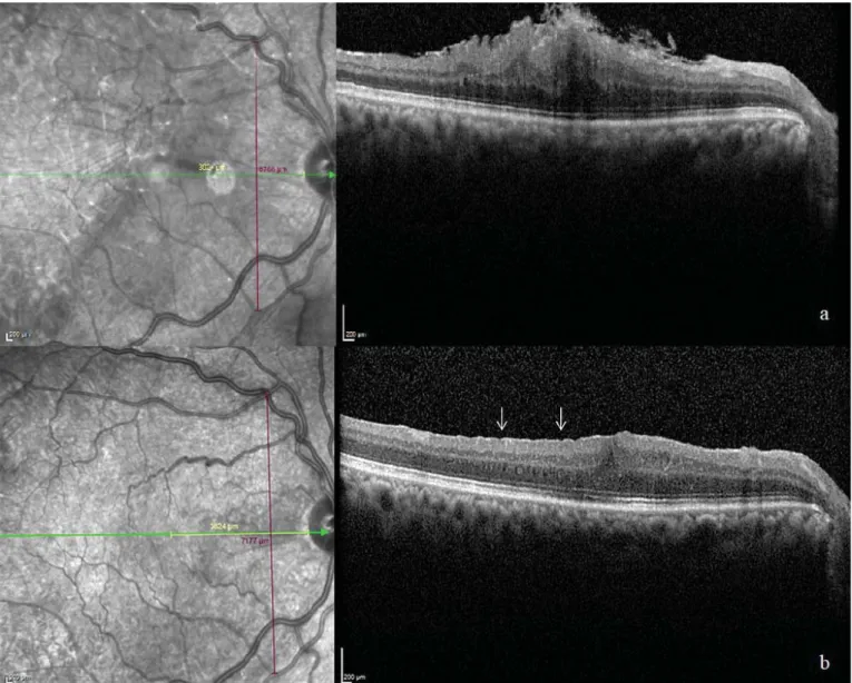
Figure 2. Right eye, idiopathic epiretinal membrane spontaneously detached as a flap: A) Color fundus photograph, B) B-scan spectral domain optical coherence tomography (SD-OCT)
Objectives: To investigate the agreement between optical coherence tomography (OCT) and OCT-based angiography in estimating retinal nerve fiber layer thickness (RNFLT) and evaluate
This case report documents for the first time treatment of sub-ILM hemorrhage in the premacular area with pneumatic tamponade in prone position leading to rapid and complete
21,22 The present study was planned with the belief that the lack of correlation between reduced macular thickness and functional gains is due to the heterogeneous macular
Optic Nerve Avulsion and Retinal Detachment After Penetrating Ocular Trauma: Case Report..
