Definitive Brachytherapy in Inoperable Endometrial Cancer
Received: January 08, 2019 Accepted: February 19, 2019 Online: April 05, 2019 Accessible online at: www.onkder.org
Cem ÖNAL
Department of Radiation Oncology, Başkent University, Faculty of Medicine, Adana-Turkey
SUMMARY
In elderly patients with medical comorbidities that increase perioperative risks, surgery cannot be per-formed, and definitive radiotherapy (RT) is a therapeutic option. Although staging for endometrial cancer is based on surgical findings, clinical and radiological imaging has not been standardized yet. For a radiological evaluation of inoperable endometrial cancer, magnetic resonance imaging (MRI) is preferred for local tumor infiltration, and positron emission tomography is accepted as the most ac-curate modality for assessing lymph node and distant metastases. External RT alone is inadequate, and intracavitary brachytherapy with or without external RT is recommended depending on the disease stage in patients with inoperable endometrial cancer. However, there is currently no standard treatment approach for patients with inoperable endometrial cancer. For patients with inoperable endometrial cancer treated with definitive RT, there are some problems including the RT technique, treatment vol-umes, and systemic treatment. Based on clinical practice, image-based brachytherapy using computed tomography and magnetic resonance imaging is highly recommended.
Keywords: Brachytherapy; endometrial cancer; inoperable; radiotherapy. Copyright © 2019, Turkish Society for Radiation Oncology
Introduction
Endometrial cancer is the most common gynecologic cancer worldwide, with an increasing incidence and mortality since the year 2000, which is mainly due to rising obesity rates.[1] The risk of endometrial can-cer rises by 50% for every 5-unit increase in the body mass index.[2] The fat tissue allows for increased conversion of androgen to estrogen, which functions as a pro-proliferative agent.[3] Morbid obesity not only increases the risk of developing endometrial cancer, but it also complicates treatment. In addition, obese women are also predisposed to other diseases, such as hypertension or diabetes. Another risk factor is an advanced age, with a mean age at diagnosis of 60 years.[4]
The standard initial treatment strategy for en-dometrial cancer is total hysterectomy and bilateral
salpingo-oophorectomy, together with pelvic and/ or paraaortic lymph node dissection.[5] However, in some instances, for example, in elderly patients with medical comorbidities that increase periopera-tive risks, surgery may not be performed. Definiperiopera-tive radiotherapy (RT) is therapeutic option for patients that are medically inoperable or patients who refuse radical surgery. In addition, systemic chemotherapy or hormonotherapy may be another treatment op-tion. However, there is currently no standard treat-ment approach for patients with inoperable endome-trial cancer.
For patients with inoperable endometrial cancer treated with definitive RT, there are some problems including the RT technique, treatment volumes, and systemic treatment. This review encompasses the prob-lematic issues in definitive RT for patients with inoper-able endometrial cancer.
Dr. Cem ÖNAL
Başkent Üniversitesi Tıp Fakültesi, Radyasyon Onkolojisi,
Anabilim Dalı, Adana-Turkey
(USG). In addition, FDG-PET/CT is accepted as the most accurate modality for assessing lymph node and distant metastases.[13]
Brachytherapy Application
Before brachytherapy application, a detailed gyneco-logical examination is required in the lithotomy po-sition. Pain control and sedation or anesthesia during the applicator insertion are essential for patient com-fort both during and after the procedure. For patients treated with high-dose-rate (HDR), low-dose-rate [LDR], or pulsed-dose-rate techniques, there are a vari-ety of applicators available for vaginal cuff brachyther-apy. These can include a single tandem with ovoids or a vaginal cylinder. Dual and triple tandem applicators with or without a vaginal cylinder are also available. It is important for patients with endometrial cancer that the tandem extends to the uterine fundus to ensure that the entire endometrial lining is treated. Modified Heyman capsules used with remote afterloading, and plastic catheters can also be used within the uterus in an umbrella configuration. The available tandem and ring applicators have fixed-length tandems that may not extend to the tip of the uterine fundus. Having a variable tandem length gives the operator the ability to place the tandem to the tip of the fundus.
Brachytherapy Planning
After the applicator insertion, the brachytherapy plan-ning is performed for delivering the prescribed doses to the target volume. However, there is still no consen-sus in treatment planning generated by point-based planning or target volume definition.
Recently, the American Brachytherapy Society (ABS) guidelines for HDR brachytherapy treatment of endometrial cancer defined the target volume for inop-erable endometrial cancer as the entire uterus, cervix, and upper 3–5 cm of the vagina.[14] In the ABS guide-lines, CT, MRI, or US to determine the uterine wall thickness is recommended. In another consensus state-ment, MRI is recommended; however, CT may be used if MRI is unavailable for volume-based brachytherapy planning.
The target volumes and organs at risk (OAR) defi-nitions are summarized in Table 2. If MR is available, the tumor within the endometrium is contoured as a gross tumor volume (GTV), which is defined as a vis-ible lesion on T2-weighted MRI. Although specific contouring guidelines do not exist, the CTV should in-clude the entire uterus, cervix, and upper 1–2 cm of the vagina. For OAR, the bladder, rectum, sigmoid, bowel, and vagina that are not included in the CTV should be contoured.
Staging
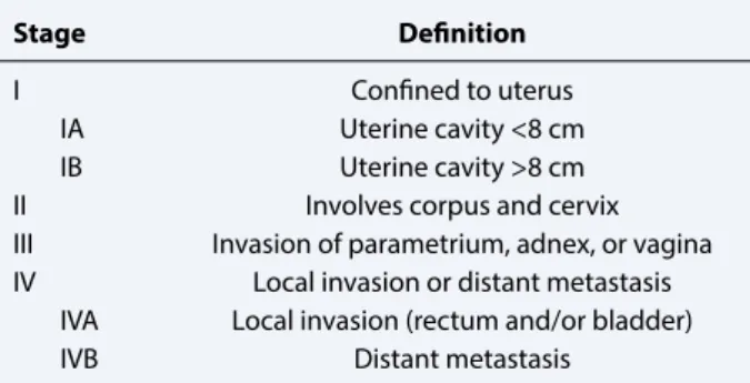
The tumor stage is one of the most important prognos-tic factors in most of the cancer types, and also in en-dometrial cancer. The staging for enen-dometrial cancer is surgical according to the International Federation of Gynecology and Obstetrics. However, the clinical stag-ing system can be used for inoperable patients (Table 1).[6] Although the clinical staging system relies on the results of a complete pelvic examination to determine the disease extent, radiological and functional imaging modalities can indicate the local extension of tumor, regional and or distant lymph node metastasis, and dis-tant organ metastasis.
For distant metastasis evaluation, computed to-mography [CT] can be used. However, the sensitiv-ity of CT for assessing distant metastasis is not high. Magnetic resonance imaging (MRI) can be used to as-sess local invasion, especially for evaluating the depth of myometrial invasion.[7] In a pooled analysis of prospective MRI studies, the negative predictive values for identifying deep myometrial involvement was 85%. [8] Thus, MRI may be best used to rule out deep my-ometrial invasion in medically inoperable patients with organ-confined disease. The deep myometrial invasion demonstrated in MRI can be combined with tumor grade on biopsy and used to estimate probability of lymph node involvement.[9] Besides a local invasion, regional lymph node evaluation is another important task in the workup of medically inoperable endome-trial cancer patients. Recent studies demonstrated that CT is less accurate than 18F-flouro-deoxyglucoseep-ositron emission tomography (FDG-PET/CT) or MRI for identifying nodal metastasis.[10,11] In a meta-analysis, the reported sensitivity and specificity rates for FDG-PET/CT in detection of pelvic and paraaor-tic lymph node metastasis in endometrial cancer were 63% and 95%, respectively.[12] In conclusion, for ra-diological evaluation for inoperable endometrial can-cer, MRI was preferred over CT and ultrasonography
Table 1 Clinical staging for patients with inoperable endometrial cancer
Stage Definition
I Confined to uterus
IA Uterine cavity <8 cm IB Uterine cavity >8 cm II Involves corpus and cervix III Invasion of parametrium, adnex, or vagina IV Local invasion or distant metastasis
IVA Local invasion (rectum and/or bladder) IVB Distant metastasis
In cases where image-based brachytherapy ning is not available, point-based brachytherapy plan-ning may be used. However, point-based planplan-ning has several limitations: the inability to define target volume doses and not being able to measure the OARs doses appropriately.
Historically, film-based planning for the LDR brachytherapy treatment of endometrial cancer re-quired the use of standard loadings similar to cervical cancer, using approximately 5 mg RaEq per centimeter of uterine tandem with up to 10 mg RaEq per centime-ter in the upper 2 cm of the tandem to increase dose to the endometrial cavity. The doses of bladder and rectum could be evaluated according to the Interna-tional Commission on Radiation Units and Measure-ments (ICRU) 38 definitions. In 2000, the ABS guide-lines recommended defining a dose specification point at 2 cm from the central axis at the midpoint along the Y-shaped uterine applicator, where the isodose is widened at the uterine fundus and was optimized to a 0.5 cm depth within the vaginal mucosa (the mucosa being defined as the surface of a vaginal cylinder).[14] Similarly, the bladder and rectal dose reference points were defined as specified in ICRU 38.
Previously, the Madison system was used to de-fine certain points for inoperable endometrial cancer brachytherapy planning (Fig. 1). The Madison system uses a central reference point M that is defined accord-ing to the applicators and the patients’ anatomy. Point
M lies 2 cm laterally to the center of the uterine canal and 2 cm cephalad from a line joining the center dwell position of the vaginal sources. The Point S is defined at about two-thirds thickness of the fundus, superior to the tip of tandem, and the Points W are located at 2 cm inferior to the tip of the tandem and two-thirds of the thickness of the uterine wall, laterally to the tandem. The lateral pelvic wall point E is a point near the pelvic wall at the intersection of a horizontal line tangential to the cephalad-most point of the acetabula and a line per-pendicular to that passing through the medial aspect of the acetabula. The vaginal surface point is identified on the surface adjacent to the center source dwell position. In the Madison system, dose is specified in four regions around a vaginal cylinder paired with either one or two tandems, where in the latter, the tandems are pointed away from each other, and the tips fall into the corn.
Outcomes
There is no consensus in treatment modalities for pa-tients with operable endometrial cancer. To the best of our knowledge, there are no prospective randomized studies; however, most studies evaluating the efficacy of image-based brachytherapy for patients with inop-erable endometrial cancer are retrospectively designed (Table 3).[15-19]
In a dosimetric study comparing point-based and volume-based treatment plans with single, dual, and triple tandem applicators, it was demonstrated that op-timal coverage was dependent on the type of applicator and the anatomy.[20] After insertion, the applicator can be secured in a stable position with a perineal bar,
Table 3 Published studies for image-based brachthera-py for patients with inoperable endometrial cancer
Study (year) n Stage D90 GTV D90CTV Outcomes
Weitmann 16 I-III 129.6 40.8 4y LC %100 Okhubo 10 I-II 88 45.9 5y LC %100 Gill 38 I 160 59.9 2y LC %91 Archaya 43 I-III 34.7 2y LC %81 Jordan 15 I 96 51.6 4y LC %93.4 Table 2 Recommended target volumes and organs at risk used during image-based brachytherapy
Volume Imaging modality Definition
GTV T2W-MRI Visible lesion
CTV MRI/CT Entire uterus, cervix, upper half of vagina
OAR MRI/CT Sigmoid, rectum, bladder, bowel, lower half of vagina
Abbreviations: GTV, gross tumor volumes; CTV, clinical target volume; OAR, organ at risk; MRI, magnetic resonance imaging; CT, computed tomography
Fig. 1. Points defined in Madison system for
brachyther-apy planning. Pt S Pt W Pt M 2 cm 2 cm 2 cm Vaginal surface Small Uterus x x Fundus Superior myometrium Paracervical region Pt W Pt M Pt S 2 cm 2 cm 2 cm Vaginal surface Large Uterus x x x
external base plate and clamp, sutures, or an alternative fixation device. Additionally, X-ray, CT, or MRI should be taken for treatment planning of each insertion. With CT and MR, a CTV can be defined to help guide dose specification. CT imaging taken for every application may be performed to ensure that there is no applicator movement requiring repositioning or revised planning. Nguyen et al.[21] found an increase in acute and late complication rates in 36 patients with Stage I en-dometrial cancer treated with HDR brachytherapy alone for a total dose of 35 Gy to Point W (serosal point). In a pilot study including 16 patients with inop-erable endometrial cancer using image-based (CT or MR) planning and optimization for HDR brachyther-apy for endometrial cancer, 12 patients had complete remission without severe acute or late side.[17]
After a median follow-up of 47 months, 5 patients were alive without evidence of disease, and 7 patients died with disease after a mean duration of 21 months. In 38 medically inoperable endometrial cancer pa-tients treated with image-guided brachytherapy, Gill et al.[15] reported a 2-year local control rate of 90.6% and an overall survival rate of 94.4%, after a median follow-up of 15 months. In this study, 20 patients were treated with brachytherapy alone with a median dose of 37.5 Gy delivered in five or six fractions, and 18 pa-tients were treated with a combination of 45 Gy exter-nal beam RT and 25 Gy brachytherapy delivered in four to five fractions. Only one patient developed bleeding after the applicator insertion requiring blood transfu-sion, and no other Grade 3 or greater acute or late toxic-ities were reported. Acharya et al.[19] reported a 2-year pelvic and distant recurrence rates of 8.3% and 13.5%, in 43 patients with inoperable endometrial cancer treated with brachytherapy alone (28 patients) and combination of brachytherapy and external RT (15 patients). No se-vere toxicity was reported in this study. In another study, 10 medically inoperable patients with T1–2 endome-trial carcinoma were treated with radiotherapy alone. [18] The authors concluded that Stage I–II endometrial cancer was well-controlled locally with minimum late toxicity by HDR brachytherapy alone. Jordan et al.[16] showed only one recurrence in 15 patients treated with BRT, with no severe acute and late toxicities.
Although these retrospective studies with a limited patient number reported excellent local control rates without high toxicity rates, there is no published guide-line for treatment of patients with inoperable endome-trial cancer.
Treatment Recommendations
Recently, in a “Systematic Review and National Cancer Database” analysis that included a total of 1261
pa-tients with inoperable endometrial cancer, papa-tients treated with external RT only (405 patients, 32%), brachytherapy only (152 patients, 12%), and combina-tion of external RT and brachytherapy (215 patients, 17%) demonstrated that an advanced age, clinical T2 tumor, and the absence of brachytherapy were negative prognosticators for overall survival.[22] There was no significant difference in overall survival between pa-tients treated with external RT and papa-tients not receiv-ing any treatment. This study implies that the external RT alone is not a sufficient treatment approach.
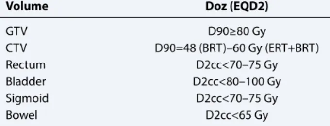
In 2015, a consensus statement for brachytherapy for the treatment of medically inoperable endometrial cancer was published.[23] According to consensus panel, patients with Stage I endometrial cancer should receive an EQD2 of at least 48 Gy for brachytherapy alone and at least 65 Gy for the combination of exter-nal beam plus brachytherapy to 90% of the (D90) CTV volume encompassing the whole uterus, depending on tumor specific and patient-specific factors (Table 4). Although data for maximum tolerated doses to the OAR specifically in medically inoperable endometrial cancer are lacking, the panel encourages to limit doses to the OAR as low as possible. The best available data recommend that the D2cc applied to the sigmoid and rectum be limited to 70–75 Gy, and D2cc applied to the bladder of 80–100 Gy.[24] Additionally, a dose limit of 65 Gy to the D2cc to the bowel may be considered.
For clinical Stage I, Grade I–II inoperable endome-trial cancer patients, brachytherapy alone can be used. The GTV includes a visible lesion in T2-weighted MRI, and CTV encompasses entire uterus. The EQD2 for GTV and CTV should be 80–90 Gy and 48–62.5 Gy, respectively. Since Stage I, Grade I–II patients can be treated with vaginal brachytherapy alone, the OARs doses should be kept within recommended dose limits. The recommended dose and fractionations for Stage I, Grade I–II inoperable endometrial cancer patients are summarized in Table 5.
For clinical Stage I disease with deep myometrial invasion, a combination of external RT and
brachyther-Table 4 Recommended target volume and organs-at-risk doses
Volume Doz (EQD2)
GTV D90≥80 Gy CTV D90=48 (BRT)–60 Gy (ERT+BRT) Rectum D2cc<70–75 Gy Bladder D2cc<80–100 Gy Sigmoid D2cc<70–75 Gy Bowel D2cc<65 Gy
apy is recommended. The GTV includes visible lesion in T2-weighted MRI, and CTV encompasses entire uterus and the upper 1–2 cm of the vagina. The goal is to deliver EQD2 of 80–90 Gy to the GTV and 65–75 Gy to the CTV. The acceptable dose and fractionation schemes for this patient group based on the published literature and the clinical experience are summarized in Table 6.
In patients with Stage II disease (cervical involve-ment), a combination of external RT and brachyther-apy is recommended. During external RT, entire uterus, cervix, and pelvic lymphatics should be treated. The GTV will encompass any gross residual disease, the endometrial lining, and the cervix, and CTV will encompass the whole uterus, cervix, and the upper 1–2 cm of the vagina. The goal will be to deliver an EQD2
of 80–90 Gy to the GTV, and 70–75 Gy to the CTV. The recommended dose and fractionations for clinical Stage II disease are summarized in Table 7.
Conclusion
Although the incidence of inoperable endometrial can-cer is ~10%, the probability will increase with advanced age, obesity, and multiple comorbidities. There are no guidelines for the treatment of patients with inoperable endometrial cancer, and intracavitary brachytherapy is an essential component of the treatment. For patients with Stage I disease, vaginal brachytherapy alone is adequate. However, for Stage I disease with deep my-ometrial invasion or extensive stages, external RT with brachytherapy is highly recommended. To increase local control and improve treatment outcomes, image-based brachytherapy is essential. To demonstrate the efficacy of image-guided brachytherapy, studies with a larger patient population and a longer follow-up are required.
Peer-review: Externally peer-reviewed.
Conflict of Interest: There is no conflict of interest. Financial Support: There is no financial support. References
1. Siegel RL, Miller KD, Jemal A. Cancer statistics, 2018. CA Cancer J Clin 2018;68(1):7–30.
2. Aune D, Navarro Rosenblatt DA, Chan DS, Vingeliene S, Abar L, Vieira AR, et al. Anthropometric factors and endometrial cancer risk: a systematic review and dose-response meta-analysis of prospective studies. Ann Oncol 2015;26(8):1635–48.
3. Taghian A, Pernot M, Hoffstetter S, Luporsi E, Bey P. Radiation therapy alone for medically inoperable pa-tients with adenocarcinoma of the endometrium. Int J Radiat Oncol Biol Phys 1988;15(5):1135–40.
4. Jick H, Walker AM, Rothman KJ. The epidemic of en-dometrial cancer: a commentary. Am J Public Health 1980;70(3):264–7.
5. SGO Clinical Practice Endometrial Cancer Working Group, Burke WM, Orr J, Leitao M, Salom E, Gehrig P, Olawaiye AB, et al. Endometrial cancer: a review and current management strategies: part I. Gynecol Oncol 2014;134(2):385–92.
6. Mikuta JJ. International Federation of Gynecology and Obstetrics staging of endometrial cancer1988. Cancer 1993;71(4 Suppl):1460–3.
7. Beddy P, Moyle P, Kataoka M, Yamamoto AK, Joubert I, Lomas D, et al. Evaluation of depth of myometrial
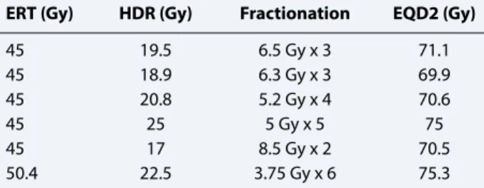
in-Table 6 Dose and fractionation schemes for inoperable endometrial cancer in clinical stage I disease with deep myometrial invasion
ERT (Gy) HDR (Gy) Fractionation EQD2 (Gy)
45 19.5 6.5 Gy x 3 71.1 45 18.9 6.3 Gy x 3 69.9 45 20.8 5.2 Gy x 4 70.6 45 25 5 Gy x 5 75 45 17 8.5 Gy x 2 70.5 50.4 12 6.0 Gy x 2 65.6 50.4 22.5 3.75 Gy x 6 75.3
Table 7 Dose and fractionation schemes for inoperable endometrial cancer in clinical stage II disease with cervical involvement
ERT (Gy) HDR (Gy) Fractionation EQD2 (Gy)
45 19.5 6.5 Gy x 3 71.1 45 18.9 6.3 Gy x 3 69.9 45 20.8 5.2 Gy x 4 70.6 45 25 5 Gy x 5 75 45 17 8.5 Gy x 2 70.5 50.4 22.5 3.75 Gy x 6 75.3
Table 5 Dose and fractionation schemes for inoperable clinical stage I, Grade I–II endometrial cancer
Total dose (Gy) Fractionation EQD2 (Gy)
36 6 Gy x 6 48
38.4 6.4 Gy x 6 52.5
36.5 7.3 Gy x 5 52.6
34 8.5 Gy x 4 52.4
vasion and overall staging in endometrial cancer: com-parison of diffusion-weighted and dynamic contrast-enhanced MR imaging. Radiology 2012;262(2):530–7. 8. Wu LM, Xu JR, Gu HY, Hua J, Haacke EM, Hu J.
Pre-dictive value of T2-weighted imaging and contrast-en-hanced MR imaging in assessing myometrial invasion in endometrial cancer: a pooled analysis of prospec-tive studies. Eur Radiol 2013;23(2):435–49.
9. Creasman WT, Morrow CP, Bundy BN, Homesley HD, Graham JE, Heller PB. Surgical pathologic spread pat-terns of endometrial cancer. A Gynecologic Oncology Group Study. Cancer 1987;60(8 Suppl):2035–41. 10. Crivellaro C, Signorelli M, Guerra L, De Ponti E,
Pirovano C, Fruscio R, et al. Tailoring systematic lymphadenectomy in high-risk clinical early stage endometrial cancer: the role of 18F-FDG PET/CT. Gynecol Oncol 2013;130(2):306–11.
11. Antonsen SL, Jensen LN, Loft A, Berthelsen AK, Costa J, Tabor A, et al. MRI, PET/CT and ultrasound in the preoperative staging of endometrial cancer - a multi-center prospective comparative study. Gynecol Oncol 2013;128(2):300–8.
12. Chang MC, Chen JH, Liang JA, Yang KT, Cheng KY, Kao CH. 18F-FDG PET or PET/CT for detection of metastatic lymph nodes in patients with endometrial cancer: a systematic review and meta-analysis. Eur J Radiol 2012;81(11):3511–7.
13. Lee JH, Dubinsky T, Andreotti RF, Cardenes HR, De-jesus Allison SO, Gaffney DK, et al. ACR appropriate-ness Criteria® pretreatment evaluation and follow-up of endometrial cancer of the uterus. Ultrasound Q 2011;27(2):139–45.
14. Nag S, Erickson B, Parikh S, Gupta N, Varia M, Glas-gow G. The American Brachytherapy Society recom-mendations for high-dose-rate brachytherapy for car-cinoma of the endometrium. Int J Radiat Oncol Biol Phys 2000;48(3):779–90.
15. Gill BS, Kim H, Houser C, Olsen A, Kelley J, Edwards RP, et al. Image-based three-dimensional conformal brachytherapy for medically inoperable endometrial carcinoma. Brachytherapy 2014;13(6):542–7.
16. Jordan SE, Micaily I, Hernandez E, Ferriss JS, Miyamoto CT, Li S, et al. Image-guided
high-dose-rate intracavitary brachytherapy in the treatment of medically inoperable early-stage endometrioid type endometrial adenocarcinoma. Brachytherapy 2017;16(6):1144–51.
17. Weitmann HD, Pötter R, Waldhäusl C, Nechvile E, Kirisits C, Knocke TH. Pilot study in the treatment of endometrial carcinoma with 3D image-based high-dose-rate brachytherapy using modified Hey-man packing: clinical experience and dose-volume histogram analysis. Int J Radiat Oncol Biol Phys 2005;62(2):468–78.
18. Ohkubo Y, Kato S, Kiyohara H, Tsuruoka I, Tamaki T, Noda SE, et al. Dose volume analysis of radiotherapy for inoperable patients with stage I-II endometrial car-cinoma. J Radiat Res 2011;52(5):666–73.
19. Acharya S, Esthappan J, Badiyan S, DeWees TA, Tan-derup K, Schwarz JK, et al. Medically inoperable en-dometrial cancer in patients with a high body mass index (BMI): Patterns of failure after 3-D image-based high dose rate (HDR) brachytherapy. Radiother Oncol 2016;118(1):167–72.
20. Johnson SB, Zhou J, Jolly S, Guo B, Young L, Priscian-daro JI. The dosimetric impact of single, dual, and triple tandem applicators in the treatment of intact uterine cancer. Brachytherapy 2014;13(3):268–74. 21. Nguyen TV, Petereit DG. High-dose-rate
brachyther-apy for medically inoperable stage I endometrial can-cer. Gynecol Oncol 1998;71(2):196–203.
22. Dutta SW, Trifiletti DM, Grover S, Boimel P, Showalter TN. Management of elderly patients with early-stage medically inoperable endometrial cancer: System-atic review and National Cancer Database analysis. Brachytherapy 2017;16(3):526–33.
23. Schwarz JK, Beriwal S, Esthappan J, Erickson B, Feltmate C, Fyles A, et al. Consensus statement for brachytherapy for the treatment of medically inoperable endometrial cancer. Brachytherapy 2015;14(5):587–99.
24. Georg P, Lang S, Dimopoulos JC, Dörr W, Sturdza AE, Berger D, et al. Dose-volume histogram parameters and late side effects in magnetic resonance image-guided adaptive cervical cancer brachytherapy. Int J Radiat Oncol Biol Phys 2011;79(2):356–62.