Original Article
Evaluation of Caco-2 cell permeability of ritonavir
nanosuspensions
Alptuğ Karaküçük
1, Naile Öztürk
2, Nevin Çelebi
1,31Gazi University, Faculty of Pharmacy, Department of Pharmaceutical Technology, Ankara, Turkey 2Inonu University, Faculty of Pharmacy, Department of Pharmaceutical Technology, Malatya, Turkey 3Baskent University, Faculty of Pharmacy, Department of Pharmaceutical Technology, Ankara, Turkey
ORCID IDs of the authors: A.K. 0000-0002-9061-2427; N.Ö. 0000-0002-7617-8433; N.Ç. 0000-0002-6402-5042
Cite this article as: Karakucuk, A., Ozturk, N., & Celebi, N. (2020). Evaluation of Caco-2 cell permeability of ritonavir nanosuspen-sions. Istanbul Journal of Pharmacy, 50(3), 251-255.
ABSTRACT
Background and Aims: Poor aqueous solubility limits drug absorption through intestinal mucosa. Nanosuspensions with nanometer range particle size provides enhanced aqueous solubility and hence permeability. The objective of this study was to investigate the cytotoxicity and in vitro cell permeability through human adenocarcinoma (Caco-2) cells of ritonavir (RTV) nanosuspensions.
Methods: The Microfluidization method was used to prepare nanosuspensions. Particle size (PS), polydispersity index (PI) and zeta potential (ZP) values were measured as characterization. MTT test was applied to evaluate the cytotoxic effect. Caco-2 cell lines were used for transport studies with RTV coarse powder, physical mixtures and nanosuspension.
Results: Approximately 600 nm PS, 0.4 PDI and 22 mV ZP values were observed for nanosuspensions. The sample groups showed no cytotoxicity on the cell lines in any RTV concentration. However, significant cytotoxic effect was determined in groups with high amounts of sodium dodecyl sulfate. The transported RTV in nanosuspension formulation enhanced by 5.3-fold and 1.5-5.3-fold in comparison with RTV coarse powder and physical mixture, respectively. Rate of the transportation and also the amount of the transported RTV were improved with nanosuspension formulation.
Conclusion: Particle size reduction of RTV into nanometer size and preparing nanosuspension system was found effective to obtain enhanced cell permeability.
Keywords: Ritonavir, nanosuspension, Caco-2, permeability
Address for Correspondence:
Nevin ÇELEBI, e-mail: [email protected]
This work is licensed under a Creative Commons Attribution 4.0 International License.
Submitted: 13.04.2020 Revision Requested: 03.06.2020 Last Revision Received: 07.06.2020 Accepted: 07.07.2020
INTRODUCTION
The solubility in intestinal fluids and permeability from intes-tinal membrane effect the drug absorption efficiency. Poor drug solubility limits dissolution and hence lower concentra-tion gradient occurs across the intestinal mucosa (Lenhardt, Vergnault, Grenier, Scherer, & Langguth, 2008). Nanosuspen-sions are colloidal systems with a particle size below 1 µm. The increased surface area of particles by reducing their particle size under nano meter range improves the saturation solubility and dissolution velocity (Xie, Luo, Liu, Ma, Yue, & Yang, 2019). Hence, the poorly soluble drug may have sufficient therapeu-tic effects with reduced side effects and better transportation to cells of the drug molecules Karakucuk, Alptug, Teksin,
Ero-glu, & Celebi, 2019). The conventional precipitation method or top-down methods are used to prepare nanosuspensions. More preferred approaches are the top-down methods such as media milling or high pressure homogenization, which downsize the particles by high energy creating cavitation and shear forces (Tashan, Karakucuk, & Celebi, 2019). Nanosuspen-sions are thermodynamically unstable system and stabilization process by using surfactant or polymer is mandatory. Steric or electrostatic stabilization can be achieved in the system with surfactant and/or polymers.
Cell and tissue culture studies are used to investigate the trans-port of drug from delivery system into specific cells and across specific barriers. Caco-2 cell lines demonstrate the
morpho-logical and biochemical features of adult differentiated entero-cytes and goblet cells, which bring benefit to study intestinal epithelial differentiation and function (Wilson, 1990). The well-established Caco-2 cell monolayer system allows direct mea-surement of the drug fluxes across an epithelium (Lenhardt et al., 2008). They also, indicate P-gp and metabolizing enzymes to predict the absorption of orally administrated compounds ( Matsumoto, Kaifuchi, Mizuhara, Warabi, & Watanabe, 2018). The in vitro (Karakucuk, Celebi, & Teksin, 2016) and in vivo stud-ies (Karakucuk et al., 2019) were evaluated in previous studstud-ies. Therefore, this study mainly focused on the investigation of cy-totoxicity and Caco-2 cell permeability of RTV nanosuspensions. Possible P-gp effect of the formulation was also examined.
MATERIALS AND METHODS Materials
Ritonavir (RTV) was kindly gifted by Mylan Pharmaceuticals (India) and hydroxypropyl methyl cellulose (HPMC) (3 cps) was donated by Colorcon Limited (Istanbul, Turkey). Sodium dodecyl sulfate (SDS) was purchased from Merck (Darmstadt, Germany). Human epithelial colorectal adenocarcinoma cell (Caco-2, ATCC HTB 37) was obtained from ATCC. Methyl-thiazo-lyl tetrazolium (MTT), Hank’s balanced salt solution (HBSS), and Dulbecco’s modified eagle medium (DMEM) were purchased from Sigma-Aldrich (St. Louis, Missouri, USA). Other chemicals were HPLC or analytical grade.
Preparation of ritonavir nanosuspensions
RTV nanosuspensions were prepared according to previous study (Karakucuk et al., 2016). Briefly, the stabilizer solution, which was consisted of 4% HPMC 3 cps and 0.8% SDS was prepared. 2% w/w of RTV was dispersed this solution and pre-homogenized for 10 minutes at 10 000 rpm using ultraturrax. The pre-treatment step allowed to reduce particle size below 84 µm in regard to d99. It is because 84 µm Z-type chamber was used to perform interaction of particles on microfluidiza-tion. Then, the coarse suspension was processed by jet stream homogenizer (Microfluidizer® Processors, Westwood, Massa-chusetts, USA) under high pressure (30 000 psi). After 20 cycles of homogenization nanosuspensions were obtained. The pro-cess continued with lyophilization, and nanosuspensions were freeze-dried to fill in the capsules as dosage form and also im-prove physical stability. For the lyophilization process approxi-mately 2 mL of the nanosuspensions were frozen at−80 °C for 2 h. Freeze-dry process was performed at −50 °C, 0.021 mbar for 40 h using Christ Alpha 1-2 LD Freeze Dryer.
Cell culture studies
The Caco-2 cells were used to investigate permeability of coarse RTV and compare permeability values with nanosuspensions. The Caco-2 cells were cultured using DMEM with L-glutamine, supplemented with fetal bovine serum (10%, v/v) and penicil-lin/ streptomycin (50 IU/mL and 50 µg/mL, respectively) in a humidified incubator at 37ºC in air containing 5% CO2.
In vitro cytotoxicity
Before performing permeability studies, the effect of the for-mulations on cell viability of Caco-2 cells was investigated by
MTT assay. Cells were seeded at a density of 5x10³ cells per well
to 96-well plate and incubated overnight and then the drug solution and formulations (RTV coarse powder, physical mix-tures, stabilizer mixtures and nanosuspension) were added to the wells at different concentrations and incubated for 4 hours. DMSO was used to solubilize the drug; therefore, DMSO solu-tion (0.25-4 %) was also applied as control. After incubasolu-tion pe-riod, 25 µL MTT solution was added to wells and incubated for 4 hours. Then the medium was removed and 200 µL of DMSO was added to wells to solubilize the formazan crystals and op-tical density of wells was measured at 570 nm.
In vitro permeability studies
For permeability studies, cell monolayers were prepared by seeding harvested cells onto inserts (12 mm Snapwell Insert with 0.4 μm pore) at a density of 7x10⁴ cells per well. After 21
days of culturing (changing the medium every other day), TEER values were measured to determine monolayer integrity. Apical to basolateral transport studies were conducted after the ap-plication of formulations in Hank’s Balanced Salt Solution (HBSS pH 7.4) to the donor compartment (0.5 mL). RTV bulk powder, nanosuspension, and physical mixtures of stabilizers with or without RTV were used as samples (RTV concentration was 50 mg/mL in all RTV containing samples, n=6). At predetermined time intervals (30, 60, 90,120 and 240 mins), 0.5 mL sample was withdrawn from basolateral compartment and then fresh me-dium (HBSS) was added to the wells. Samples were isolated from the receiver compartment (1.5 mL) 4 hours after the in-cubation. The apparent permeability coefficients (Papp,cm/sn)
were calculated according to equation below (Eq. 1):
. . (Eq. 1)
Where dQ/dt (µmol/L.min) is the cumulative amount of RTV which has been transported over the membrane, A (1.12 cm2)
is the surface area of the inserts and C0 (µmol/L) is the initial
concentration of the RTV on the donor site.
The HPLC method was used to analyze of the samples at 239 nm, which was validated according to accuracy, precision, re-peatability, specificity, detection limit, quantitation limit, linear-ity, and range. The HPLC instrument was HP 1050 series and the C18 RP column (250 mm x 4.6 mm, 5 µm) was equipped as an analytical column. The mobile phase was consisted of ace-tonitrile: 0.05 M phosphoric acid (55:45 v/v). Injection volume was 25 µL and flow rate was 1 mL/min. Retention time of RTV was 11-12 minutes.
Statistical analysis
Statistical evaluation was performed with one-way ANOVA fol-lowed Tukey HSD Post hoc test at the significance level of 0.05. Mean ± standard deviation was provided for all data.
RESULTS AND DISCUSSION
Preparation of ritonavir nanosuspensions
The main process factors for the microfluidization method that effect quality of final nanosuspensions are the chamber type and size, temperature, homogenization cycle, and pressure
(Salazar, Heinzerling, Müller, & Möschwitzer, 2011). Additionally, formulation parameters such as physicochemical properties of the drug, amount of the drug, type and amount of the stabi-lizers should be considered and optimized (Van Eerdenbrugh, Van den Mooter, & Augustijns, 2008). In this study, 84 µm Z-type chamber was used to obtain nanosuspensions, which is the appropriate size and type for microfluidization. Tem-perature was controlled during the study. 20 homogenization cycles were applied. The well-known stabilizers, which were HPMC and SDS were used to stabilize the nanosuspensions. 2% of RTV was added in the formulations considering the final oral dose for capsule dosage form. The nanosuspensions were obtained approximately 600 nm PS, 0.4 PDI and -22 mV ZP values. The final RTV nanosuspensions were found physically stable for 30 days of storage time at room temperature. Also, physicochemical properties of RTV were preserved according to differential calorimetry and X-ray diffraction results which is shown in previous study (Karakucuk et al., 2016).
Caco-2 cell viability and permeability
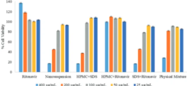
Monolayer integrity of Caco-2 cell was shown with TEER values of 500-1000 ohm.cm2. MTT assay was conducted to
under-stand the in vitro toxicity of the RTV in different formulations. Cell viability (%) was observed after the interaction between Caco-2 cells and RTV formulations for 4 hours. Cytotoxicity was not observed by using RTV coarse powder even in the highest dose which was 400 µg. The cytotoxicity was resulted from the formulations with SDS either in the existence of RTV or not. In the findings from different concentrations, the increased use of SDS caused toxicity on Caco-2 cells. Nanosuspension and the other formulations had no cytotoxic effect at the 50 µg dose used in the permeability study (Figure 1). Cell viability re-sults were above 100% for some treatment groups, especially for coarse RTV powder. MTT is converted to formazan by viable cells with active metabolism so MTT reduction indicates viable cell metabolism rather than cell proliferation (Riss et al., 2016). Thus our results suggest that increasing RTV concentration, in-creases cellular metabolic activity of Caco-2 cells.
The Permeability value of coarse RTV and physical mixture were determined 1.57x10⁻⁵ cm/s and 5.75x10⁻⁵ cm/s, respectively
(Figure 2). The permeability value of coarse RTV was found similar to the literature which is at about 2x10⁻⁵ cm/s (Alsenz,
Steffen, & Alex, 1998; Holmstock, Annaert, & Augustijns, 2012). As the permeability value of RTV was found above 1x10⁻⁵ cm/s,
it was concluded that the RTV has high permeability property which means RTV belongs to BCS Class II drug with this
per-meability value. The literature data have also proven this result (Chowdary, Annamma Devi, & Dhanalakshmi, 2012; Sinha, Ali, Baboota, Ahuja, Kumar, & Ali, 2010).
The permeability value of nanosuspension was increased and was found 8.38x10⁻⁵ cm/s (Figure 2). The permeability value of
nanosuspension was higher than the other physical mixtures (p<0.05). The increment of the permeability value of nanosus-pension can be resulted from decreasing particle size under micrometer scale. Beside the nanometer particle size values, nanosuspension was prepared by SDS and it is known that SDS can inhibit P-glycoprotein (P-gp) which can result in permeabil-ity enhancement (Koga, Ohyashiki, Murakami, & Kawashima, 2000; Miller, Batrakova, & Kabanov, 1999; Rege, Yu Lawrence, Hussain, & Polli, 2001). It is also known that RTV is p-gp substrate by itself Schmitt, Kaeser, Riek, Bech, & Kreuzer, 2010) and stud-ies showed that RTV induced protein expression of p-gp and increased cellular drug exclusion ( Perloff, Von Moltke, March-and, & Greenblatt, 2001). Using RTV as drug efflux transporter is a strategy to improve intracellular concentration of drug mol-ecules (Janneh, Jones, Chandler, Owen, & Khoo, 2007).
Cumulative transported RTV amount to the basolateral mem-brane is given in Figure 3. No further drug transportation was observed after 120 minutes by nanosuspension exposure. Even the nanosuspension showed faster drug transport, the cumulative drug transported at the end of the assay was simi-lar among nanosuspension, physical mixture, and RTV with SDS formulations. SDS was provided P-gp inhibition which was resulted in an increase in RTV amount in basolateral
Figure 1. Cell viabilities (%) for the sample groups (n=6).
Figure 2. Permeability values of different groups (n=6).
membrane. However, RTV coarse powder showed slower and lower drug transport to the basolateral membrane even in comparison with RTV and HPMC mixture. As a polymer, HPMC possessing p-gp inhibiting activity (Rehman et al., 2017), this could also explain that HPMC might be caused P-gp inhibition. It is know that inhibition or induction of functionality of P-gp can affect the pharmacokinetics, efficacy, safety, or tissue levels (Matsumoto et al., 2018).
CONCLUSION
The microfluidization method was found an effective method to develop nanosuspension formulation regarding to fewer pro-cess parameters and easy for scale-up. Neither nanometer range RSV nor using stabilizer combination did not show cytotoxic effect on Caco-2 cells at desired concentration. The nanosus-pensions with particle size below 1000 nm showed enhanced permeability through Caco-2 cells due to increased saturation solubility. It could also be said that stabilizers showed P-gp ef-fect, which resulted in improved cumulative drug transport. It is concluded that beside nanosuspensions being beneficial to im-prove saturation solubility, dissolution and bioavailability, they also increase the cell permeability of poorly soluble drugs.
Peer-review: Externally peer-reviewed.
Informed Consent: Written consent was obtained from the partici-pants.
Author Contributions: Conception/Design of Study- A.K., N.Ö., N.Ç.; Data Acquisition- A.K., N.Ö., N.Ç.; Data Analysis/Interpretation- A.K., N.Ö., N.Ç.; Drafting Manuscript- A.K., N.Ö., N.Ç.; Critical Revision of Manuscript- A.K., N.Ö., N.Ç.; Final Approval and Accountability- A.K., N.Ö., N.Ç.; Techni-cal or Material Support- A.K., N.Ö., N.Ç.; Supervision- A.K., N.Ö., N.Ç. Conflict of Interest: The authors have no conflict of interest to declare. Financial Disclosure: This study was supported by a grant from The Scientific and Technological Research Council of Turkey. (Project No: 113S842, TUBITAK)
REFERENCES
• Alsenz, J., Steffen, H., & Alex, R. (1998). Active apical secretory efflux of the HIV protease inhibitors saquinavir and ritonavir in Caco-2 cell monolayers. Pharmaceutical Research, 15(3), 423–428. https://doi.org/10.1023/A:1011924314899
• Chowdary, K. P. R., Annamma Devi, D. G., & Dhanalakshmi, K. (2012). A factorial study on enhancement of solubility and dis-solution rate of ibuprofen by hydroxy propyl β cyclodextrin and solutol hs15. International Journal of Pharmaceutical Sciences Re-view and Research, 2(4), 1–7.
• Holmstock, N., Annaert, P., & Augustijns, P. (2012). Boosting of HIV protease inhibitors by ritonavir in the intestine: The relative role of cytochrome P450 and P-glycoprotein inhibition based on Caco-2 monolayers versus in situ intestinal perfusion in mice. Drug Metabolism and Disposition, 40(8), 1473–1477. https://doi. org/10.1124/dmd.112.044677
• Janneh, O., Jones, E., Chandler, B., Owen, A., & Khoo, S. H. (2007). Inhibition of P-glycoprotein and multidrug resistance-associated proteins modulates the intracellular concentration of lopinavir in cultured CD4 T cells and primary human lymphocytes. Jour-nal of Antimicrobial Chemotherapy, 60(5), 987–993. https://doi. org/10.1093/jac/dkm353
• Karakucuk, A., Celebi, N., & Teksin, Z. S. (2016). Preparation of ritonavir nanosuspensions by microfluidization using polymeric stabilizers: I. A Design of Experiment approach. European Journal of Pharmaceutical Sciences, 95, 111–121. https://doi.org/10.1016/j. ejps.2016.05.010
• Karakucuk, Alptug, Teksin, Z. S., Eroglu, H., & Celebi, N. (2019). Eval-uation of improved oral bioavailability of ritonavir nanosuspen-sion. European Journal of Pharmaceutical Sciences, 131(February), 153–158. https://doi.org/10.1016/j.ejps.2019.02.028
• Koga, K., Ohyashiki, T., Murakami, M., & Kawashima, S. (2000). Mod-ification of ceftibuten transport by the addition of non-ionic sur-factants. European Journal of Pharmaceutics and Biopharmaceu-tics, 49(1), 17–25. https://doi.org/10.1016/S0939-6411(99)00059-4 • Lenhardt, T., Vergnault, G., Grenier, P., Scherer, D., & Langguth, P.
(2008). Evaluation of nanosuspensions for absorption enhance-ment of poorly soluble drugs: In vitro transport studies across intestinal epithelial monolayers. AAPS Journal, 10(3), 435–438. https://doi.org/10.1208/s12248-008-9050-7
• Matsumoto, T., Kaifuchi, N., Mizuhara, Y., Warabi, E., & Watanabe, J. (2018). Use of a Caco-2 permeability assay to evaluate the effects of several Kampo medicines on the drug transporter P-glyco-protein. Journal of Natural Medicines, 72(4), 897–904. https://doi. org/10.1007/s11418-018-1222-x
• Miller, D. W., Batrakova, E. V, & Kabanov, A. V. (1999). Inhibition of multidrug resistance-associated protein (MRP) functional activity with pluronic block copolymers. Pharmaceutical Research, 16(3), 396–401. https://doi.org/10.1023/A:1018873702411
• Perloff, M. D., Von Moltke, L. L., Marchand, J. E., & Greenblatt, D. J. (2001). Ritonavir induces P-glycoprotein expression, multi-drug resistance-associated protein (MRP1) expression, and multi-drug transporter-mediated activity in a human intestinal cell line. Jour-nal of Pharmaceutical Sciences, 90(11), 1829–1837. https://doi. org/10.1002/jps.1133
• Rege, B. D., Yu Lawrence, X., Hussain, A. S., & Polli, J. E. (2001). Effect of common excipients on Caco-2 transport of low-permeability drugs. Journal of Pharmaceutical Sciences, 90(11), 1776–1786. https://doi.org/10.1002/jps.1127
• Rehman, S., Nabi, B., Fazil, M., Khan, S., Bari, N. K., Singh, R., Ahmad, S., Kumar, V., Baboota, S., & Ali, J. (2017). Role of P-Glycoprotein In-hibitors in the Bioavailability Enhancement of Solid Dispersion of Darunavir. BioMed Research International, 2017, 1–17. https://doi. org/10.1155/2017/8274927
• Riss, T. L., Moravec, R. A., Niles, A. L., Duellman, S., Benink, H. A., Worzella, T. J., & Minor, L. (2016). Cell viability assays. In Assay Guid-ance Manual [Internet]. Eli Lilly & Company and the National Cen-ter for Advancing Translational Sciences.
• Salazar, J., Heinzerling, O., Müller, R. H., & Möschwitzer, J. P. (2011). Process optimization of a novel production method for nanosus-pensions using design of experiments (DoE). International Jour-nal of Pharmaceutics, 420(2), 395–403. https://doi.org/10.1016/j. ijpharm.2011.09.003
• Schmitt, C., Kaeser, B., Riek, M., Bech, N., & Kreuzer, C. (2010). Ef-fect of saquinavir/ritonavir on P-glycoprotein activity in healthy volunteers using digoxin as a probe. International Journal of Clini-cal Pharmacology and Therapeutics, 48(3), 192–199. https://doi. org/10.5414/CPP48192
• Sinha, S., Ali, M., Baboota, S., Ahuja, A., Kumar, A., & Ali, J. (2010). Solid dispersion as an approach for bioavailability enhancement of poorly water-soluble drug ritonavir. AAPS PharmSciTech, 11(2), 518–527. https://doi.org/10.1208/s12249-010-9404-1
• Tashan, E., Karakucuk, A., & Celebi, N. (2019). Optimization and in vitro evaluation of ziprasidone nanosuspensions produced by a top-down approach. Journal of Drug Delivery Science and Technol-ogy, 52(March), 37–45. https://doi.org/10.1016/j.jddst.2019.04.024
• Van Eerdenbrugh, B., Van den Mooter, G., & Augustijns, P. (2008). Top-down production of drug nanocrystals: Nanosuspension sta-bilization, miniaturization and transformation into solid products. International Journal of Pharmaceutics, 364(1), 64–75. https://doi. org/10.1016/j.ijpharm.2008.07.023
• Wilson, G. (1990). Cell culture techniques for the study of drug transport. European Journal of Drug Metabolism and Pharmacoki-netics, 15(2), 159–163. https://doi.org/10.1007/BF03190199
• Xie, J., Luo, Y., Liu, Y., Ma, Y., Yue, P., & Yang, M. (2019). Novel re-dispersible nanosuspensions stabilized by co-processed nano-crystalline cellulose–Sodium carboxymethyl starch for enhanc-ing dissolution and oral bioavailability of baicalin. International Journal of Nanomedicine, 14, 353–369. https://doi.org/10.2147/ IJN.S184374