https://doi.org/10.1177/1120672119865688 European Journal of Ophthalmology 2020, Vol. 30(6) 1272 –1277 © The Author(s) 2019 Article reuse guidelines: sagepub.com/journals-permissions DOI: 10.1177/1120672119865688 journals.sagepub.com/home/ejo
EJO
European Journal of OphthalmologyIntroduction
Myopia correction can be managed by a variety of proce-dures. Although there are newer techniques, laser-assisted in situ keratomileusis (LASIK) is still one of the most commonly performed refractive surgery procedures world-wide.1 In the LASIK method, a corneal flap is created with
the help of a femtosecond laser and then an excimer laser is applied to the corneal stroma. Changes in refraction are created by changing the curvature of the central part of the cornea with ablation.
Visual quality following LASIK surgery is a major concern for myopic patients. While low-order optical aberrations such as astigmatism and defocus can be
corrected by LASIK surgery, high-order aberrations are often induced.2 Refractive surgery applied for myopia
and myopic astigmatism can cause poor perceived qual-ity of vision by increasing high-order aberrations in sco-topic (low-light) conditions and with low-contrast visual acuity test. Previous studies investigating radial keratotomy (RK), photorefractive keratectomy (PRK)
The effect of different optical zone
diameters on the results of high-order
aberrations in femto-laser-assisted in
situ keratomileusis
Kemal Özülken
1and Ahmet Kaderli
2Abstract
Purpose: To evaluate the postoperative high-order aberration differences of femto-LASIK surgery in 6.5 and 7 mm
optic zones.
Patients and Methods: We retrospectively reviewed 80 eyes of 40 patients with myopia or myopia with astigmatism
who underwent femtosecond LASIK surgery. Q values, z3, 3 (h. trefoil), z3, –3 (v. trefoil), z3, 1 (h. coma), z3, –1 (vertical coma), z4, 0 (spherical aberration), z5, –1 (second other v. coma), aberration coefficients were evaluated 3 months after surgery. Central corneal thicknesses, intraocular pressures, patient ages and genders, optical zone diameters and ablation depths are collected from patients’ medical records.
Results: The mean age was 28.4 ± 0.69 years (range, 20–47 years). Lower z4, 0 spherical aberrations and aberration
coefficient values were associated with larger optical zones (7 mm) (z4, 0 spherical aberrations = 1.25, p = 0.01; coefficient value = –1.21, p < 0.01). Although a smaller optical zone (6.5 mm) was associated with an increase in most of the wave-front aberration variables, measurements were not statistically different between the two groups other than z4, 0 spherical aberrations and aberration coefficients.
Discussion: LASIK treatment with 6.5 and 7 mm optical zones is safe and effective for correcting myopia and myopic
astigmatism and has statistically similar visual outcomes. Moreover, larger optical zone (7 mm) was found to be associated with lower spherical aberration induction and smaller aberration coefficient values compared to 6.5 mm optical zone. This can be important for decision-making in femto-LASIK surgery for better postoperative results.
Keywords
High-order aberrations, laser in situ keratomileusis, optical zones, refractive surgery
Date received: 20 October 2018; accepted: 4 July 2019
1TOBB ETU, Ankara, Turkey
2Muğla Sıtkı Koçman Üniversitesi, Muğla, Turkey Corresponding author:
Ahmet Kaderli, Muğla Sıtkı Koçman Üniversitesi, Kötekli, Marmaris Yolu No:48, 48000 Menteşe/Muğla, Turkey.
Email: [email protected] Original research article
and LASIK operations have shown that after these opera-tions, naturally occurring third-order (coma-like) and fourth-order (spherical-like) high-order aberrations are markedly increased. In addition, it has been demonstrated that coma-like aberrations occur as a result of surface asymmetry and forms a lateral blur. Spherical-like aber-rations occur as a result of the focal plane focusing on the central and peripheral light rays, and it creates a blur along the entire visual axis.
Dysphotopic symptoms such as glare, halo, starburst and disturbances in night vision are associated with high-order aberrations. Recent developments in aberrometry allow us to incorporate the measurement of higher-order aberrations (HOAs) into corneal refractive surgery.3
There are many parameters, such as initial and residual refractive error and pupil size, that affect visual quality after LASIK surgery.4 Optical ablation zone diameter is
one of the crucial parameters to achieve satisfactory refrac-tive and visual results.5
Optical zone refers to the area treated in the cornea. Many authors recommend reducing the increase in HOAs using a wider optical zone in refractive surgery.5 Endl
et al.6 have shown that using a wider optical zone and
peripheral blend zone decreases HOAs in scotopic conditions.
The aim of this study was to evaluate and compare the effects of 6.5 and 7 mm optical zone diameters on the results of HOAs of femtosecond LASIK surgery for cor-recting myopia and myopia with astigmatism.
Subjects and methods
Patients
Eighty eyes of 40 patients with myopia or myopia with astigmatism who underwent femtosecond LASIK from February 2017 to December 2017 were retrospectively enrolled in the study. All patients underwent a full oph-thalmologic examination before and after 3 months of surgery, consisting of measurements of uncorrected vis-ual acuity (UCVA), best-corrected visvis-ual acuity (BCVA), slit-lamp biomicroscopy, tonometry, corneal pachymetry, corneal topography, aberrations and funduscopy. Patients wearing rigid contact lenses were instructed to stop wear-ing them at least 4 weeks prior and patients wearwear-ing soft contact lenses were instructed to stop wearing them at least 2 weeks prior to the surgery. All patients in the study were between 18 and 45 years old with stable refraction (a change of ±0.50 D or less) for at least 1 year prior to surgery. In addition, the inclusion criteria according to the patients’ refractive errors were 0.50–8.00 dioptres (D) for spherical myopia, astigmatism between 0.0 and 3.00 D and the maximum manifest spherical equivalent of 9.00 D. In all cases (for both groups), the transition zone value was set as 1 mm.
Preoperative and postoperative evaluation
All LASIK surgeries were performed by one surgeon (K.O.) at one centre with a wave-front-optimized photoab-lation profile using the WaveLight® EX500 (Alcon Laboratories, Fort Worth, TX, USA). WaveLight FS200 Femtosecond Laser (Alcon Laboratories, Ft Worth, TX, USA) was used to perform a flap thickness of 120 μm and a flap diameter of 9 mm with a 70° angled sidecut. Aberration measurements and corneal topography were performed using the WaveLight®Oculyzer II (Pentacam, Germany). To achieve reliable measurements, ocular aber-rations were measured under a scotopic condition with 10 min of dark adaptation in advance. Q values, z3, 3 (h. trefoil), z3, –3(v. trefoil), z3, 1 (h. coma), z3, –1 (vertical coma), z4, 0 (spherical aberration), z5, –1 (second other v. coma), and aberration coefficients were evaluated. The Topolyzer performs a Zernike analysis on measured height data. It calculates for each Zernike polynomial a coeffi-cient that describes the contribution of that polynomial to the height data. The Zernike coefficients can be viewed as ‘Z separate’ or ‘Z vectors’ modes. The relative contribu-tion of each Zernike polynomial (tilt, astigmatism, focus, trefoil, coma, spherical aberration, etc.) is displayed in numerical values. The aberration coefficient is calculated from the value of the Zernike polynomial coefficients used to reconstruct the anterior corneal surface. If there is no abnormal corneal abnormality, the aberration coefficient is close to 0.0; otherwise, it will be 1.0 or greater depending on the degree of deviation.7 These measurements were
taken preoperatively and again 3 months after surgery. Central corneal thicknesses, intraocular pressures, patient ages and genders, optical zone diameters and ablation depths were collected from patients’ medical records.
Statistical analysis
Descriptive statistics for each variable were calculated and presented as mean ± standard error of the mean. Prior to hypothesis testing, data were examined with the Shapiro– Wilk test for normality and Levene test for homogeneity of variances as parametric test assumptions. Differences in wave-front measurements between two groups (ablation Zone: 6.5 vs 7.0) were evaluated using Student’s t test. A two-factor mixed design analysis of variance (ANOVA) using a general linear model for repeated measures proce-dure was used to evaluate the difference between ablation zone categories (6.5 vs 7) for pre- and postoperative pachymetry, spheric equivalent and intraocular pressure measurements. Each model included ‘group (6.5 vs 7)’ and ‘time (pre- vs postoperative)’ as the main effects and ‘group × time’ as an interaction effect. The Tukey test was performed as a post hoc procedure. A probability value of less than 0.05 was considered significant unless otherwise noted. All statistical analyses were performed using SPSS
14.01 statistical software. This retrospective study was approved by the Review Board of TOBB ETU Medical School Hospital and adhered to the provisions of the Declaration of Helsinki for research involving human subjects.
Results
Of the 40 patients, 27 (67.5%) patients were men and 13 (32.5%) patients were women. The mean age was 28.4 ± 0.69 years (mean SE; range, 20–47 years). In half of the eyes (n = 20) 6.5 mm optic zone was selected to treat (Group 1) and in the other half (n = 20) 7 mm optic zone was selected (Group 2). There were no statistically signifi-cant differences between the two groups (>0.05) in preop-erative spherical equivalent, intraocular pressures (Table 1). All surgeries were successful, without flap-related complications. No flap decentration occurred. No compli-cation was observed in both the 6.5- and 7-mm optical zone groups during surgery and in postoperative controls.
The UCVA increased, pachymetry and postoperative spheric equivalent measurements decreased at 3 months
visit in both groups. There was no statistical significance between the two groups in the increase of UCVA and decrease of pachymetry and spheric equivalents (p > 0.05,
p > 0.05 respectively) (Table 1).
The mean scotopic pupil measurements were 6.47 ± 0.85 mm preoperatively and 6.04 ± 0.44 mm postopera-tively in the 6.5 mm optical zone group and 6.45 ± 0.87 mm preoperatively and 6.11 ± 0.40 mm in the 7 mm optical zone group. There was no statistically significant difference between the two groups in terms of preoperative and postoperative pupil size (p > 0.05).
The results of analysis of wave-front aberrations with 6.5 and 7 mm zones are presented in Table 2. Lower z4, 0 spherical aberrations and aberration coefficient values were associated with larger optical zones (z4, 0 spherical aberrations = 1.25, p = 0.01; coefficient value = –1.21,
p < 0.01, respectively) (Figures 1 and 2). Although smaller optical zone (6.5 mm) was associated with an increase in most of the wave-front aberration variables, measurements were not statistically different between the two groups other than z4, 0 spherical aberrations and aberration coef-ficients (Table 2).
Table 1. The spheric equivalents, intraocular pressure, and pachymetry measurements pre- and postoperatively in two ablation
zone groups.
Variable Time Ablation zone (mm) p-value
6.5 7
Mean ± SEM Mean ± SEM Group Time Group × time
Spheric equivalent Pre-op −3.68 ± 0.29 −3.58 ± 0.27 0.321 <0.001 0.615
Post-op −0.24 ± 0.06 0.06 ± 0.03
Intraocular pressure Pre-op 15.35 ± 0.38 14.78 ± 0.24 0.326 <0.001 0.614
Post-op 11.25 ± 0.43 11.05 ± 0.44
Pachymetry Pre-op 534.05 ± 3.09 536.48 ± 3.37 0.583 <0.001 0.187
SEM: standard error of the mean.
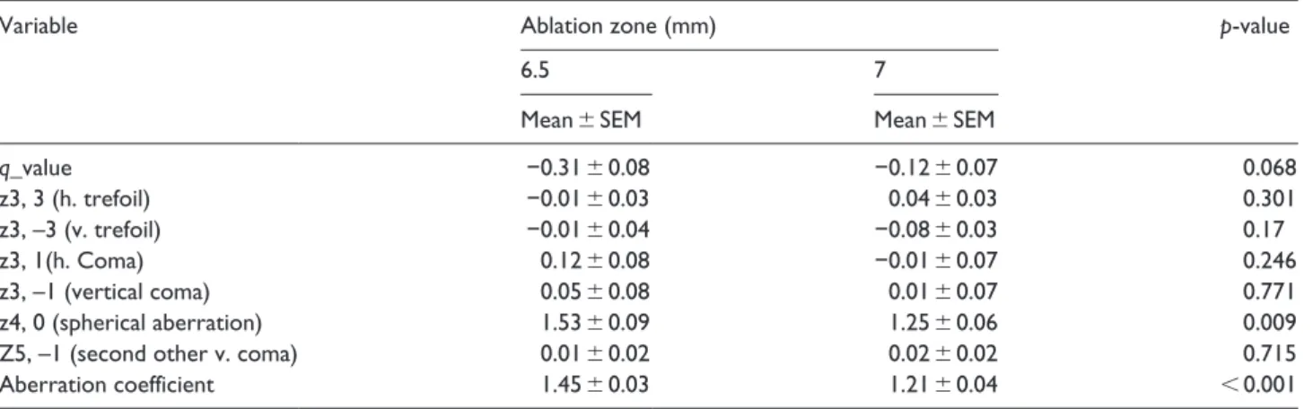
Table 2. Wavefront measurement outcomes of the comparison between two groups.
Variable Ablation zone (mm) p-value
6.5 7
Mean ± SEM Mean ± SEM
q_value −0.31 ± 0.08 −0.12 ± 0.07 0.068 z3, 3 (h. trefoil) −0.01 ± 0.03 0.04 ± 0.03 0.301 z3, –3 (v. trefoil) −0.01 ± 0.04 −0.08 ± 0.03 0.17 z3, 1(h. Coma) 0.12 ± 0.08 −0.01 ± 0.07 0.246 z3, –1 (vertical coma) 0.05 ± 0.08 0.01 ± 0.07 0.771 z4, 0 (spherical aberration) 1.53 ± 0.09 1.25 ± 0.06 0.009
Z5, –1 (second other v. coma) 0.01 ± 0.02 0.02 ± 0.02 0.715
Aberration coefficient 1.45 ± 0.03 1.21 ± 0.04 < 0.001
Discussion
Excimer laser ablation for myopia correction disrupts nor-mal corneal asphericity. Naturally, the prolate cornea is reshaped with an excimer laser to centrally flatter oblate cornea. Therefore, in corneas having deteriorated aspheric-ity coma-like and spherical-like high-order aberrations are induced after surgery. Despite continuous improvement in
refractive surgery techniques, these induced aberrations continue to be one of the most important causes of poor perceived quality of vision in postoperative patients.
This study evaluated the outcomes of femto-LASIK surgery using 6.5 and 7 mm optical zones in comparison. We found that both smaller and larger optical zone treat-ments are safe and effective in correcting myopia and myopia with astigmatism, with no significant differences
Figure 1. Postoperative z4, 0 (spherical aberrations) in 6.5 and 7 mm optical zones.
in UCVA, pachymetry decrease and postoperative spheric equivalents during the follow-up period. A novel result that was obtained in this study is that despite most of the high-order aberration parameters showing no significant difference between the two groups, aberration coefficient values and z4, 0 spherical aberrations were found to be lower in the larger-ablation-zone group. Although there are similar visual outcomes in both groups for refractive error correction, larger optic zones seem to be more advan-tageous than smaller optic zones due to lower aberration coefficient and less spherical aberration. In contrast, Gershoni et al.8 reported worse outcomes in terms of safety
and efficacy in larger optical zones with trans-photorefrac-tive keratectomy surgery; they found no difference between optical zone diameter groups (6, 6.5 and 7 mm) with femto-LASIK. Other than this, large optical zones with increased stromal tissue consumption were thought to be unsafe and not suitable for all patients.9 However, in a
recent study, Milivojevic et al.10 supported that diameter
enlargement of the treated optical zone from 6.5 to 7 mm does not threaten the stability of the cornea structure and improves outcomes.
In many publications, it has been shown that the aber-rations induced by PRK and LASIK in the postoperative period were continuously decreased for 18 months and the greatest decrease was in the first 2 months after the operation. This change in optical aberration is attributed to the wound healing reaction in the first 2 months after surgery. Therefore, we performed aberration measure-ments 3 months after the operation.
The difference between high-order aberrations is related to the relationship between pupil size and optical zone size. In our study, preoperative and postoperative mean pupil diameters were not statistically different between both groups in natural scotopic condition. In light of this result, we can say that the effects of natural pupil diameters on postoperative measurements between the two groups are minimal. Alarcon et al.4 showed that
pupil size had a negative effect on the retinal image only when it was greater than the diameter of the optical zone. In our study, the pupil diameters were smaller than the optical zones in both groups.
High-order aberrations after refractive surgery are known to be the fourth-order dominance (spherical-like aberration) in larger pupils. In our study, induction of spherical-like aberrations in scotopic condition measure-ments was statistically significantly higher in the 6.5 mm optical zone group than in the 7-mm optical zone group.
In the past, operational definitions of the optical zones have been validated by test ablations on flat plastic.11 Now,
it is validated by direct wave-front measurements of the cornea. Previous studies showed that high-order aberra-tions are increased in myopic patients after refractive sur-geries.3,12,13 High-order aberration changes after laser
refractive surgery are one of the main factors affecting the
visual quality after refractive operations.13,14 Seo et al.15
and Endl et al.6 indicated that wave-front aberrations after
refractive surgery with a larger ablation zone are less pro-nounced and closer to physiological level than those with a regular ablation zone in LASEK and PRK, consecutively. Consistent with previous studies,3,6 we found that a smaller
optical zone (6.5 mm) is associated with a greater increase in aberration coefficient and z4, 0 spherical aberrations in femto-LASIK surgery in myopic patients. Light passing through the area connecting the ablation zone and the tran-sition zone under a smaller optical zone and larger pupil diameter, which increases the aberration and reduces the contrast of the retinal image, can be an explanation for the similar results. This may be the cause of glare and halo encountered at night in patients with smaller ablation zones and larger pupil diameters. It can be concluded that a larger optical zone design prevents a significant increase in HOAs, which would, in turn, decrease visual quality.
To our knowledge, there are few studies that have investigated high-order aberration parameters in different optical zones in femto-LASIK surgery.
One of the limitations of this study is that our patients were not grouped as low, moderate and high myopia. Many studies have shown that the amount of induced high-order aberrations is correlated with the amount of refrac-tion value planned to be corrected. If the grouping was done for myopia, it could have enabled us to obtain more valuable results in our study. Another limitation of our study is that eliminating all HOAs could probably not be the best strategy to optimize visual function. For example, some controlled aberrations can improve the depth of focus with minimal degradation of image quality. Benard et al.16 showed that adding some high-order aberration to
the eye by means of an artificial system or refractive sur-gery can spread the concentration of rays along the visual axis producing a multifocality that could increase the depth of focus. Another limitation is that the influence of accom-modation on ocular aberration is well documented;17,18
therefore, an unstable accommodation status would cause declining measurement repeatability of aberration.
Declaration of conflicting interests
The author(s) declared no potential conflicts of interest with respect to the research, authorship and/or publication of this article.
Funding
The author(s) received no financial support for the research, authorship and/or publication of this article.
References
1. Sugar A, Rapuano CJ, Culbertson WW, et al. Laser in situ keratomileusis for myopia and astigmatism: safety and effi-cacy: a report by the American Academy of Ophthalmology.
2. Bailey MD, Mitchell GL, Dhaliwal DK, et al. Patient satis-faction and visual symptoms after laser in situ keratomileu-sis. Ophthalmology 2003; 110: 1371–1378.
3. Oshika T, Klyce SD, Applegate RA, et al. Comparison of corneal wavefront aberrations after photorefractive kera-tectomy and laser in situ keratomileusis. Am J Ophthalmol 1999; 127(1): 1–7.
4. Alarcon A, Rubino M, Peeerez-Ocon F, et al. Theoretical analysis of the effect of pupil size, initial myopic level, and optical zone on quality of vision after corneal refractive sur-gery. J Refract Surg 2012; 28(12): 901–906.
5. Carones F, Brancato R, Morico A, et al. Evaluation of three different approaches to perform excimer laser photorefrac-tive keratectomy for myopia. Ophthalmic Surg Lasers 1996; 27(5 Suppl): S458–S465.
6. Endl MJ, Martinez CE, Klyce SD, et al. Effect of larger ablation zone and transition zone on corneal optical aber-rations after photorefractive keratectomy. Arch Ophthalmol 2001; 119(8): 1159–1164.
7. Murta J and Martins Rosa A. Measurement and topogra-phy guided treatment of irregular astigmatism. In: Goggin M (ed.) Astigmatism – optics, physiology and management. Rijeka: InTech, 2012, pp. 247–266.
8. Gershoni A, Knaneh S, Mimouni M, et al. Effect of treat-ment zone diameter on the clinical results of femtosecond laser-assisted in situ keratomileusis and trans-photorefrac-tive keratectomy for the correction of myopia. Cli Exp Vis
Eye Res J 2018; 1: 1–6.
9. Gatinel D, Hoang-Xuan T and Azar DT. Volume estimation of excimer laser tissue ablation for correction of spherical myopia and hyperopia. Invest Ophthalmol Vis Sci 2002; 43(5): 1445–1449.
10. Milivojevic M, Petrovic V, Vukosavljevic M, et al. The assessment of the stability of the corneal structure after
LASIK correction of myopia by different optical zone diam-eters. Vojnosanit Pregl 2016; 73(6): 572–576.
11. Roberts CW and Koester CJ. Optical zone diameters for photorefractive corneal surgery. Invest Ophthalmol Vis Sci 1993; 34(7): 2275–2281.
12. Almahmoud T, Munger R and Jackson WB. Effects of advanced surface ablations and intralase femtosecond LASIK on higher order aberrations and visual acuity out-come. Saudi J Ophthalmol 2011; 25(3): 275–280.
13. Moreno-Barriuso E, Lloves JM, Marcos S, et al. Ocular aberrations before and after myopic corneal refractive sur-gery: LASIK-induced changes measured with laser ray trac-ing. Invest Ophthalmol Vis Sci 2001; 42(6): 1396–1403. 14. Mrochen M, Kaemmerer M, Mierdel P, et al. Increased
higher-order optical aberrations after laser refractive sur-gery: a problem of subclinical decentration. J Cataract
Refract Surg 2001; 27(3): 362–369.
15. Seo KY, Lee JB, Kang JJ, et al. Comparison of higher-order aberrations after LASEK with a 6.0 mm ablation zone and a 6.5 mm ablation zone with blend zone. J Cataract Refract
Surg 2004; 30(3): 653–657.
16. Benard Y, Lopez-Gil N and Legras R. Optimizing the sub-jective depth-of-focus with combinations of fourth- and sixth-order spherical aberration. Vision Res 2011; 51(23– 24): 2471–2477.
17. Gabriel C, Klaproth OK, Titke C, et al. Repeatability of topographic and aberrometric measurements at different accommodative states using a combined topographer and open-view aberrometer. J Cataract Refract Surg 2015; 41(4): 806–811.
18. Zhu D, Shao Y, Peng Y, et al. Real-time measurement of dynamic changes of anterior segment biometry and wave-front aberrations during accommodation. Eye Contact Lens 2016; 42(5): 322–327.