Can We Differentiate Pyelonephritis and Cystitis without
99mTc-Dimercaptosuccinic Acid Scan in Children?
Çocuklarda
99mTc-Dimerkaptosüksinik
Asid
ile
Sintigrafik
Görüntü
Yapılmaksızın Piyelonefrit ve Sistit Tanısı Ayırt Edilebilir mi?
Buket Kılıçaslan1, Aytül Noyan2
, Nurcan Cengiz2, Çagla Sarıtürk3, Gönül Parmaksız2, Esra Baskın4
1
Baskent University, Faculty of Medicine, Adana Teaching and Medical Research Center, Department of Pediatrics, 2Department of Pediatric Nephrology, 3Division of Bioistatistics, ADANA
4
Baskent University, Faculty of Medicine, Department of Pediatric Nephrology, ANKARA
Cukurova Medical Journal 2015;40(3):495-503.
ABSTRACT
Purpose: Urinary tract infection is one of the most common infections in childhood. Because of the long term sequelae, differentiation of pyelonephritis from cystitis is important. The aim of this study is to determine the value of biomarkers such as C-reactive protein and procalcitonin and whether preferred to predict pyelonephritis in children without 99mTc-Dimercaptosuccinic Acid scan.
Material and Methods: Fifty children aged 3 months to 16 years with a first urinary tract infection were included in this retrospective observational study. The medians, sensitivity, specificity, and cut-off values of serum C - reactive protein and procalcitonin to predict pyelonephritis were determined.
Results: Thirty-two (64%) patients were diagnosed with pyelonephritis and 18 (36%) were diagnosed with cystitis. The cut-off value for C - reactive protein was 34 mg/L to predict pyelonephritis, with 69% sensitivity and 61% specificity. The cut-off value for procalcitonin was 0.23 ng/mL to predict pyehlonephritis, with 69% sensitivity and 66% specificity. In combination, these biomarkers were 63% sensitive and 78% specific to predict pyelonephritis.
Conclusion: Using a combination of procalcitonin and C-Reactive Protein is preferred to predict pyelonephritis in children, instead of the Dimercaptosuccinic Acid scan. Because of its disadvantages, the 99mTc-Dimercaptosuccinic Acid scan should be avoided in children.
Key words: Procalcitonin, Pyelonephritis, C - reactive protein
ÖZET
Amaç: İdrar yolu enfeksiyonu, çocukluk çağının en sık görülen enfeksiyonlarından biridir. Piyelonefritin uzun dönemde görülen sekelleri nedeniyle sistitten ayırımı önemlidir. Bu çalışmanın amacı, çocuklarda 99mTc-Dimerkaptosüksinik Asid ile sintigrafik görüntüleme yapılmaksızın piyelonefrit ayırımını yapmakta prokalsitonin ve C-Reaktif Protein gibi belirteçlerin tanısal değerini saptamak, 99mTc-Dimerkaptosüksinik Asid ile sintigrafik görüntüleme yapılacak hastalar konusunda yol gösterici olup olmayacağını belirlemektir.
Materyal ve Metod: Bu çalışmaya, 3 ay ile 16 yaş arasında, ilk kez idrar yolu enfeksiyonu geçiren 50 çocuk dahil edilmiştir. Retrospektif gözleme dayalı bir çalışmadır. Piyelonefrit tanısı için serum C-Reaktif Protein ve prokalsitonin düzeylerinin ortanca değeri, duyarlılık, özgüllük ve cut-off değerleri saptandı.
Bulgular: Hastaların 32’si (%64) piyelonefrit tanısı alırken, 18 (%36) hasta sistit tanısı aldı. Piyelonefrit tanısı için C-Reaktif Protein cut-off değeri %69 duyarlılık ve %61 özgüllük ile 34 mg/L iken, prokalsitonin için bu değer %69 duyarlılık
ve %66 özgüllük ile 0,23 ng/mL saptandı. Her iki belirteçin birlikte kullanılması ise %63 duyarlılık ve %78 özgüllük ile piyelonefrit tanısını destekledi.
Sonuç: Çocuklarda piyelonefrit tanısını koymakta serum prokalsitonin ve C-Reaktif Proteinin birlikte değerlendirilmesi, 99mTc-Dimerkaptosüksinik Asid ile sintigrafik görüntüleme yapılacak hastalarda yol gösterici olabilir. Böylece çocuklar 99mTc-Dimerkaptosüksinik Asid ile sintigrafik görüntülemenin dezavantajlarından uzak tutulabilir.
Anahtar Kelimeler: Prokalsitonin, Piyelonefrit, C-Reaktif Protein
INTRODUCTION
Urinary tract infections (UTIs) are common in childhood. Symptoms may vary with age and include vomiting, growth retardation, irritability, dysuria, diarrhea, constipation, urgency, and abdominal pain. Fever may be the only symptom, especially in infants1,2.
The differentiation between upper and lower UTIs is important because of long term sequelae. When UTI is limited to the bladder, it is known as cystitis; if it spreads to the kidneys it may cause pyelonephritis. Pyelonephritis, unlike cystitis, increases the risk of renal scarring, which may eventually lead to hypertension, proteinuria, and chronic renal disease in the long term period1,3. In Turkish children, vesicoureteral reflux and related urological problems initially presenting as UTIs, and are the predominant underlying causes of chronic kidney disease4.
The diagnosis of acute pyelonephritis is generally based on symptoms such as fever, flank pain associated with pyuria, and a positive urine culture5. However, the gold standard for diagnosis is 99mTc-dimercaptosuccinic acid (DMSA) scan6. The disadvantages of DMSA include high cost, radiation exposure, and the fact that it is not available in all centers7.
C-reactive protein (CRP) is also accepted as an alternative confirmation of the presence of pyelonephritis. High CRP levels are an indicator of tissue injury or inflammation6. Another indicator of infection with systemic inflammation is procalcitonin (PCT), a propeptide of calcitonin, described in 19938. Several studies have demonstrated the importance of PCT and CRP, and each one is more valuable to determine the level of UTI.
The primary aim of this study was to determine the diagnostic value of PCT and CRP as markers of pyelonephritis.
MATERIAL and METHODS
We have retrospectively reviewed the medical records of children admitted to Department of Pediatric Nephrology of Baskent University, Faculty of Medicine, Adana Teaching and Medical Research Center, in southern Turkey, during the past 6 months. We enrolled 50 children aged 3 months to 16 years old with first UTI. Clinical findings such as fever, feeding problems, irritability, vomiting, abdominal pain, malodorous urine, dysuria, pollacuria, urinary incontinence, hematuria, constipation, and flank pain; and laboratory features including urinalysis, urine culture, white blood cell count, CRP, PCT and DMSA scan were recorded. Renal and bladder functions were normal. Urine samples for urinalysis were obtained by the midstream method in toilet-trained children and by a urine bag in infants and small children. If culture was required, bladder catheterization or suprapubic aspiration was performed in infants. UTI was defined by the presence of ≥100,000 colonies per milliliter of a single bacterial species on a specimen obtained by the midstream method; ≥50,000 colonies for catheterization and ≥1000 colonies was definitive of infection in suprapubic aspiration. PCT was determined by the electrochemieluminescence immunoassay method. A PCT of greater than 0.05 ng/ml was considered abnormal. CRP was determined by the nepholometric method, according to the manufacturer's instructions. A CRP of greater than 6 mg/L was considered abnormal. In our study, patients with a potential of
raising the CRP and PRC values nonspecifically, patients with surgical, traumatic or any other kind of infection were excluded. The DMSA scan was performed on every child with a positive urine culture within 7 days of admission. Patients were planarily screened with "GE medical systems infinia israel" and "SIEMENS symbia true point SPECT CT USA " devices. Four hours after the patients were given 3 mCi (111 mBq) 99mTc-DMSA intravenously, patients were screened in anterior, posterior, right posterior oblique, and left posterior oblique positions. Pyelonephritis was defined as focal or diffuse areas of decreased or absent DMSA uptake.
Statistical analysis was performed using the SPSS software version 17.0. For each continuous variable, normality was checked by the Kolmogorov Smirnov and Shapiro-Wilk tests and by histograms. Comparisons between groups were performed using the Mann-Whitney U test for data not normally distributed. Receiver operating characteristic curves (ROC curves), areas under the curve (AUC), sensitivity, and specificity were calculated. A p-value of less than 0.05 was considered as statistically significant. Cut off values for CRP and PCT were determined by ROC analysis by taking the highest common values of sensitivity and specificity values.
RESULTS
Fifty children were diagnosed with UTI. Mean age was 56 months old (range 3 months to 16 years). Eighty four percent of cases were female. There were 31 (62%) toilet trained children in the cohort. The most frequent symptoms were fever (72%), abdominal pain (32%), vomiting (30%), irritability (22%), incontinence (20%), dysuria (16%), flank pain (16%), and feeding problems (16%). The most commonly isolated microorganism from urine samples was
Escherichia coli (82%). Other microorganisms
were Klebsiella (6%), Proteus (6%), and
Enterococcus fecalis (6%).
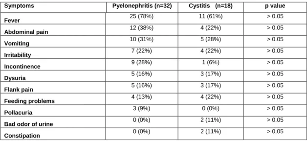
The results of the DMSA scan for 32 (64%) children revealed pyelonephritis. Eighteen (36%) were diagnosed as cystitis with a normal DMSA scan. Four boys (4/8; 50%) and 28 girls (28/42; 67%) were diagnosed with pyelonephritis. The median age of children with pyelonephritis and cystitis were 74 and 19 months, respectively. Comparison of age and gender of children with cystitis and pyelonephritis showed no statistically significant difference (p=0.069 and p=0.436, respectively). Among those with pyelonephritis, 69% (22/32) of them were toilet-trained; while 50% (9/18) with cystitis were toilet-trained (p=0.233). No statistically significant difference was detected between groups in case of fever existence (≥38ºC axillary) (p=0.325; Table 1).
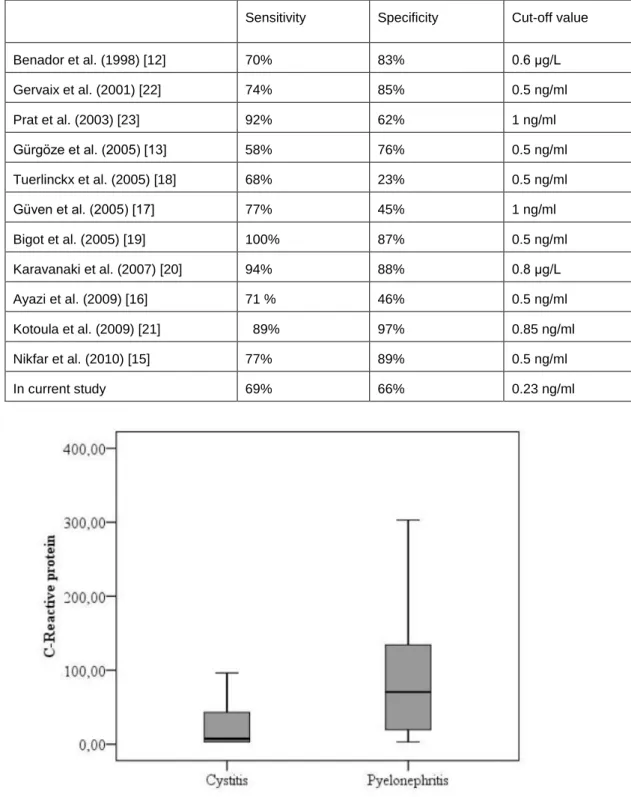
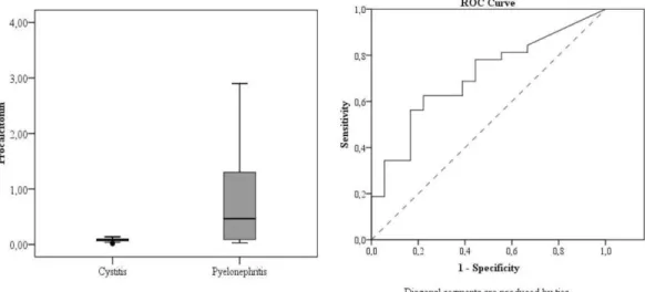
The median CRP of children with pyelonephritis and cystitis were 70.5 mg/L (3-303) and 9.8 mg/L (3-144) respectively. The median PCT of children with pyelonephritis and cystitis were 0.64 ng/ml (0.03-21.1), and 0.1 ng/ml (0.02-14.4) respectively. The median CRP and PCT values were found to be statistically significant between groups (p=0.016 and p=0.030, respectively) (Fig. 1)
The cut-off value for CRP was 34 mg/L to predict pyelonephritis, with 69% sensitivity and 61% specificity (AUC=0.71; Figure-2a); while the cut-off value for PCT was 0.23 ng/ml to predict pyelonephritis, with 69% sensitivity and 66% specificity (AUC=0.69; Fig. 2b). When CRP and PCT were combined, the sensitivity was 63% and the specifity was 78% to predict pyelonephritis at their cut-off values.
In our study, positive predictive value is 75.9% (60.3-91.4, 95% CI) for CRP over 34 mg/L and the negative predictive value is 52.4% (31.0-73.7, 95% CI). The positive predictive value for PCT over 0.23 ng/ml was found as 78.6% (63.4-93.8, 95% CI) and the negative predictive value as 54.5% (33.7-75.4). In the situation of CRP and PCT together being stated as high, then the positive predictive value was found as 83.3%
(68.4-98.2, 95% CI) and the negative predictive value was 53.8% (34.7-73, 95% CI).
Table 1. Symptoms of the patients with pyelonephritis and cystitis in our study
Symptoms Pyelonephritis (n=32) Cystitis (n=18) p value
Fever 25 (78%) 11 (61%) > 0.05 Abdominal pain 12 (38%) 4 (22%) > 0.05 Vomiting 10 (31%) 5 (28%) > 0.05 Irritability 7 (22%) 4 (22%) > 0.05 Incontinence 9 (28%) 1 (6%) > 0.05 Dysuria 5 (16%) 3 (17%) > 0.05 Flank pain 5 (16%) 3 (17%) > 0.05 Feeding problems 4 (13%) 4 (22%) > 0.05 Pollacuria 3 (9%) 0 (0%) > 0.05 Bad odor of urine 0 (0%) 2 (11%) > 0.05 Constipation 0 (0%) 2 (11%) > 0.05 Table 2. Sensitivity and specificity of C-reactive protein to predict pyelonephritis
Sensitivity Specificity Cut-off values
Benador et al. (1998) [12] 100% 26% 10 mg/L Biggi et al. (2001) [11] 64% 68% 87.5 mg/dl Gürgöze et al. (2005) [13] 94% 58% 20 mg/L Tuerlinckx et al. (2005) [18] 94% 39% 34 mg/L Bigot et al. (2005) [19] 94% 30% 20 mg/L Güven et al. (2005) [17] 67% 67% 2 mg/dl Karavanaki et al. (2007) [20] 73% 70% 50mg/L Kotoula et al. (2009) [21] 81% 90% 3.5 mg/dl Ayazi et al. (2009) [16] 96 % 4% 10 mg/L Nikfar et al. (2010) [15] 80% 65% 20 mg/L In current study 69% 61% 34 mg/L
Table 3. Sensitivity and specificity of procalcitonin to predict pyelonephritis
Sensitivity Specificity Cut-off value
Benador et al. (1998) [12] 70% 83% 0.6 µg/L Gervaix et al. (2001) [22] 74% 85% 0.5 ng/ml Prat et al. (2003) [23] 92% 62% 1 ng/ml Gürgöze et al. (2005) [13] 58% 76% 0.5 ng/ml Tuerlinckx et al. (2005) [18] 68% 23% 0.5 ng/ml Güven et al. (2005) [17] 77% 45% 1 ng/ml Bigot et al. (2005) [19] 100% 87% 0.5 ng/ml Karavanaki et al. (2007) [20] 94% 88% 0.8 µg/L Ayazi et al. (2009) [16] 71 % 46% 0.5 ng/ml Kotoula et al. (2009) [21] 89% 97% 0.85 ng/ml Nikfar et al. (2010) [15] 77% 89% 0.5 ng/ml In current study 69% 66% 0.23 ng/ml
Figure 1. The median serum C-reactive protein (1a) and procalcitonin (1b) concentrations of the patients with urinary tract infection (p=0.016 and p=0.030, respectively)
Figure 2. ROC curve areas for C-reactive protein (2a) and procalcitonin (2b) to predict pyelonephritis
DISCUSSION
Urinary tract infections are one of the most frequent infections of childhood, and can be diagnosed as pyelonephritis or cystitis. The differentiation between pyelonephritis and cystitis is important because of the long-term sequela of renal scarring that may occur following an episode of pyelonephritis9. Chronic renal disease or hypertension may be the consequence of renal scarring in future follow-up10.
In order to distinguish the site of UTI, fever has usually been identified as the classic symptom of pyelonephritis; and the absence of feverindicates cystitis. However, some studies have shown that fever is not enough for the diagnosis of pyelonephritis, as a sole indicator9,11. Similar to these reports, fever was not a significant clinical sign of pyelonephritis in our study. In our cohort, fever was present in 78% of children with pyelonephritis and in 61% of children with cystitis.
We concluded that fever is not the diagnostic clinical criteria to distinguish pyelonephritis from cystitits.
The DMSA scan is the gold standard for the diagnosis of pyelonephritis. Besides, we included biomarkers such as CRP and PCT to determine the presence of pyelonephritis. In our study, median CRP level was significantly higher in children with pyelonephritis compared to those with cystitis. Although CRP had been reported as a significant biomarker of pyelonephritis in previous studies9,11-15 controversy about this data had also been revealed16-18. The median PCT level in our experience was also higher in children with pyelonephritis, as confirmed by other studies12-15. However, the significance of PCT as a predictor in pyelonephritis was not shown by several studies in children17,18.
The sensitivity and specificity of CRP in pyelonephritis has been reported between 64-100% and 4-90%, respectively11-21. In the English literature, the lowest sensitivity to predict pyelonephritis for CRP was reported by Biggi et al. and the highest sensitivity was reported by Benador et al.11,12. In pyelonephritis, to predict the sensitivity of CRP in diagnosis, cut-off values of 2 mg/dl, 5 mg/dl, and 10 mg/dl were associated with 67%, 67%, and 73% sensitivity respectively, by Güven et al.17
. In current study, a cut-off CRP value of 34 mg/L was found to be 69% sensitive to predict pyelonephritis, similar to the studies of both Biggi et al.11 (64% sensitivity) and Güven et al.17
(67% sensitivity). Previously, the lowest specificity for CRP was detected by Ayazi et al.16 while the highest specificity was reported by Kotoula et al.21. Specificity of CRP was similar to the other studies of Biggi,11 Nikfar15 and Güven et al.17
(68%, 65% and 67% specificity, respectively) (Table 2). In some studies different cut-off values were given for CRP (Table 2). However, if the units used are standardised, such differences disappear. The most significantly different cut-off value was 87.5 mg/dl, stated by Biggie et al.11. Although this study has similarities with ours in terms of population,
age, and location, there are differences in the gender, race and number of the patients. The difference in the cut off value may be related to the differences in the race, gender and the number of the patients as well as the difference in the method.
Since 1993, PCT has been used to demonstrate the severity of infection in several studies in children; in a few studies, PCT and CRP levels have been compared as predictors of pyelonephritis. In previous studies, the lowest and highest sensitivities for PRC were reported as 58% and 100%12-22. The lowest sensitivity for PCT to predict pyelonephritis was reported by Gürgöze13
while the highest sensitivity was reported by Bigot19. Sensitivity rates for PCT according to the different cut-off values of 0.5 ng/ml, 1 ng/ml, and 2 ng/ml were declared as 65%, 77%, and 100%, respectively by Güven et al17
. In our study, at a cut-off value of 0.23 ng/ml, PCT was 69% sensitive to predict pyelonephritis. This result was similar to Tuerlinckx et al.[18] (68% sensitivity). In the previous studies, the lowest and highest specificities for PCT were 23% and 97%, respectively21,18. The lowest specificity was reported by Tuerlinckx18 at a cut-off value of 0.5 ng/ml.Different cut-off values of 0.5 ng/ml, 1 ng/ml, and 2 ng/ml for PCT were declared by Güven et al17 with 38%, 45%, and 43% specificity to predict pyelonephritis, respectively. Another report of low specificity for PCT was reported by Ayazi et al. as 46%6 In our study, PCT showed higher specificity as 66%, similar with the study of Prat (62%).23. The highest specificity for PCT was reported by Kotoul et al.21. (Table 3).
It is very important to follow patients with the risk of renal scarring in UTIs with advanced screening methods. On the other hand, children with lower UTIs and with low risk of renal scarring shouldn't be exposed to unnecessary advanced screening methods. In deciding whether to use advanced screening methods or not, DMSA is still accepted as the gold standard, however, less invasive biochemical parameters with no radiation
exposition are still needed. In our study it has been proved that combined use of PCT and CRP is rather efficient in predicting pyelonephritis. In patients with high CRP and PCT values, pyelonephritis has been proved to occur 5.8 times more. In addition to this, in case of patients with high PCT and CRP values, the ratio of pyelonephritis has been found as 83.3% (positive predictive value). Having the high specifity of 78% when using CRP and PCT together has been proved to be efficient in defining patients with low risk of renal scarring.
CONCLUSION
We conclude that, CRP and PCT may be useful parameters to predict pyelonephritis when they are used in combination, and may help to determine whether DMSA scanning is necessary or not. Keeping in mind the possible disadvantages of DMSA scanning in children, this evaluation may be advantageous.
REFERENCES
1. American Academy of Pediatrics. Practice parameter: the diagnosis, treatment, and evaluation of the initial urinary tract infection in febrile infants and young children. Pediatrics. 1999;103:843-52.
2. Ammenti A, Cataldi L, Chimenz R, et al. Febrile urinary tract infections in young children: recommendations for the diagnosis, treatment and follow-up. Acta-Paediatr. 2012;10:451-7.
3. Glauser MP, Lyons JM, Braude AI. Prevention of chronic experimental pyelonephritis by suppression of acute suppuration. J Clin Invest. 1978;61:403-7. 4. Bek K, Akman S, Bilge I, et al. Chronic kidney
disease in children in Turkey. Pediatr Nephrol. 2009;24:797-806.
5. Jodal U, Lindberg U. Guidelines for management of children with urinary tract infection and vesico-ureteral reflux. Recommendation from Swedish state of the art conference. Acta Paediatr. 1999;431:87-9. 6. Rushton HG. The evaluation of acute pyhelonephritis
and renal scarring with technetium
99m-concepts and future directions. Pediatr Nephrol. 1997;11:108-20.
7. Pecile P, Mirion E, Romanello C, et al. Procalcitonin: A marker of severity of acute pyelonephritis among children. Pediatrics. 2004;114: 249-54.
8. Assicot M, Gendrel D, Carsin H, et al. High serum procalcitonin concentrations in patients with sepsis and infection. Lancet. 1993;341:515-8.
9. Garin EH, Olavarria F, Araya C, et al. Diagnostic significance of clinical and laboratory findings to localize site of urinary infection. Pediatr Nephrol. 2007;22:1002-6.
10. Jacobsson SH, Eklof O, Eriksson CG, et al. Development of hypertension and uremia after pyelonephritis in childhood:27 year follow-up. BMJ. 1989;299:703-6.
11. Biggi A, Dardanelli L, Pomero G, et al. Acute renal cortical scintigraphy in children with a first urinary tract infection. Pediatric Nephrol. 2001;16:733-8. 12. Benador N, Siegrist CA, Gendrel D, et al.
Procalcitonin is a marker of severity of renal lesions in pyelonephritis. Pediatrics. 1998;102:1422-5. 13. Gürgöze MK, Akarsu S, Yılmaz E, et al.
Proinflammatory cytokines and procalcitonin in children with acute pyelonephritis. Pediatr Nephrol. 2005;20:1445-8.
14. Smolkin V, Koren A, Raz R, et al. Procalcitonin as a marker of acute pyelonephritis in infants and children. Pediatr Nephrol. 2002;17:409-12.
15. Nikfar R, Khotaee G, Ataee N, Shams S. Usefulness of procalcitonin rapid test for the diagnosis of acute pyelonephritis in children in the emergency department. Pediatr Int. 2010;52:196-8.
16. Ayazi P, Mahyar A, Hashemi HJ, et al. Comparison of procalcitonin and C-reactive protein tests in children with urinary tract infection. Iran J Pediatr. 2009;19:381-6.
17. Güven AG, Kazdal HZ, Koyun M, et al. Accurate diagnosis of acute pyelonephritis: How helpful is procalcitonin? Nucl Med Commun. 2006;27:715-21. 18. Tuerlinckx D, Borght TV, Glupczynski Y, et al. Is
procalcitonin a good marker of renal lesion in febrile urinary tract infection? Eur J Pediatr. 2005;164:651-2.
19. Bigot S, Leblond P, Foucher C, et al. Usefulness of procalcitonin for the diagnosis of acute pyelonephritis in children. Arch Pediatr. 2005;12:1075-80.
20. Karavanaki K, Haliotis FA, Sourani M, et al. DMSA scintigraphy in febrile urinary tract infections could be omitted in children with low procalcitonin levels. Infect Dis Clin Prac. 2007;15:377-81.
21. Kotoula A, Gardikis S, Tsalkidis A, et al.
Comparative efficacies of procalcitonin and conventional inflammatory markers for prediction of renal parenchymal inflammation in pediatric first urinary tract infection. Urology. 2009;73:782-6.
22. Gervaix A, Galetto-Lacour A, Gueron T, et al. Usefulness of procalcitonin and C-reactive protein rapid tests for the management of children with urinary tract infection. Pediatr Infect Dis J. 2001;20:507-11.
23. Prat C, Dominguez J, Rodrigo C, et al. Elevated serum procalcitonin values correlate with renal scarring in children with urinary tract infection. Pediatr Infect Dis J. 2003;22:438-42.
Yazışma Adresi / Address for Correspondence: Dr. Buket Kılıçaslan
Başkent University Faculty of Medicine Adana Teahcing and Medical Research Center Department of Pediatrics ADANA Tel: 0 532 313 72 02 +90 322 458 68 68 Fax: +90 322 459 26 22 E-mail: [email protected] Geliş tarihi/Received on : 22.01.2015 Kabul tarihi/Accepted on: 20.02.2015