Ankara Üniv Vet Fak Derg, 52, 7-12, 2005
Mast cells in the dog skin: distribution, density, heterogeneity and
influence of fixation techniques
Reşat Nuri AŞTI1, Arif KURTDEDE2, Nevin KURTDEDE1, Emel ERGÜN3, Murat GÜZEL4
1 Department of Histology and Embryology, 2Department of Internal Disease, Faculty of Veterinary Medicine, University of Ankara; 3Department of Histology and Embryology, Faculty of Veterinary Medicine, University of Kırıkkale; 4Department of Internal
Disease, Faculty of Veterinary Medicine, University of Mustafa Kemal, Hatay.
Summary: This study was conducted with the aim of examining the location, the morphology, and numerical distribution of
mast cells in the different areas of the healthy adult dog skin. Skin samples taken from the cheek, pinna, thorax and thigh areas of 10 healthy dogs were used as the study material. The results show the existence of mast cell heterogeneity in dog skin with respect to staining with alcian blue / safranin O and formaldehyde fixation sensitivity. Mast cells characterized by four combination of these parameters were observed: Mast cells containing only alcian blue-positive granules which were formalin-sensitive, mast cells containing only alcian blue-positive granules which were formalin-resistant, mast cells containing only safranin O-positive granules which were formalin resistant and mast cells containing a mixture of alcian blue-positive and safranin O-positive granules which were formalin resistant. With toluidine blue staining method, it was founded that IFAA fixed tissues were contained more mast cells than those tissues fixed with the 10% formaldehyde in all four regions. The difference in average number of mast cells per mm² between the IFAA and the 10% formaldehyde fixed tissues was statistically significant (p<0.001). In the cheek area, whilst the difference between superficial dermis and deeper dermis in terms of the average mast cell count per mm² was not statistically significant (p>0.05), in the other three areas, statistically significant differences (p<0.001) were found. In the pinna, thorax and thigh areas, superficial dermis contained more mast cells than deeper dermis. In both fixations the highest number of mast cells was observed in the skin of the pinna (p<0.001).
Key words: Distribution, dog skin, heterogeneity, mast cell.
Köpek derisindeki mast hücreleri: dağılım,yoğunluk, heterojenite ve tespit tekniklerinin etkisi
Özet: Bu araştırma sağlıklı erişkin köpeklerde farklı deri bölgelerindeki mast hücrelerinin yerleşim yerlerini, morfolojilerini
ve sayısal dağılımlarını incelemek amacıyla yapıldı. Çalışmada materyal olarak 10 adet sağlıklı köpeklerin yanak, kulak kepçesi, kaburga ve but bölgelerinden alınan deri örnekleri kullanıldı. Bulgularımız, alcian blue / safranin O ile boyanmasına ve formaldehit tespitine duyarlılığına göre köpek derisinde mast hücre heterojenitesini gösterdi. Mast hücreleri bu parametrelerin dört kombinasyonu ile tanımlandı: Formole duyarlı sadece alcian blue-pozitif granüller içeren mast hücreleri, formole dirençli sadece alcian blue-pozitif granüller içeren mast hücreleri, formole dirençli sadece safranin O-pozitif granüller içeren mast hücreleri ve formole dirençli hem safranin O-pozitif hem de alcian blue-pozitif mikst granüller içeren mast hücreleri. Toluidin blue ile boyanmış kesitlerde dört bölgede de IFAA tespitli dokuların % 10 formolle tespit edilenlere göre daha fazla mast hücresi içerdiği dikkati çekti. Bu iki tespit arasında mm²’de mast hücre sayı ortalamalarındaki farklılığın istatistiksel olarak önemli (p<0.001) olduğu saptandı. Yanak bölgesinde yüzlek ve derin dermis katmanlarında mm²’de ortalama mast hücre sayıları istatistiksel olarak önemsiz (p>0.05) iken, diğer üç bölgede istatistiksel fark önemli (p<0.001) bulundu. Kulak kepçesi, kaburga ve but bölgelerinde yüzlek katmanda derin katmana göre daha fazla mast hücresi bulunduğu saptandı. Her iki tespitte de en fazla mast hücresi kulak kepçesi derisinde gözlendi (p<0.001).
Anahtar sözcükler: Dağılım, heterojenite, köpek derisi, mast hücresi
Introduction
The mast cells which play role in both physiological and pathological events have been reported to secrete biologically active substances like heparin, histamine, serotonin, platelet activating factors, neutrophil chemotactic factors, proteolytic enzymes, leukotrienes and prostaglandins in response to mechanical, chemical and immunological stimuli (29). The dermal mast cell mediators that take part in events preceding inflammation
and the ability to respond quickly to allergens are considered the first line of defense protecting in the skin against antigens (4). In view of their location close to the nerves and blood vessels, they have also been reported to play a role in the regulation of blood flow to the skin by means of neuropeptides when stimulated (4,17). Mast cell are the principal effector cells in IgE- dependent hypersensitivity reactions. Mast cells are capable of synt-hesising and responding nerve growth factor (NGF) (27).
When morphological, biochemical and physiologi-cal differences are considered, mast cells are identified in two types as mucosal mast cells (MMC) and connective tissue mast cells (CTMC) (7,13,14). It has been shown that the formalin sensitive granules of the MMC stain AB(+) (14) in the combined AB/SO stain (8,13, 26) whilst those of the CTMC granules stain as formalin resistant (8,13, 26) SO(+) granules (14). For the fixation of the formalin sensitive MMC the use of any one of the solutions as isotonic formaldehyde-acetic acide (IFAA), Carnoy’s and Mota is recommended (13). While the CTMC are designated typical mast cells, the MMC are designated atypical mast cells (2,13). These two subtypes of mast cells differ in their glucosaminoglycans, protease and histamine contents. The response of these cells to their stimulators and inhibitors are also different (5). Histamine contained granules of mast cells have been linked with the classic allergic reactions (4). Marshall and Bienenstock (20) have reported that the mucosal mast cell granules to be rich in chondroitin sulphate whereas the connective tissue mast cell granules are rich in heparin. It has been suggested that whilst the atypical mast cells are responsible for the early phase response against antigens in the skin of the dog, the typical mast cells may be responsible for the late phase response (3). Mast cell play roles in type I hypersensitivity reactions (17, 20) and in skin diseases like contact dermatitis and fibrosis (25). These type I hypersensitivity reactions have a role in the formation of atopic dermatitis in the dog (9, 25).
The present study was carried out to determine distribution of mast cell numbers and subtypes in the dog according to skin location, staining and fixation method and this help to evaluate their role in skin diseases.
Materials and Methods
In the study, skin samples taken from the cheek, pinna, thorax and thigh areas of 10 healthy dogs were used as the study material. Punch biopsies were obtained from each site.The specimens were divided into two portions. One portion of the specimen was fixed in 10% formaldehyde and rinsed in top water later. The remaining portion was fixed in isotonic formaldehyde-acetic acide (IFAA, pH 2,9) solution for twelve hours and kept for another twelve hours in 70 % alcohol. Both portions were then passed through a series of varying degrees of alcohol, methylbenzoate and benzol and finally blocked in paraplast (13). Then, took 6 µm serial sections.
Sections were stained with 5% toluidine blue (prepared in a buffer of Mc Ilvaine’s citric acid disodium phosphate, pH 4), and with a combined alcian blue / safranine O (AB/SO) method (14): from the above mentioned serial sections, 10 sections (each 6 µm thick) were selected, i.e. sections 1 and 2 (next 5 sections
omitted), sections 8 and 9 ( next 5 sections omitted), sections 15 and 16 (next 5 sections omitted), sections 22 and 23 (next 5 sections omitted), sections 29 and 30. Thus, 5 pairs of sections were yielded, with a distance of 30 µm between each of the pairs. Each of the pair sections was fixed in 10% formaldehyde and IFAA were placed on the same slide and stained in an alcian blue / safranin O staining procedure (14).
The skin and intestine of rat used as the control. For to determine the numerical distribution of the mast cells in the toluidin blue stained sections, cell counts per ocular 100 squares micrometer (eyepiece graticule) was used. Under the 40x magnification objective, the number of cells per unit area in the 100 squares were counted. In each section, the number of cells from ten different areas selected at randomly in both superficiale dermis and deeper dermis were counted (6). Later, all the numerical data were converted to number of mast cells per unit area (mm²).
The difference in the average mast cell count between fixations and between the layers of the dermis were determined by the Wilcoxon analysis (19) in each region. However, for every fixation, the difference among regions was analyzed by the Kruskal-Wallis test (19).
Results
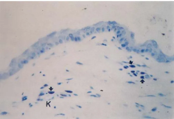
The mast cells from the cheek, pinna, thorax and thigh areas of the dog skin were easily identified because of their exhibition of metachromasia upon staining with toluidine blue after fixation with IFAA and 10% formaldehyde (Figures 1, 2, 3 arrows). Mast cells from the IFAA fixed tissue (Figures 1, 3), were noted to be more preserved and their granules stained dark than those from the tissue fixed with 10% formaldehyde (Figure 2).
Mast cells were observed to be oval, round, or spindle-shaped with different sizes. Because the cytoplasm of the cells were homogeneously stained, the
Figure 1. The appearance of the mast cells (arrows) in cheek skin by fixed IFAA. K: Blood vessel. Toluidin blue stain.x425. Şekil 1. IFAA ile tespit edilmiş yanak derisindeki mast hücreleri (oklar). K: Kan damarı. Toluidin blue boyaması.x425.
granules could not be individually identified (Figures 1, 2, 3 arrows). In some cells, the nuclei were also observed to be covered by this homogeneous mass.
Mast cells (Figures 1, 2, 3 arrows) are found abundantly especially around blood vessels (K), nerves, sebaceous gland (Figure 3 Y), sweat gland (T) and hair follicles (F) and also immediately underneath the epidermis of the skin.
In all the areas, tissues fixed with both the IFAA and the 10% formaldehyde, mast cells granules that stained blue as AB(+), (Figure 4, arrow), red as SO(+) (Figure 5, arrow), and both red and blue as AB/SO(+) mixed granules (arrow head) with the AB/SO staining were seen. The mast cell heterogeneity in dog skin respect to staining with AB/SO and formaldehyde fixation sensitivity. Mast cells characterized by four combination of these parameters were observed: Mast cells containing only alcian blue-positive granules which were formalin-sensitive, mast cells containing only alcian blue-positive granules (Figure 4, arrow) which were formalin-resistant, mast cells containing only safranin O-positive granules (Figure 5, arrow) which were
Figure 4. AB (+) mast cell (arrow) in thigh skin by fixed 10% formaldehyde. AB/SO stain.x785.
Şekil 4. %10 formolle tespit edilmiş but derisindeki AB (+) mast hücresi (ok). AB/SO boyaması.x785.
Figure 2. The appearance of the mast cells (arrows) in cheek skin by fixed 10% formaldehyde. K: Blood vessel. Toluidin blue stain.x500.
Şekil 2. %10 formolle tespit edilmiş yanak derisindeki mast hücreleri (oklar). K: Kan damarı. Toluidin blue boyaması.x500.
Figure 3. The appearance of the mast cells (arrows) in thorax skin by fixed IFAA. K: Blood vessel, Y: Sebaceous gland, T: Sweat gland, F: Hair follicle. Toluidin blue stain.x475. Şekil 3. IFAA ile tespit edilmiş kaburga derisindeki mast hücre-leri (oklar). K: Kan damarı, Y: Yağ bezi, T: Ter bezi, F: Kıl follikülü. Toluidin blue boyaması.x475.
Figure 5. SO (+) (arrow) and AB/SO (+) mixed (arrow head) mast cells in thigh skin by fixed IFAA. AB/SO stain.x780. Şekil 5. IFAA ile tespit edilmiş but derisindeki SO (+) (ok) ve AB/SO (+) mikst (ok başı) mast hücreleri. AB/SO boyaması. x780.
Table 1: In four regions the difference in average number of mast cells per mm² between the IFAA and the 10% formaldehyde fixed tissues.
Tablo 1: IFAA ve %10 formolle tespit edilmiş dört bölge arasında mm²’de ortalama mast hücre sayılarındaki farklılıklar
Area Fixation n x+ Sx Z IFAA 10 47,04 ± 2,72 Cheek 3,014* 10% Formaldehyde 10 34,24 ± 2,56 IFAA 10 105,04 ± 4,32 Pinna 5,473* 10% Formaldehyde 10 81,76 ± 3,20 IFAA 10 45,92 ± 2,56 Thorax 3,214* 10% Formaldehyde 10 32,40 ± 1,92 IFAA 10 57,76 ± 2,72 Thigh 4,825* 10% Formaldehyde 10 39,12 ± 1,92 * : p<0,001
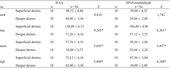
Table 2: Statistically differences between the average mast cell count per mm² area of two layers of the dermis. Tablo 2: Dermisin iki katmanında mm²’de ortalama mast hücre sayılarındaki farklılıklar.
IFAA 10%Formaldehyde Area n x+ Sx Z n x+ Sx Z Superficial dermis 10 49,12 ± 4,48 10 39,04 ± 4,32 Cheek 0,816¯ 1,742¯ Deeper dermis 10 44,96 ± 3,36 10 29,44 ± 2,88 Superficial dermis 10 138,88 ± 6,72 10 106,40 ± 4,96 Pinna 9,265* 6,361* Deeper dermis 10 71,20 ± 4,16 10 57,12 ± 3,52 Superficial dermis 10 57,76 ± 4,32 10 39,36 ± 2,88 Thorax 5,643* 3,457* Deeper dermis 10 34,08 ± 2,72 10 25,44 ± 2,24 Superficial dermis 10 73,12 ± 4,16 10 47,36 ± 3,04 Thigh 5,048* 4,388* Deeper dermis 10 42,40 ± 3,36 10 30,88 ± 2,40 ¯ : p> 0,05 * : p< 0,001
formalin resistant and mast cells containing a mixture of alcian blue-positive and safranin O-positive granules (Figure 5, arrow head) which were formalin resistant. It was also observed that tissue fixed with IFAA had a greater proportion of stained cells, in which the majority were AB(+) stained, than those cells fixed with 10% formaldehyde. However, SO(+) and AB/SO(+) stained granules were relatively rare.
Results of the cell counts and statistical analysis to determine the numerical distribution of the mast cells in the cheek, pinna, thorax and thigh areas of the dog skin are shown in tables 1-4. In the serial sections counted, it was founded that IFAA fixed tissues were contained more mast cells than those tissues fixed with the 10% formaldehyde in all four regions. The average mast cell count per mm² in the superficiale dermis and deeper dermis are similar in the cheek area. However, in the pinna, thorax and thigh regions, the mast cell count was found to be higher in the superficiale dermis than in the
deeper dermis. In the four different areas fixed with I-FAA, it was found that pinna had significantly (p< 0.001) more mast cells than the other three regions. There were no significant differences among remaining regions. However, in 10% formaldehyde fixed tissues, mast cell numbers were in the following order; pinna, thigh, cheek, thorax.
Table 3: Statistically differences among the four different body sites by fixed IFAA.
Tablo 3: IFAA ile tespit edilmiş dört bölge arasındaki istatistik-sel farklılık. Area n x ± Sx Chi-square Cheek 10 47,04 ± 2,72a Pinna 10 105,04 ± 4,32b 12,545* Thorax 10 45,92 ± 2,56a Thigh 10 57,76 ± 2,72a *: p < 0,001
a,b: The averages of groups with different letters in the same column are different.
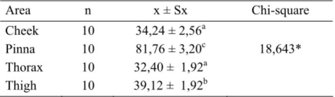
Table 4. Statistically differences among the four different body sites by fixed 10% formaldehyde.
Tablo 4: %10 formol ile tespit edilmiş dört bölge arasındaki istatistiksel farklılık. Area n x ± Sx Chi-square Cheek 10 34,24 ± 2,56a Pinna 10 81,76 ± 3,20c 18,643* Thorax 10 32,40 ± 1,92a Thigh 10 39,12 ± 1,92b *: p < 0,001
a,b,c: The averages of groups with different letters in the same column are different.
Discussion and Conclusion
Identification of the mast cells by basic dyes like toluidine blue, thionine and azure A, with which the specific granules stain metachromatically is an acceptable method (13,14, 24). It has been shown that mast cells in the IFAA fixed tissue are more preserved, more prominent (13, 14), and have much more dense stained granules (14). It has been reported that mast cells were observed especially around blood vessels, nerves, hair follicles (15, 18), sebaceous and sweat glands in the skin (2, 10, 12, 22, 28). In sections which stained with toluidine blue and examined under the light microscope, it has been shown that these metachromatic granules are not identifiable individually (13, 14) and that in some cells even the nucleus is covered with these homogeneous substance (18). The findings from this study conforms with that of the researchers (2, 10, 13, 14, 15).
In a study (21) conducted on the formalin sensitivity and staining properties of the mast cells of the human skin stained with the combined AB/SO staining, four different types of mast cells (the formalin sensitive only AB(+) and formalin resistant only AB(+), formalin resistant only SO(+), and formalin resistant AB(+) and SO(+) mixed granular cells), were observed. It is also reported that the majority of mast cells were formalin sensitive AB(+) MMC (21). Whilst in a study on rat skin, El Sayed and Dyson (11) found that tissue fixed with formaldehyde and stained with the combined AB/SO staining to have a majority of mast cells being AB(+) and a much lesser SO(+) and the least number of AB/SO(+) mixed granules, in those tissue fixed in the Carnoy’s solution and stained with the AB/SO combined staining, only mast cells containing AB(+) granules were observed. This finding was attributed to the presence of mast cells containing Carnoy’s solution sensitive formalin fixable SO(+) granules in the skin of the rat (11). This fixation and staining properties support the idea that number of mast cells in the formaldehyde fixed tissues is higher than that in the Carnoy’s fluid fixed tissues in the toluidine blue stained sections (11).
In the study, tissues fixed with both the IFAA and the 10% formaldehyde solutions were observed to have mast cells that exhibited only blue stained AB(+) granules, only red stained SO(+) granules and stained with red and blue as mixed granular AB/SO(+) when stained with the AB/SO staining. It was remarkable that IFAA fixed tissues had more stained cells in comparison to the 10% formaldehyde fixed tissues. Majority of which cells had AB(+) granules. However, the cells including SO(+) and mixed AB/SO(+) granules were in minority. The results of this study are agree with those of Marshall et al. (21). However, the results of this study differ from those of El Sayed and Dyson’s (11) in which fixaton in the Carnoy’s fluid and staining with the AB/SO stain showed mast cells containing only AB(+) granule, and the 10% formaldehyde fixed tissues compared with the Carnoy’s fluid fixed tissues showed much more mast cells. Tissues of the human (21) and dog (1, 2) skin have been shown to contain much more mast cells when fixed in the Carnoy’s or Mota fixatives than in 10% formaldehyde. The results of the study agrees with that of these researchers. IFAA fixed tissues were contained more mast cells than those tissues fixed with the 10% formaldehyde in all four regions. The difference in average number of mast cells per mm² between the IFAA and the 10% formaldehyde fixed tissues was statistically significant (p<0.001).
Mast cells have been shown to be much more abundant in the superficial dermis than in the deeper dermis in the skin of humans (9, 10, 21, 28), chicken and quails (18), and the dog (2, 23). However, it was more abundant in the deeper dermis than in the superficial dermis in rats (11, 23). In studies conducted on mast cells from the different areas of the skin, the highest number of mast cells was reported in the pinna (1,12,16). In present study, in the cheek area, whilst the difference between superficial dermis and deeper dermis in terms of the average mast cell count per mm² was not statistically significant (p>0.05), in the other three areas, statistically significant differences (p<0.001) were found. In the pinna, thorax and thigh areas, the mast cell count were higher in the superficial dermis than in the deeper dermis.
In conclusion, among the four different areas examined in present study, due to the finding of the highest number of mast cells in the pinna area in tissue fixed in both fixations, was reason for our support of the view of Auxilia and Hill (1) that the pinna is one of the predilection sites in atopic dermatitis.
References
1. Auxilia ST, Hill PB (2000): Mast cell distribution,
epidermal thickness and hair follicle density in normal ca-nine skin: possible explanations for the predilection sites of atopic dermatitis? Vet Dermatol, 11, 247-254.
2. Becker AB, Chung KF, McDonald DM, Lazarus SC, Frick OL, Gold WM (1985): Mast cell heterogeneity in
dog skin. Anat Rec, 213, 477-480.
3. Becker AB, Chung KF, McDonald DM, Lazarus SC, Frick OL, Gold WM (1986): Cutaneous mast cell
heterogeneity, response to antigen in atopic dogs. J
Allergy Clin Immunol, 78, 937-942.
4. Benyon RC (1989): The human skin mast cell. Clin Exp Allergy, 19, 375-387.
5. Bienenstock J, Befus AD, Pearce F, Denburg J, Goodacre R (1982): Mast cell heterogeneity: derivation
and function, with emphasis on the intestine. J Allergy Clin
Immunol, 70, 407-412.
6. Böck P (1989): Romeis Mikroskopische Tecknik. 17. Aufl. Urban und Schwarzenberg München.
7. Chen W, Alley MR, Manktelow BW, Davey P (1990a):
Mast cells in the ovine lower respiratory tract: heterogeneity, morphology and density. Int Arch Allergy
Appl Immunol, 93, 99-106.
8. Chen W, Alley MR, Manktelow BW, Slack P (1990b):
Mast cells in the bovine lower respiratory tract: morpho-logy, density and distribution. Br Vet J, 146, 425-435.
9. Cowen T, Trigg P, Eady RAJ (1979): Distribution of
mast cells in human dermis: development of a mapping technique. Brit J Dermatol, 100, 635-641.
10. Eady RAJ, Cowen T, Marshall TF, Plummer V, Greaves MW (1979): Mast cell population density, blood
vessel density and histamine content in normal human skin. Brit J Dermatol, 100, 623-633.
11. El Sayed S, Dyson M (1993): Histochemical
heterogeneity of mast cells in rat dermis. Biotech
Histochem, 68, 326-332.
12. Emerson JL, Cross RF (1965): The distribution of mast
cells in normal canine skin. Am J Vet Res, 26,1379-1382.
13. Enerbäck L (1966a): Mast cells in rat gastrointestinal
mucosa. 1. effects of fixation. Acta Path Microbiol
Scandinav, 66, 289-302.
14. Enerbäck L (1966b): Mast cells in rat gastrointestinal
mucosa. 2. dye-binding and metachromatic properties.
Acta Path Microbiol Scandinav, 66, 303-312.
15. Eren Ü (2000): Köpek derisinde mast hücreleri. Ankara Üniv Vet Fak Derg, 47, 167-175.
16. Foster AP (1994): A study of the number and distribution
of cutaneous mast cells with disease not affecting the skin.
Vet Dermatol, 5, 17-20.
17. Gordon JR, Parris RB, Galli JS (1990): Mast cells as a
source of multifunctional cytokines. Immunol Today, 11,
458-464.
18. Kurtdede N, Yörük M (1995): Tavuk ve bıldırcın
deri-sinde mast hücrelerinin morfolojik ve histometrik incelen-mesi. Ankara Üniv Vet Fak Derg, 42, 77-83.
19. Lehmann EL (1998): Non parametrics statistical methods
based on ranks. First edition, Prentice-Hall, Inc.
Simon&Schuster/ A Viacom Company Upper Saddle River, New Jersey.
20. Marshall JS, Bienenstock J (1994): The role of mast cells
in inflammatory reactions of the airways, skin and intestine. Curr Opin Immunol, 6, 853-859.
21. Marshall JS, Ford GP, Bell EB (1987): Formalin
sensitivity and differential staining of mast cells in human dermis. Br J Dermatol, 117, 29-36.
22. Mikhail GR, Miller-Milinska A (1964): Mast cell
population in human skin. J Invest Dermatol, 43, 249-254.
23. Persinger MA, Lapage P, Simard JP, Barker GH (1983): Mast cell number in incisional wounds in rat skin
as a function of distance, time and treatment. Br J
Derma-tol, 108, 179-187.
24. Schwartz LB, Austen KF (1984): Structure and function
of the chemical mediators of mast cells. Prog Allergy, 34,
271-321.
25. Scott DW, Miller WH, Griffin CE (1995): Muller and
Kirk's small animal dermatology. 5th Edition,W. B.
Saunders Company, Philadelphia, 488-500.
26. Shanahan F, Macniven I, Dyck N, Denburg JA, Bienenstock J, Befus AD (1987): Human lung mast cells:
distribution and abundance of histochemicaly distinct subpopulations. lnt Arch Allergy Appl Immunol, 83,
329-331.
27. Skaper SD, Pollock M, Facci L (2001): Mast cells
differentially express and release active high molecular weight neurotrophins. Brain Res Mol Brain Res, 97,
177-185.
28. Walton S, DeSouza EJ (1983): Variation in mast cell
numbers in psoriasis and lichen planus: comparisons with normal skin. Dermatologica, 166, 236-239.
29. Warton A, Papadimitriou JM, Goldie RG, Paterson JW (1986): An ultrastructural study of mast cells in the
alveolar wall of normal and asthmatic lung. Aust Exp Biol
Med Sci, 64, 435-444.
Geliş tarihi: 08.12.2003 / Kabul tarihi: 02.04.2004
Corresponding address:
Prof. Dr. Reşat Nuri Aştı
Ankara Üniversitesi Veteriner Fakültesi Histoloji-Embriyoloji Anabilim Dalı 06110 Dışkapı, Ankara