O R I G I N A L A R T I C L E
Hypoxia
‐induced endothelial cell responses – possible roles
during periodontal disease
Reila T. Mendes
1,2,3|
Daniel Nguyen
1|
Danielle Stephens
1|
Ferda Pamuk
4|
Daniel Fernandes
5|
Hatice Hasturk
1|
Thomas E. Van Dyke
1|
Alpdogan Kantarci
11
Forsyth Institute, MA, USA
2
Ponta Grossa State University, Brazil
3
Faculdade Herrero, Brazil
4
Beykent University‐ Faculty of Dentistry Department of Periodontology, Istanbul, Turkey
5
Universidade Federal de Santa Catarina, Brazil
Correspondence
Alpdogan Kantarci, Forsyth Institute, 245 First Street, Cambridge, MA 02142, USA. Email: [email protected] Funding information
CAPES/BRAZIL– Science, Grant/Award Number: 2100/13‐4; NIH/NIDCR, Grant/ Award Numbers: DE20906 and DE25030
Abstract
Background and objective Inflammatory periodontal pockets are known to be hypoxic.
Hypoxia influences vascular response to periodontal inflammation, including
angiogen-esis, which is critical for oxygen and nutrient delivery to periodontal tissues and
granulation tissue formation. Our previous work suggests that periodontal bacteria
may actively contribute to pocket hypoxia. Herein, we test the hypothesis that
Fusobacterium nucleatum actively induces low oxygen tension, which modulates
angio-genesis and endothelial cell activity. HUVEC cells were incubated in 1.5% oxygen for
(Folkman & Shing, 1992)48 hours. Cell proliferation was measured by MTT; surface
expression of CD31, CD34 and VEGF receptors (VEGFR1, VEGFR2) were analyzed
by FACS. mRNA expression of HIF isoforms, iNOS, eNOS, COX
‐2, and VEGF was
mea-sured by quantitative PCR. Supernatants were analyzed for the release of IL
‐1α, TNF‐α,
and VEGF by ELISA or multiplex immunoassays and nitric oxide was measured by
colorimetric assay. F. nucleatum actively depleted oxygen. Hypoxia resulted in a
signif-icant increase of HIF isoforms. iNOS was increased while nitric oxide was unchanged.
VEGF release was increased at 4 hours followed by an increase in VEGFR1 at 12 hours,
but not VEGFR2. CD31 expression was reduced and CD34 was increased after
48 hours (p < 0.05). IL
‐1α and TNF‐α release were decreased at 4 hours (p < 0.05),
but both increased by 24 hours; TNF
‐α increased at 24 h. The data highlight the role
of hypoxia in endothelial cell inflammatory changes. F. nucleatum, considered a bridging
species in the development of periodontopathic biofilms induces hypoxia in the
periodontium leading to angiogenic changes in periodontal disease pathogenesis.
K E Y W O R D S
endothelial cells, Fusobacterium nucleatum, inflammation, oxygen, periodontitis
1
|B A C K G R O U N D
Hypoxia and inflammation are closely related (Ahmad & Ahmed, 2004). Hypoxia regulates vascular tone and is a potent stimulus of angiogenesis (Bartruff, Yukna, & Layman, 2005). The increased vasculature enables
delivery of oxygen and nutrients to tissues and in healing wounds restores homeostasis (Cochran, 2008). Chronic exposure to hypoxia and changes in angiogenesis are closely related to pathologies including inflammatory diseases, tumors and their metastatic expansion (Cudmore, Hewett, Ahmad, et al., 2012; Darveau, 2010), ischemic diseases (Devine,
-This is an open access article under the terms of the Creative Commons Attribution License, which permits use, distribution and reproduction in any medium, provided the original work is properly cited.
©2018 The Authors. Clinical and Experimental Dental Research published by John Wiley & Sons Ltd. DOI: 10.1002/cre2.135
Marsh, & Meade, 2015) and bone and tissue injuries (Du, Lu, Petritsch, Liu, et al., 2008). Endothelial cell function is central to the interface between the blood and tissues and is the first affected by changes in oxy-gen tension (Yost, Duran‐Pinedo, Teles, Krishnan, & Frias‐Lopez, 2015). Endothelial cells respond to hypoxia through the expression of regulatory genes mediated by a variety of oxygen dependent signaling cascades (Faller, 1999; Ferrara, Gerber, & LeCouter, 2003). Hypoxia‐inducible fac-tor (HIF) is a key regulafac-tor protein of hypoxia‐mediated events (Folkman & Shing, 1992). The HIF family of transcription factors regulates the expression of multiple genes involved in processes that drive adaptation to hypoxia, including cell proliferation, apoptosis, metabolism, immune responses, genomic instability and vascularization (Folkman & Shing, 1992). HIF modulates expression of VEGF, nitric oxide synthases (NOS), and cytokine release regulating angiogenesis (Graves, 2008).
In addition to presenting an environmental change for bacterial col-onization, hypoxia may be an important regulator of inflammatory mediators in periodontal disease (Keith, Randal, & Simon, 2011). Very little is known about the impact of hypoxia on oral tissues (Keith et al., 2011). The degree of vascularization depends on the stage of inflammatory disease progression and healing; however, it is not clear how periodontal vasculature responds to hypoxia. The inflammatory process in the periodontium is a dynamic process that is characterized by tissue damage that results from a failure to eliminate the microbial biofilm leading to host‐mediated destructive events (Cochran, 2008; Koo et al., 2007). The pathogenic process in periodontal disease involves an active and reciprocal modification of the host response and biofilm during disease progression. The biofilm expands, evolves, becomes more complex and commensal microbial species become pathogenic as the pathological changes in gingival tissues and periodontal attachment to the tooth surface is destroyed (Lee et al., 2005). As the periodontal pocket deepens, more pathogenic and anaerobic bacteria dominate associated with reduced oxygen in the environment. Fusobacterium nucleatum is considered a“bridging species” that is particularly important for the onset and progression of periodontitis because it is thought to enable the colonization of other late‐colonizing periodontopathogenic species such as Porphyromonas gingivalis (Lin & Sessa, 2006).
We have recently characterized the endothelial cell response to Fusobacterium nucleatum and demonstrated its active role in promot-ing proinflammatory changes in endothelial cells, suggestpromot-ing that the pathogenic progression of periodontitis might be enhanced by modi-fied endothelial cell responses (Liu & Shi, 2012). Based on this obser-vation and literature suggesting that F. nucleatum may deplete the oxygen content in its environment (Marsh & Devine, 2011; Mendes et al., 2016), we hypothesized that F. nucleatum directly induces hyp-oxia, which modulates endothelial cell activity in periodontal disease pathogenesis. In order to test this hypothesis, we measured the impact of the F. nucleatum on hypoxia and the actions on endothelial cells and their functional regulation.
2
|M A T E R I A L S A N D M E T H O D S
2.1
|F. nucleatum Growth and Culture
F. nucleatum strain ATCC 25586 was cultured on blood agar plates in an anaerobic system under 10% H2, 80% N2 and 10% CO2 for
(Tandle et al., 2009)6 days. The cultures were then inoculated into brain‐heart infusion broth, supplemented with hemin, and incubated at 37°C for 2 days until they reached an OD540nmof 0.8,
correspond-ing to 109CFU mL−1. The bacteria were then diluted at 107CFU mL−1
corresponding to a multiplicity of infection (MOI) of 100.
2.2
|Endothelial Cell Culture
Primary Human Umbilical Vein Endothelial Cells (HUVEC) (ATCC‐PCS‐ 100‐010) were purchased from American Type Culture Collection (ATCC). Cells were cultured in vascular basal cell medium (ATCC PCS‐100‐030) supplemented with Endothelial Cell Growth Kit‐VEGF (ATCC PCS‐100‐041), penicillin, and streptomycin. Cells were cultured in 75 cm2flasks (Corning®) and maintained in an incubator with 5%
CO2 at 37°C. Cells were used from passages 4 to 8. Media was
changed every three days, in accordance with the manufacturer's recommendations. Cell characterization was accomplished through morphological analysis after reaching confluence. 2 × 10⁵ HUVEC
were placed in (Keith et al., 2011)well plates and were pre‐incubated at 37°C for 2 hours. Cells were then incubated in a hypoxia chamber (1.5% O2); cells and supernatants were collected and analyzed at
baseline up to 48 hours. Normoxia was used as the control for hypoxia conditions.
2.3
|Oxygen Content in Media in Response to F.
nucleatum
In order to measure the oxygen content in cell cultures, 5 × 10⁵ endo-thelial cells were plated in (Keith et al., 2011)well plates in 1 mL of media. The plates were divided into three groups: control, hypoxia (the plates incubated in a hypoxic chamber) and F. nucleatum. Bacteria were added (MOI = 1, 10, 100). The third group was only stimulated with the bacteria (MOI = 1, 10, 100). It was not a co‐culture model and it was incubated in normal conditions of oxygen. Oxygen levels in culture were measured using a NeoFox GT (Ocean Optics Sensors) according to the instructions by the manufacturer. Briefly, 200μL of each sample was loaded in a 96‐well plate with oxygen sensors, which operate by embedding a ruthenium or platinum‐based porphyrin dye into a sol–gel thin film and correlating fluorescence lifetime to the oxygen partial pressure. The NeoFox Phase Fluorimeter excites this chemistry with a blue LED and collects the resulting fluorescence sig-nal. Measurements were recorded in NeoFox Viewer Software v2.30.
2.4
|Endothelial Cell Proliferation in Response to
Hypoxia
In order to determine the impact of hypoxia on HUVEC cell prolifera-tion, we used the 3‐(4, (Paris et al., 2005)dimethylthiazol‐(Tandle et al., 2009)ul)‐2, (Paris et al., 2005)diphenyltetrazolium bromide (MTT) assay. HUVEC cells were seeded at 1 × 10⁴cells/well in 96
‐well plates and incubated for 24 hours at 37°C. They were then incubated under hypoxia for (Folkman & Shing, 1992)48 hours. At each time point, the media was removed; 90μL of PBS and 10 μL of MTT solution 5% were added. The plates were incubated for 3.5 hours at 37°C; 75μL of the solution was removed and 50μL of dimethyl sulfide (DMSO)
was added. The plates were incubated again at 37°C for 10 minutes. Absorbance was measured at 540 nm.
2.5
|Tube Formation Assay
As an indicator of in vitro vascularization, we investigated the role of hypoxia on endothelial cell tube formation. Forty‐five μL of Matrigel (BD Biosciences) were added to each well in 96‐well plates. The plates were incubated at 37°C for 1 hour to allow gelling. Endothelial cells were added at a concentration of 5 × 10³in each well. The plates were
then incubated at 37°C for one more hour and incubated under hyp-oxia. Images were obtained and evaluated on Image‐Pro Plus® Ver-sion 4.5.0.29 (Media Cybernetics. Silver Spring, MD, USA). The numbers of tubes were counted and the total area was measured.
2.6
|Hypoxia
‐Inducible Factor (HIF) Expression in
Endothelial Cells
In order to study the role of hypoxia on expression of alpha HIF iso-forms, which are actively translocated to the cytosol (HIF‐1α, HIF‐2α, and HIF‐3α), endothelial cells were cultured in normoxia and total RNA was extracted using TRIzol reagent. Total RNA was quantified in a spectrophotometer at an absorbance (A) of 260 nm. The RNA samples had an A260: A280 ratio close to 2.0 to guarantee high purity. One μg of total RNA from each sample was subjected to reverse transcription. Each real‐time PCR was carried out in triplicate in a total of 20μL reaction mixture. Primers used for real‐time PCR analysis were purchased from Life Technologies. The housekeeping gene, glyceraldehyde (Kimmel, Grant, & Ditata, 2016)phosphate dehydrogenase (GADPH), was amplified in each sample as control and was used for normalization. Data analysis was performed using theΔΔCt method.
2.7
|Flow Cytometry Analysis of Endothelial Cell
Surface Markers
Changes in surface markers due to hypoxia were analyzed by flow cytometry. Endothelial cells were plated in (Keith et al., 2011)well plates (1 mL of media containing 2 × 10⁵cells) and incubated under hypoxia. Cells were collected, washed twice with PBS/BSA, incubated and labeled with APC anti‐human CD31 antibody (Biolegend), APC anti‐human CD34 antibody (Biolegend), PE anti‐human VEGFR1 anti-body (Biolegend) or APC anti‐human VEGFR2 antibody (Biolegend) for 45 minutes, washed twice with PBS/BSA and analyzed by Flow Cytometry (FACScan using CellQuest software, BD Bioscience). Isotype controls for APC and PE (Biolegend) were used. Data were expressed as a percentage of positive cells to target molecules.
2.8
|VEGF and Pro
‐inflammatory Cytokine Release
by Endothelial Cells in Response to Hypoxia
The supernatants of endothelial cell cultures were tested for VEGF, IL‐1α, and TNFα using specific MILLIPLEX® Multiplex Assays using Luminex® according to the manufacturer's instructions. Results were corrected to total protein contents of the supernatant media in pg/mL.
2.9
|NOX release in supernatants, nitric oxide
synthase (NOS) and COX2 expression
In order to study the nitric oxide (NOX) release in supernatants, we measured nitric oxide metabolites of nitrite and nitrate using a specific assay kit from R&D Systems (catalog number KGE001) according to the manufacturer's instructions. Results were expressed in μmol/L. Total RNA was quantified as described above for iNOS (inducible NOS), eNOS (endothelial NOS) and COX2 quantification. Each real‐ time PCR was carried out in triplicate in a total of 20μL reaction mix-ture. Primers used for real‐time PCR analysis were purchased from Life Technologies. The housekeeping gene, glyceraldehyde (Kimmel et al., 2016)phosphate dehydrogenase (GADPH), was amplified in each sample as control and was used for normalization. Data analysis was performed using theΔΔCt method.
2.10
|Statistical Analysis
All experiments were performed in triplicate and repeated a minimum of three times. Data are expressed as mean ± SD. Since the distribu-tion of the data did not show major variadistribu-tions among each assay/sam-ple, we have chosen to use the parametric analyses. We analyzed the data using one‐way ANOVA followed by Bonferroni post hoc correc-tions for multiple comparisons. (GraphPad Prism version 5.01, San Diego, CA, USA). P < 0.05 was considered significant.
3
|R E S U L T S
3.1
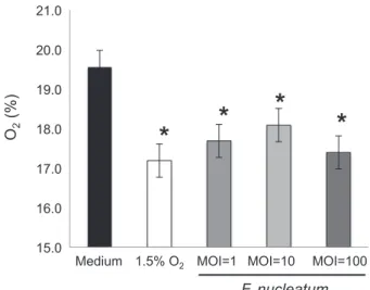
|F. nucleatum Induces Hypoxia
Endothelial cells were incubated with F. nucleatum and compared to a hypoxic atmosphere of 1.5% O2(Figure 1). The data demonstrate that
F. nucleatum reduces oxygen content creating hypoxia, which is inde-pendent of the MOI (p < 0.05).
3.2
|Hypoxia Modulates Proliferation and Tube
Formation of Endothelial Cells
The early endothelial proliferation in response to hypoxia was similar to the normoxic control; however, extended hypoxia (24 hours) resulted in significantly suppressed endothelial cell proliferation ( Figure 2A). In parallel, new vessel formation quantified as the number of tubes was reduced in response to hypoxia and the morphology of the tubes was altered (Figure 2B and C).
3.3
|Hypoxia Modulates Hypoxia Inducible Factor in
Endothelial Cells
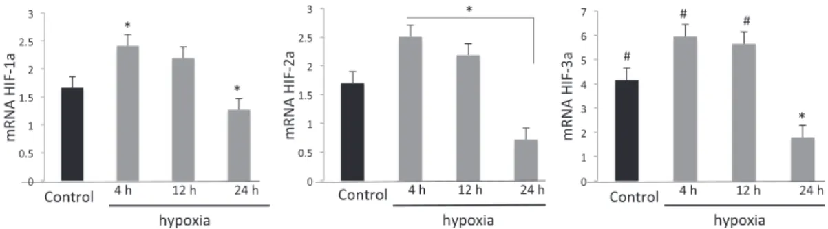
Hypoxia results in increased expression of all HIF isoforms after 4 hours of incubation (p < 0.05; Figure 3). This impact was transient and resulted in a significant decrease in HIF expression at or below the baseline levels after 24 hours.
3.4
|The impact of Hypoxia on Adhesion Molecule
Expression
While CD31 expression was constant for 24 hours, there was a reduc-tion after 48 hours (Figure 4A; p < 0.05) suggesting reduced cell–cell interaction and an increased vascular permeability. Figure 4B shows a significant decrease in CD34 expression after 4 hours of hypoxia, which increases after 48 hours of hypoxia suggesting increased undif-ferentiated endothelial cells.
3.5
|Hypoxia modulates VEGF release and VEGF
receptor expression
We next investigated downstream genes targeted by HIF. VEGF release was significantly increased after 4 hours (Figure 4C; p < 0.05). Further, we quantified VEGF receptor expression by qPCR. VEGFR1 expression was decreased at 4 hours and increased at 12 and 24 hours (p < 0.05), returning to initial levels after 48 hours (Figure 4 D). VEGFR2 expression was stable, with not statistically different changes over the 48‐hour assay (Figure 4E).
3.6
|Hypoxia Modulates COX
‐2 and Up‐regulates
iNOS
COX‐2 and NOS play key roles during the endothelial response to inflammation. Expression of COX‐2 and two isoforms of NOS was measured by qPCR. There was an initial and significant decrease in endothelial cell COX‐2 expression after 4 hours of hypoxia, after which the levels returned to normal (Figure 5A; p < 0.05). Nitric oxide release remained unchanged at all time points (data not shown). iNOS was increased after 12 hours (Figure 5B; p < 0.05) whereas mRNA for eNOS remained unchanged (Figure 5C), suggesting that only the inducible form of endothelial cell nitric oxide synthase is activated in response to hypoxia.
3.7
|Hypoxia Modulates Inflammatory Cytokine
Release
Hypoxia down‐regulates release of IL‐1α (Figure 5D) and TNF‐α (Figure 5E) in supernatants after 4 hours of incubation. IL‐1α returns to basal levels after 24 h, whereas TNF‐α returns to basal levels after 12 h and is increased almost (Tandle et al., 2009)fold after 24 h. These FIGURE 1 F. nucleatum promotes hypoxia. The oxygen level of
samples containing F. nucleatum at an MOI 1, 10 and 100 and control (media only) was measured using NeoFox GT (Ocean Optics). F. nucleatum significantly reduced oxygen levels in supernatants. Data are expressed as mean ± SD and analyzed using one‐way ANOVA followed by Bonferroni post hoc. P < 0.05 was considered significant (*p < 0.05)
FIGURE 2 Hypoxia modulates cell proliferation and tube formation. Hypoxia significantly reduces endothelial cell proliferation over time (Panel A). Representative images of tube formation after 12 h are shown in panel B. Tube formation is markedly reduced at 12 and 24 hours (Panel C). Data are expressed as mean ± SD and analyzed using one‐way ANOVA followed by Bonferroni post hoc. P < 0.05 was considered significant *p < 0.05 compared to control)
data suggest that prolonged hypoxia plays a role in progressive inflam-mation and differentiation of endothelial cells that acquire a pro‐ inflammatory phenotype.
4
|D I S C U S S I O N
Vascular changes are an important aspect of the onset of chronic peri-odontitis (Olsson, Dimberg, Kreuger, & Claesson‐Welsh, 2006; Zoellnor & Hunter, 1991) as well as other inflammatory lesions, cancer metastases, wound healing, atherosclerosis, tumor growth and angio-genesis (Paris et al., 2005). Hypoxia, which is a master regulator of angiogenesis, indirectly signals VEGF, (Rodriguez‐Morata et al., 2008) and a selective mitogen for endothelial cells (Salim, Nacamuli, Morgan,
Giaccia, & Longaker, 2004). Recently, we demonstrated that F. nucleatum exerts direct actions on endothelial cells, such as decrease in cell proliferation associated with a reduced tube formation; lower expression of CD31 accompanied by higher expression of CD34 representing an increase in cell permeability and a higher number of undifferentiated cells suggesting that F. nucleatum leads to impairment on tissue vascularization during inflammation (Liu & Shi, 2012). Based on this work, we hypothesized that F. nucleatum directly contributes to a hypoxic environment modulating endothelial cell function in the pathogenesis of periodontal disease. Collectively, the data suggest that F. nucleatum can impact the endothelial cell function directly and indirectly through the hypoxic changes in the environment.
Human endothelial cells incubated under hypoxic conditions dem-onstrated reduced proliferation and suppressed and irregular FIGURE 4 Hypoxia modulates endothelial cell surface marker expression, changes VEGF release in supernatants and VEGF receptor expression. The percentage of endothelial cells expressing CD31 (Panel A) or CD34 (Panel B) at baseline and after 4, 12, 24 or 48 hours of hypoxic incubation was analyzed by flow cytometer. Hypoxia increases VEGF release in supernatants (pg/mL) after 4 hours (Panel C) and modulates VEGF receptors 1 (Panel D) and 2 (Panel E) shown as a percentage of expression of endothelial cells at baseline and after 4, 12, 24 or 48 hours of hypoxia. Data are expressed as mean ± SD and analyzed using one‐way ANOVA followed by Bonferroni post hoc. P < 0.05 was considered significant (*p < 0.05 compared to control; #p < 0.05 compared to control)
FIGURE 3 Hypoxia modulates the Hypoxia Inducible Factor Family of Proteins. Hypoxia activates HIF isoforms 1α, 2αand 3α as early as 4 hours of incubation. Data are expressed as mean ± SD and analyzed using one‐way ANOVA followed by Bonferroni post hoc. P < 0.05 was considered significant (*p < 0.05 compared to control; #p < 0.05 compared to the 24 h group)
angiogenesis mediated by HIF isoforms. Downstream targets of HIF including iNOS activation, VEGF release, and VEGFR1 appear to medi-ate these changes that were associmedi-ated with an endothelial cell pro‐ inflammatory phenotype shift. The data demonstrate that prolonged hypoxia results in endothelial dysfunction and increased inflammation. In addition, functional changes of endothelial cells were characterized by alterations in adhesion molecules (CD31 and CD34) that are asso-ciated with a loss of cell–cell adhesion and a significant increase in the number of undifferentiated cells.
Endothelial cell proliferation was decreased with reduced tube formation suggesting defective and disorganized vascular activity, which may be a critical characteristic of inflammatory vascularization. A previous study showed that a (Sluimer, Gasc, van Wanroji, et al., 2008)hour exposure to hypoxia stimulated cell proliferation (Ferrara et al., 2003). We did not measure the (Sluimer et al., 2008)hour time point but our closest data point at 4 hours showed no difference between normoxic and hypoxic conditions. We observed a stable hyp-oxia‐mediated response as of (Keith et al., 2011) hour time point, which lasted throughout the observation period. This finding suggests that a“chronic” exposure to hypoxia is critical for changes in cell func-tion. Persistent hypoxia also changed the expression of surface markers on endothelial cells linked to function. Decreased expression of CD31 and an increase in CD34 suggests an increased potential for vascular permeability of undifferentiated cells, both of which are critical signs of inflammation. CD31 serves as an adhesion molecule on endothelial cells (Sluimer et al., 2008) and is involved in the regula-tion of leukocyte detachment, T‐cell activation (Tandle et al., 2009; Taylor, 2008), and platelet activation and angiogenesis (Taylor, 2008). CD31 activation may be dependent on inflammatory cytokines (Taylor, 2008) and a reduction in CD31 expression may be associated
with increased T cell activation (Tandle et al., 2009). These findings are consistent with the chronicity of inflammation induced by hypoxia and implicate endothelial cells as playing an active role in the induction of inflammation.
Hypoxia had a direct impact on VEGF expression and actions pro-moting a switch in the VEGFR1: VEGFR2 ratio, as well as an initially increased VEGF‐A release. Numerous studies emphasize that VEGF and its receptors are critically important regulators of endothelial cell and vessel formation (Ferrara et al., 2003; Torimoto, Rothstein, Dang, Schlossman, & Morimoto, 1992). Under physiological conditions, endothelial cells express approximately 10 times less VEGFR1 than VEGFR2 (Unger, Krump‐Konvalinkova, Peters, & Kirkpatrick, 2002; Ve Val & Black, 2009). VEGFR1 mainly modulates VEGF activity creat-ing heterodimers with VEGFR2 (Wigerup, Påhlman, & Bexell, 2016). VEGFR1 negatively regulates cell proliferation through VEGFR2, and its increase reduces angiogenesis (Wirthlin & Hussain, 1992). Here, we demonstrate that early hypoxia reduces VEGFR1 expression, but this is reversed and increased over time returning to baseline by 48 hours. These observations are consistent with our observed decrease in cell proliferation induced by hypoxia but it may also high-light a limitation of the in vitro testing of exposure to hypoxia and hyp-oxia‐mediated events.
The most striking alteration induced by hypoxia was the transcrip-tional activation of the Hypoxia Inducible Factor (HIF) family of pro-teins as early as 4 hours after exposure to hypoxia. HIFs coordinate the first steps in vessel development through a paracrine mechanism. HIF activation correlates with the presence of more differentiated cells (Zhao et al., 2012; Zoellnor & Hunter, 1991), which taken together with our findings, suggest that CD34‐mediated differentia-tion of endothelial cells was regulated by HIF in response to hypoxia. FIGURE 5 Impact of Hypoxia on COX‐2 expression, cytokine release, iNOS, and eNOS. Hypoxia modulates COX‐2 mRNA (shown as fold increase) by endothelial cells (Panel A). After 12 hours iNOS mRNA was higher than after 24 h (Panel B), while eNOS mRNA remained unchanged (Panel C). Hypoxia decreases inflammatory cytokines in supernatants characterized by IL‐1α (pg/mL) (Panel D) and TNF‐α (pg/mL) (Panel E) after 4 hours. IL‐1α returns to basal levels after 24 h, whereas TNF‐α returns to basal levels after 12 h and is increased almost 2‐fold after 24 h. Data are expressed as mean ± SD and analyzed using one‐way ANOVA followed by Bonferroni post hoc. P < 0.05 was considered significant (*p < 0.05 compared to control; **p < 0.05 comparing groups 4 h and 24 h)
HIF‐mediated signaling is linked to VEGF generation and inflammatory cytokine production (Folkman & Shing, 1992). Endothelial cells showed a (Sluimer et al., 2008) fold increase in VEGF with hypoxia in 4 hours followed by TNF‐α increase suggesting a phenotypic change in endothelial cells towards becoming more pro‐inflammatory. In summary, the data highlights the role of hypoxia in the induc-tion of endothelial cell inflammainduc-tion that can be induced by F. nucleatum, a periodontal organism long thought to be involved in the development of biofilm dysbiosis. The shift between normoxia ‐hyp-oxia and different gradients of oxygen content at any given time and any site in the periodontal pocket and biofilm is highly dynamic and very complex. Our study addresses only an angle of this dynamic sys-tem, which may be mechanistically relevant to solve this complexity. There are obvious limitations of in vitro studies and reductionist approaches using cell culture systems and artificial environments. Yet, the demonstration of the direct actions of F. nucleatum taken together with elucidation of downstream impact on endothelial cell phenotype and function may provide an important mechanistic insight for our understanding of hypoxia‐induced angiogenic changes in peri-odontal disease pathogenesis.
A C K N O W L E D G E M E N T S
Supported by NIH/NIDCR grants DE20906 and DE25030, and a grant from CAPES/BRAZIL– Science without borders.
C O N F L I C T O F I N T E R E S T
The authors declare no conflicts of interest.
O R C I D
Reila T. Mendes http://orcid.org/0000-0003-0030-8649
Daniel Fernandes http://orcid.org/0000-0002-8935-4176
R E F E R E N C E S
Ahmad, S., & Ahmed, A. (2004). Elevated placental soluble vascular endo-thelial growth factor receptor‐1 inhibits angiogenesis in preeclampsia. Circulation Research, 95, 884–891.
Bartruff, J. B., Yukna, R. A., & Layman, D. L. (2005). Outer membrane ves-icles from Porphyromonas gingivalis affect the growth and function of cultured human gingival fibroblasts and umbilical vein endothelial cells. Journal of Periodontology, 76, 972–979.
Cochran, D. L. (2008). Inflammation and bone loss in periodontal disease. Journal of Periodontology, 79, 1569–1576.
Cudmore, M. J., Hewett, P. W., Ahmad, S., et al. (2012). The role of heterodimerization between VEGFR‐1 and VEGFR‐2 in the regulation of endothelial cell homeostasis. Nature Communications, 24, 972. Darveau, R. P. (2010). Periodontitis: a polymicrobial disruption of host
homeostasis. Nature Reviews. Microbiology, 8, 481–490.
Devine, D. A., Marsh, P. D., & Meade, J. (2015). Modulation of host responses by oral commensal bacteria. Journal of Oral Microbiology, 6, 26941. https://doi.org/10.3402/jom.v7.26941
Du, R., Lu, K. V., Petritsch, C., Liu, P., et al. (2008). HIF1alpha induces the recruitment of bone marrow‐derived vascular modulatory cells to reg-ulate tumor angiogenesis and invasion. Cancer Cell, 13, 206–220. Faller, D. V. (1999). Endothelial cell responses to hypoxic stress. Clinical
and Experimental Pharmacology & Physiology, 26, 74–84.
Ferrara, N., Gerber, H. P., & LeCouter, J. (2003). The biology of VEGF and its receptors. Nature Medicine, 9, 669–676.
Folkman, J., & Shing, Y. (1992). Angiogenesis. The Journal of Biological Chemistry, 267, 10931–10934.
Graves, D. (2008). Cytokines that promote periodontal tissue destruction. Journal of Periodontology, 79(8), 1585–1591.
Keith, B., Randal, S. J., & Simon, M. C. (2011). HIF1a and HIF2a: sibling rivalry in hypoxic tumor growth and progression. Nature Reviews. Can-cer, 12, 9–22.
Kimmel, H. M., Grant, A., & Ditata, J. (2016). The presence of oxygen in wound healing. Wounds, 28, 264–270.
Koo, T. H., Jun, H. O., Bae, S. K., Kim, S. R., Moon, C. P., Jeong, S. K.,… Bae, M. K. (2007). Porphyromonas gingivalis, periodontal pathogen, lipopoly-saccharide induces angiogenesis via extracellular signal‐regulated kinase ½ activation in human vascular endothelial cells. Archives of Pharmacal Research, 30, 34–42.
Lee, C. N., Cheng, W. F., Chang, M. C., Su, Y. N., Chen, C. A., & Hsieh, F. J. (2005). Hypoxia‐induced apoptosis in endothelial cells and embryonic stem cells. Apoptosis, 10, 887–894.
Lin, M. I., & Sessa, W. C. (2006). Vascular endothelial growth factor signal-ing to endothelial nitric oxide synthase: more than a FLeeTsignal-ing moment. Circulation Research, 99, 666–668.
Liu, L., & Shi, G. P. (2012). CD31: beyond a marker for endothelial cells. Cardiovascular Research, 94, 3–5.
Marsh, P. D., & Devine, D. A. (2011). How is the development of dental biofilms influenced by the host? Journal of Clinical Periodontology, 38, 28–35.
Mendes, R. T., Nguyen, D., Stephens, D., Pamuk, F., Fernandes, D., van Dyke, T. E., & Kantarci, A. (2016). Endothelial Cell Response to Fusobacterium nucleatum. Infection and Immunity, 84, 2141–2148. Olsson, A. K., Dimberg, A., Kreuger, J., & Claesson‐Welsh, L. (2006). VEGF
receptor signaling—in control of vascular function. Nature Reviews. Molecular Cell Biology, 7, 359–371.
Paris, S., Denis, H., Delaive, E., Dieu, M., Dumont, V., Ninane, N., … Michiels, C. (2005). Up‐regulation of 94‐KDa glucose‐regulated protein by hypoxia‐inducible factor‐1 in human endothelial cells in response to hypoxia. FEBS Letters, 579, 105–114.
Rodriguez‐Morata, A., Garzon, I., Alaminos, M., Garcia‐Honduvilla, N., Sanchez‐Quevedo, M. C., Bujan, J., & Campos, A. (2008). Cell viability and prostacyclin release in cultured human umbilical vein endothelial cells. Annals of Vascular Surgery, 22, 440–448.
Salim, A., Nacamuli, R. P., Morgan, E. F., Giaccia, A. J., & Longaker, M. T. (2004). Transient changes in oxygen tension inhibit osteogenic differ-entiation and Runx2 expression in osteoblasts. The Journal of Biological Chemistry, 279, 40007–40016.
Sluimer, J. C., Gasc, J. M., van Wanroji, J. L., et al. (2008). Hypoxia, hypoxia‐ inducible transcription factor, and macrophages in human atheroscle-rotic plaques are correlated with intraplaque angiogenesis. Journal of the American College of Cardiology, 51, 1258–1265.
Tandle, A. T., Calvani, M., Uranchimeg, B., Zahavi, D., Melillo, G., & Libutti, S. K. (2009). Endothelial monocyte activating polypeptide‐II modulates endothelial cell responses by degrading hypoxia‐inducible factor‐ 1alpha trough interaction with PSMA7, a component of the protea-some. Experimental Cell Research, 315, 1850–1859.
Taylor, C. T. (2008). Interdependent roles for hypoxia inducible factor and nuclear factor‐kB in hypoxic inflammation. The Journal of Physiology, 586, 4055–4059.
Torimoto, Y., Rothstein, D. M., Dang, N. H., Schlossman, S. F., & Morimoto, C. (1992). CD31, a novel cell surface marker for CD4 cells of suppres-sor lineage, unaltered by state of activation. Journal of Immunology, 148, 388–396.
Unger, R. E., Krump‐Konvalinkova, V., Peters, K., & Kirkpatrick, C. J. (2002). In vitro expression of the endothelial phenotype: comparative study of primary isolated cells and cell lines, including the novel cell line HPMEC‐ST1.6R. Microvascular Research, 64, 384–397.
Ve Val, S., & Black, B. L. (2009). Transcriptional control of endothelial cell development. Developmental Cell, 16, 180–195.
Wigerup, C., Påhlman, S., & Bexell, D. (2016). Therapeutic targeting of hyp-oxia and hyphyp-oxia‐inducible factors in cancer. Pharmacology & Therapeutics, 164, 152–169.
Wirthlin, M. R., & Hussain, M. Z. (1992). Clinical and light microscopic observations of gingivitis and early ligature‐induced periodontitis in the Cynomolgus monkey. Journal of Periodontology, 63, 533–539. Erratum toYost, S., Duran‐Pinedo, A. E., Teles, R., Krishnan, K., & Frias‐
Lopez, J. (2015). Functional signatures of oral dysbiosis during periodon-titis progression revealed by microbial metatranscriptome analysis. Genome Med, 7, 111. https://doi.org/10.1186/s13073‐015‐0231‐6 Zhao, L., Wu, Y., Xu, Z., Wang, H., Zhao, Z., Li, Y.,… Wei, X. (2012).
Involve-ment of COX‐2/PGE2 signaling in hypoxia‐induced angiogenic
response in endothelial cells. Journal of Cellular and Molecular Medicine, 16, 1840–1855.
Zoellnor, H., & Hunter, N. (1991). Vascular expansion in chronic periodon-titis. Journal of Oral Pathology & Medicine, 20, 433–437.
How to cite this article: Mendes RT, Nguyen D, Stephens D, et al. Hypoxia‐induced endothelial cell responses – possible roles during periodontal disease. Clin Exp Dent Res. 2018;4:241–248.https://doi.org/10.1002/cre2.135