Correlation of Carotid Intima Media Thickness and Aortic Stiffness
Index With Androgenetic Alopecia
Andogenetik Alopesililerde Karotid İntima Media Kalınlığı Ve Aortik Sertlik İndeksi İlișkisi
Mustafa Fatih Erkoç
1,
Emine Çölgeçen
2, Hüseyin Ede
3, Yurdanur Akyüz
1, Ali Rıza Erbay
3 1 Bozok University School of Medicine, Department of Radiology,Yozgat, Turkey
2 Bozok University School of Medicine, Department of Dermatology,
Yozgat, Turkey
3 Bozok University School of Medicine, Department of Cardiology,
Yozgat, Turkey
Aim: Androgenetic alopecia (AGA) is a common health problem which is well associated with hair loss in both male and female subjects by the effect of androgens under the presence of genetic predisposition. Recent studies showed that there is an increased risk for coronary ar-tery disease in AGA patients. The aortic stiffness index (ASI) and the carotid intima media thickness (CIMT) are accepted as a strong marker for the presence of atherosclerosis. The aim of this study is to determine the possible correlation of CIMT and ASI with presence of AGA. Materials and methods: A total of 159 male asymptomatic AGA patients between 18-55 years old without any history of chronic disease enrolled to the study. Patients were classified in the means of their AGA stage as group I (stage 1-2) (n=49), group II (stage 3-5) (n=71) and group III (stage 6) (n=39) according to the Hamilton scale. Echocardiography was used to determine the elastic properties of aorta assessed by ASI and aortic distensibility (AD) parameters. CIMT was measured by color Doppler ultrasonography. ASI, AD and mean CIMT were calculated. The data including the patient’s metabolic profile and anthropometric measurements were rec-orded.
Results: There was no significant difference between the groups in the means of age, height, body mass index, and left ventricle ejection fraction. The values of mean ASI, CIMT and systolic blood pressures were significantly higher in group III than group I and group II statistically (p<0.001 for all three parameters). On the other hand, AD was significantly higher in group I compared with group II and III statistically (p<0.001).
Conclusions: ASI and AD are parameters reflecting the aortic elastic properties. In case of an aortic elasticity deterioration, ASI value increases while AD value decrease. This study con-cluded that the patients at higher stages of AGA had higher CIMT and more deteriorated aor-tic elasaor-ticity which are indicators of atherosclerosis. Thus, we assumed that patients with ad-vanced AGA stages may have subclinical atherosclerosis more commonly compared to the patients with beginning stages of AGA.
Key Words: Atherosclerosis, Androgenetic alopecia, arterial stiffness index, aortic disten-sibility, carotid intima media thickness.
Amaç: Androgenetik alopezi (AGA), hem erkeklerde hem de kadınlarda genetic yatkınlık varlığında androgenlerin etkisiyle saç kaybı ile giden yaygın bir sağlık sorunudur. Yeni çalıșma-lar AGA’lı hastaçalıșma-larda artmıș koroner arter hastalığı riski ile ilișkili olduğunu göstermiștir. Aortik sertlik indeksi (ASİ) ve karotid intima media kalınlığı (KİMK) ateroskeroz varlığı için güçlü belir-teçler olarak kabul edilir. Bu çalıșmanın amacı, AGA varlığında ASİ ve KİMK arasındaki olası ilișkiyi belirlemektir.
Gereç ve Yöntem: Her hangi bir kronik hastalığı olmayan 18-55 yaș arası toplam 159 asemp-tomatik AGA hastası çalıșmaya dahil edildi. Hastaların Hamilton skalasına göre AGA seviyeleri grup I (seviye 1 ve 2) (n=49); grup II (seviye 3-5) ve grup III (seviye 6) olarak sınıflandırıldı. ASİ ve aortik distensibilite (AD) parametreleri ile değerlendirilen aortun elastik özelliklerini belirlemek için ekokardiyografi kullanıldı. KİMK, renkli Doppler ultrasonografi kullanılarak ölçüldü. ASİ, AD ve ortalama KİMK hesaplandı. Hastaların metabolik profillerini ve antropometrik ölçümlerini içeren veriler kaydedildi.
Bulgular: Gruplar arasında yaș, boy, vücut kitle indeksi ve sol ventrikül ejeksiyon fraksiyonu açısından anlamlı fark saptanmadı. İstatiksel olarak III. grubun ortalama ASİ, KİMK ve sistolik kan basıncı değerleri I. ve II. gruba nazaran daha yüksekti (her üç parametre için p<0.001). Buna karșın AD değeri anlamlı olarak I. grupta, II. ve III. gruba göre daha yüksekti (p<0.001). Sonuç: ASİ ve AD aortik elastik özellikleri gösteren parametrelerdir. Aortik elastisitenin bo-zulması durumunda, AD değeri düșerken ASİ değeri yükselir. Bu çalıșma, ileri așamada AGA’sı olan hastalarda ateroskleroz göstergesi olan daha yüksek KİMK ve daha fazla bozulmuș aortik elastisite olduğu sonucuna ulașılmıștır. Bu nedenle, ilerlemiș AGA’li hastalar bașlangıç așamasındaki AGA’li hastalara nazaran daha yaygın olarak subklinik ateroskleroza sahip olabilirler.
Anahtar Sözcükler: Ateroskleroz, Androgenetik Alopesi, Artepiyel sertlik indeksi, Aortik distensibilite, Karotid intima media kalınlığı
Received: 04.04.2015 Accepted: 01.06.2015 Corresponding author:
Yrd. Doç. Dr. Hüseyin Ede
GSM: 0 505 671 11 04 Fax : 0 354 212 37 39 E-mail : [email protected]
Bozok University School of Medicine, Department of Cardiology,6200 Yozgat, Turkey
Androgenetic alopecia (AGA) is a com-mon health problem which is well associated with hair loss especially in male patients. There is an in-creased androgenic activity in these patients (1). In the literature it is known that androgenic activity causes coronary artery disease (CAD) (2) and AGA patients can face with this health problem in rest of their life. Early diagnosis of this pathological course will solve possible problems that can disap-pear in future. Since aortic stiffness index ASI and aortic distensibility AD show the aortic elastic proper-ties, our hypothesis is that evolu-tion of ASI, AD, and carotid inti-ma media thickness (CIMT) can show the beginning stage of CAD. The aim of this study is to deter-mine the possible correlation of CIMT with ASI in AGA patients.
MATERIALS AND METHODS
Study population:
Subjects were collected from the male patients who were referred to der-matology department with com-plaints of hair loss in five months period (June-October 2014), pro-spectively. All patients were ques-tioned for the presence history of chronic disease such as hyperten-sion, CAD, diabetes mellitus, thy-roid, or adrenal gland disease and chronic renal failure. To rule out the left ventricular systolic dysfunc-tion; patients with a left ventricular ejection fraction value less than 50% excluded from the study. Any current or previous treatment for alopecia, present use of any drugs that can change the androgen pro-file such as anti-androgen, insulin sensitizing drugs or glucocorticoids and smokers were excluded also. After exclusion, a total of 159 male patients were included to the study. Patients’ metabolic profile and an-thropometric measurements such as age, height, and weight were recorded. Body mass index of all subjects were calculated. The ethi-cal approval and patients’ consent form from each patient obtained
for the study and the investigation was performed with obeying the principles outlined in the Declara-tion of Helsinki.
Assessment of AGA
AGA was classified according to the Hamilton-Norwood baldness scale (3, 4). A well trained physician ob-served the participant’s head from two angles (side and top), com-pared the natural hair pattern with series of 12 figures, and chose the best matching figure. We modified the 12 categories of the Hamilton scale into six by collapsing the con-secutive categories as Doğramacı et all. (5) reported before and classi-fied the patients in the means of their AGA stage as group I (stage 1-2) (n=49), group II (stage 3-5) (n=71), and group III (stage 6) (n=39).
Assessment of arterial stiffness and ca-rotid intima-media thickness meas-urements:
Echocardiographic examination was performed on an ultrasound ma-chine (Prosound alpha 7, IPF 1701 Model, 2009; Hitachi Aloka Medi-cal, Ltd. Tokyo, Japan) with a 2.5-MHz transducer by a cardiologist blinded to the study. Standard 2-dimensional measurements were performed as advised by the Amer-ican Society of Echocardiography (6). Following the echocardio-graphic examination of heart, at parasternal long axis M-mode im-ages, the systolic (Asd) and diastol-ic (Add) aortdiastol-ic diameters of as-cending aorta from anterior to pos-terior wall were measured 3 cm dis-tal to the aortic valve level, differ-entiating diastole and systole by us-ing simultaneous electrocardio-graphic recordings. The blood pressure (BP) levels were obtained from both arms of the subjects in a sitting position by one trained ob-server blind to the study in the echocardiography laboratory. BP was measured twice with five minutes interval. The systolic BP (SBP) and diastolic BP (DBP) were recorded at the first and fifth
Korotkoff phases respectively us-ing a mercury sphygmomanometer. The average of the four BP meas-urements was used for analysis. The difference of SBP and DBP was accepted as the pulse pressure (PP). Aortic stiffness index is cal-culated by using ASI= ln(SBP/DBP)/[(Asd-Add)/Add] while aortic distensibility is ob-tained by using AD [1/(10³xmmHg)] = 2x [(Asd-Add) /Add]/PP formulas (7).
Carotid Doppler ultrasonography (US) was performed with an Aloca prosound A6 (Hitachi Aloka Medi-cal, America) equipped with a 7.5 MHz linear array imaging probe. All measurements were performed by the same radiologist blinded with the status of the patient’s clin-ic with the patient lying supine, the head directed away from the side of interest and the neck extended slightly. To maximize the lumen diameter, transducer located in longitudinal plane. At a location of 1 cm proximal to the carotid bifur-cation the images were magnified to achieve a higher resolution of detail. CIMT of the far wall was evaluated as the distance between the lumen–intima interface and the media-adventitia interface. Meas-urements were obtained from five contiguous sites at 1-mm intervals bilaterally, and the average of the all measurements of the patient was used for the statistical analyses. CIMT values more than 1 mm was accepted as abnormal (8).
Statistical analysis:
All statistical analyses were carried out using the SPSS software program (ver 15.0). Continuous variables were expressed in mean± standard deviation and categorical variables were shown as frequencies (%). Except CIMT, ASI, and total cho-lesterol, the other continuous vari-ables did not show normal distri-bution according to Kolmogorov-Smirnov test. Categorical variables were compared using the chi-square test or Fisher’s exact test accordingly. Pearson or Spearman
Figure 1: Comparison of the groups in respect to average aortic stiffness index
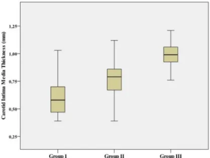
Figure 2: Comparison of the groups in respect to average carotid intima media thickness
simple correlation analyses were performed to search the associa-tion between continuous parame-ters accordingly while Student’s t
test, Mann-Whitney U test, and Kruskal-Wallis tests were per-formed to compare groups accord-ingly. A p value of less than 0.05
was considered to show statistically significant result.
RESULTS
A total of 159 patients included to the study after the presided exclusion criteria. The group I with mean age of 40±7 years was composed of 49 subjects, the group II with mean age of 40±7 years included 71 subjects, and the group III with mean age of 40±5 years had 39 patients. Age, height, weight, BMI, the left ventri-cle ejection fraction values were sta-tistically similar across the groups (p values 0.879, 0.298, 0.913, 0.452, and 0.299 respectively). Although SBP was the highest for the group III (p <0.001), DBP did not differ between the groups (p<0.436).
ASI values of the groups were 2.53±0.37, 3.06±0.29, and 3.67±0.32 respective-ly (Figure 1). Average ASI value of the group II was statistically higher than group I (p<0.001) and signifi-cantly lower than the group III (p<0.001); inversely average AD val-ue of group II was significantly low-er than group I (p<0.05 with z score= -2.362) but higher than the group III (p <0.01 with z score = -2.724).
Average CIMT values of the groups were 0.61±0.17, 0.76±0.15, and 0.98±0.14 respectively (Figure 2). Similar to the ASI values; CIMT value of the group II was statistically higher than the group I (p<0.001) but lower than the group III (p<0.001). Also frequency of abnormal CIMT values was high-est in group III, then group II and the least in group I (2%, 6%, and 46% respectively). Although we could not find any significant differ-ence between the group I and the group II in respect to frequency of abnormal CIMT, the group III had
higher frequency of abnormal CIMT than both the group I (p <0.001) and the group II (p <0.001). Rest of demographic and ultrasonographic findings of the subjects was expressed in Table 1 and 2. In correlation analysis, we found significant positive correlation between ASI and CIMT (r=0.668; p <0.001) and inverse correlation between AD and CIMT (r= -0.687; p<0.001). BMI was well correlated to ASI (r= 0.156; p<0.05), CIMT (r=0.372; p<0.001), and age (r=0.283; p<0.001). In our study, age did not correlate to parameters reflecting aortic elasticity but had significant statistical correlation with CIMT (r=0.218; p<0.01). Glucose, creatinine, TSH values and lipid profile of the groups did not differ from each other as shown in Table 3.
Table 1: Demographic findings of the subjects.
Group I (n=49) Group II (n=71) Group III (n=39) p value
Age (year) 40±7 40±7 40±5 0.879
Height (cm) 174±7 173±7 172±5 0.298
Weight (kg) 85±11 85±8 85+9 0.813
BMI (kg/m²) 27.9±2.7 28.2+2.2 28.8+3.0 0.452
(BMI: Body mass index)
Table 2: Ultrasonographic findings of the subjects
Group I (n=49) Group II (n=71) Group III (n=39) p value
SBP (mmHg) 113±9 120±9 126±8 <0.001
DBP (mmHg) 75±4 76±6 76±7 0.436
LVEF (%) 63±2 63±3 62±3 0.299
Aortic Stiffness Index 2.53±0.37 3.06±0.29 3.67±0.32 <0.001
Aortic Distensibility 7.04±2.91 3.61±1.20 1.82±0.61 <0.001
CIMT (mm) 0.61±0.17 0.76±0.15 0.98±0.14 <0.001
CIMK > 1 mm (%) 1 (2) 4 (6) 18 (46) <0.001
CIMT: Carotid intima media thickness; DBP: Diastolic blood pressure; LVEF: Left ventricular ejection fraction; SBP: Systolic blood pressure; Aortic distensibility showed as [1/(10³xmmHg)]
Table 3: Laboratory findings of the subjects.
Group I (n=49) Group II (n=71) Group III (n=39) p value
Hemoglobin (gr/dl) 15.4±1.0 15.3±1.2 14.8±1.5 0.753 Glucose (mg/dl) 101±11 100±17 101±13 0.13 Creatinine (mg/dl) 0.94±0.14 0.94±0.11 0.99±0.14 0.516 Total Cholesterol (mg/dl) 196±40 204±41 207±72 0.336 Triglyceride (mg/dl) 192±78 189±86 201±77 0.8 HDL (mg/dl) 38±7 40±8 39±8 0.184 LDL (mg/dl) 124±34 131±32 131±26 0.154 TSH (µIU/mL) 1.58±1.07 1.32±0.54 1.39±0.98 0.749
HDL: High density lipoprotein. LDL: Low density lipoprotein. TSH: Thyroid stimulating hormone
DISCUSSION
AGA is described as recession of frontal hair line resulting as balding of the scalp cortex in which the Hamil-ton-Norwood baldness scale (3,4) is used to determine the classifica-tion. Several studies discussed the effect of androgens in AGA, re-garding that young male AGA pa-tients shows a higher dihydro-teststerone-teststerone ratio com-pared with healthy population (1,9). The effects of androgens on formation of atherosclerosis is
ex-plained by the possible harmful af-fects combined with vasocon-striction caused by androgens (2,10). Fujimoto et al. showed that serum androgen causes increased atherosclerotic changes in rats by stimulating the proliferation of vascular smooth muscles (11). Since Cotton et al. (12) first de-scribed the association between AGA and CAD, the literature faced with efforts to find an an-swer to solve the effects of this possible correlation (13-16). Lesko et al (13) found a positive
correla-tion between risks of myocardial infarction with severity of alopecia. Latufo et al (14) showed that ver-tex pattern AGA is a marker for increased risk for CAD. The au-thors proved that pathogenesis of carotid atherosclerosis are similar with coronary atherosclerosis; presence of any atheromatous plaque located in carotid arteries predicts a strongly possible coro-nary artery disease (17). Since the CIMT is an early predictor of CAD (18), by the postulate based on cor-relation of AGA with CAD, color
Doppler US can be used to deter-mine the early effect of AGA on CAD due to its sensitivity on imag-ing premature atheroma. Normal value of CIMT ranges from 0,25 mm to 1 mm and values greater than 1mm accepted as abnormal (8). The correlation of AGA score with CIMT value is discussed be-fore but results are controversial. Some authors (5, 19) found a posi-tive correlation between AGA score compared with CIMT value; while others report no significant correlation (20). We found a signif-icant correlation as AGA scores crease, the mean CIMT values in-crease in contrast with the reports of Agac et al. (20) and this discord-ance is due to the result of their exclusion criteria in selection of study population. They included relatively younger ages of patients who are not in the range of athero-sclerotic border yet.
Aortic elasticity was measured via aortic stiffness index and aortic distensi-bility. Both parameters are inverse-ly associated with each other and they are reflecting aging and ather-osclerosis (21). Presence of the
cardiovascular risk factors fastens atherosclerotic process; therefore reduce aortic elasticity meaning in-crement in ASI and reduction in AD. Also impairment in aortic elasticity was found to be related to extent of coronary artery disease (22). But mostly, the carotid-femoral pulse wave velocity (PWV) method was used to measure vas-cular stiffness in these studies (20-22). However the arterial stiffness was evaluated by echocardiography in our study. It is known that aorta reflects central hemodynamics bet-ter than femoral arbet-tery since femo-ral artery is a muscular vessel (23, 24). Additionally, the effect of ath-erosclerosis on muscular vessels are less than elastic great vessels such as the aorta. Difficulty in groin exposure especially in obese patients and uncorrectly estimated distance between two recording points (resulting overestimation among obese patients) are other limitations to PWV method which impairs its diagnostic accuracy (24). Echocardiography offers an easy way to assess arterial stiffness and can be alternative to PWV method in presence of these limitations.
ASI and AD shows the aortic elastic properties as mentioned before. In case of an aortic elasticity deterio-ration, ASI value increases with a decrement of AD value. Since the arterial stiffness may precede the early pathologic intimal changes (20).Our hypothesis is that AGA might be an indicator of arterial stiffness and early-onset of athero-sclerosis when combined with CIMT values. We found that as AGA score increases; the values of aortic elastic properties are nega-tively affected by increment in ASI value and decrement in AD value along with increment of mean CIMT value.
To conclude; as increased androgenic activity causes atherosclerotic changes, AGA patients should alert the clinicians on the means of CAD and should be closely fol-lowed up with their CIMT and ASI values. Increment of these values in AGA patients indicating early onset atherosclerosis should induce the physician for possible CAD. This approach gives chance to pre-vent AGA patients from future ad-verse cardiovascular events. REFERENCES
1- Sreekumar GP, Pardinas J, Wong CQ et
al. Serum androgens and genetic linkage analysis in early onset androgenetic alo-pecia. J Invest Dermatol 1999;113:277-279.
2- Dunajska K, Milewicz A, Szymczak J et
al. Evaluation of sex hormone levels and some metabolic factors in men with coronary atherosclerosis. Aging Male 2004;7:197–204.
3- Hamilton JB. Male hormone stimulation
is prerequisite and an incitantin com-mon baldness. Am J Anat 1942:71:451-480.
4-. Norwood OT. Male pattern baldness:
Classification and incidence. South Med J 1975;68:1359-1365.
5- Dogramaci AC, Balci DD, Balci A, et
al.Is androgenetic alopecia a risk for atherosclerosis?J Eur Acad Dermatol Venereol. 2009;23:673-677.
6- Lang RM, Bierig M, Devereux RB, et al.
Chamber Quantification Writing Group, American Society of Echocardiog-raphy’s Guidelines and Standards Committee, European Association of Echocardiography.Recommendations for chamber quantification: a report from the American Society of Echocar-diography’s Guidelines and Stand-ardsCommittee and the Chamber Quan-tification Writing Group, developed in conjunction with the European Associa-tion of Echocardiography, a branch of the European Society of Cardiology. J Am SocEchocardiogr 2005;18:1440– 1463.
7- Erdogan D, Calıskan M, Yıldırım I. Effect of normal blood pressure, prehy-pertension and hyprehy-pertension on left ven-tricular diastolic function and aortic elastic properties. Blood Pressure 2007;16:114-121.
8- Katakami N, Kaneto H, Shimomura I. Carotid ultrasonography: A potent tool for better clinical practice in diagnosis of atherosclerosis in diabetic patients. J Diabetes Investig. 2014;5:3-13. 9- Nyholt DR, Gillespie NA, Heath AC, et
al. Genetic basis of male pattern bald-ness. J Invest Dermatol 2003;121:1561-1564.
10- Wynne FL, Khalil RA. Testosterone and coronary vascular tone: implications in coronary artery disease. J Endocrinol Invest 2003;26:181-186.
11- Fujimoto R, Morimoto I, Morita E et al. Androgen receptors, 5 alphareductase activity and androgen-dependent prolif-eration of vascular smooth muscle cells. J Steroid Biochem Mol Biol 1994;50:169-174.
12- Cotton SG, Nixon JM, Carpenter RG , et al. Factors discriminating men with coronary heart disease from healthy controls. Br Heart J. 1972;34:458–464.
13- Lesko SM, Rosenberg L, Shapiro S. A case-control study of baldness in rela-tion to myocardial infarcrela-tion in men.JAMA 1993; 269:998-1003.
14- Lotufo PA, Chae CU, Ajani UA et al. Male pattern baldness and coronary heart disease: the physicians’ health study. Arch Intern Med 2000;160:165-171.
15- Herrera CR, D’Agostino RB, Gerstman BB et al. Baldness and coronary heart disease rates in men from the Framing-ham Study. Am J Epidemiol 1995;142:828-833.
16- Miric D, Fabijanic D, Giunio L et al. Dermatological indicators of coronary risk: a case-control study. Int J Cardiol 1998; 67:251-255
17- Sharrett AR, Sorlie PD, Chambless LE, et al. Relative importance of various risk factors for asymptomatic carotid
atherosclerosis versus coronary heart disease incidence: The Atherosclerosis Risk in Communities Study. Am J Epi-demiol. 1999;149:843-852.
18- van der Meer IM, Bots ML, Hofman A et al. Predictive value of noninvasive measures of atherosclerosis for incident myocardial infarction: the Rotterdam Study. Circulation 2004; 109:1089-1094.
19- Arias-Santiago S, Gutiérrez-Salmerón MT, Castellote-Caballero L, et al. And-rogenetic alopecia and cardiovascular risk factors in men and women: a com-parative study. J Am Acad Dermatol. 2010;63:420-429.
20- Agac MT, Bektas H, Korkmaz L, et al. Androgenetic alopecia is associated with increased arterial stiffness in asymptomatic young adults. J Eur Acad Dermatol Venereol. 2015;29:26-30.
21- Sethi S, Rivera O, Oliveros R, et al.. Aortic stiffness: pathophysiology, clini-cal implications, and approach to treat-ment. Integr Blood Press Control. 2014;7:29-34.
22- Roos CJ, Djaberi R, Schuijf JD, et al. Relationship between vascular stiffness and stress myocardial perfusion imaging in asymptomatic patients with diabetes. Eur J Nucl Med Mol Imaging. 2011;38:2050-2057.
23- Safar ME, Levy BI. Studies on arterial stiffness and wave reflections in hyper-tension. Am J Hypertens. 2015;28:1-6. 24- Cavalcante JL, Lima JA, Redheuil A, et
al. Aortic stiffness: current understand-ing and future directions. J Am Coll Cardiol. 2011;57:1511-1522.