© 2018 Journal of Medical Ultrasound | Published by Wolters Kluwer - Medknow 107
i
ntroduCtionSplenic torsion is a rare condition happening due to the weakening or malformation of the splenic ligaments and is mostly seen in children and young women. It is usually associated with the wandering spleen disease. Whereas, in the present case, the spleen was in its normal anatomical position which is exceedingly rare and is mentioned in only one case report.[1] Lack of symptoms except abdominal pain may lead to difficulty in diagnosis of torsion. This issue may be more applicable to patients with partial splenic torsion in normal anatomical position, like in the present case. Early surgical intervention is crucial for prevention of splenic infarction.
C
AsEr
EportA 20-year-old female presented with complaints of severe left upper quadrant pain of 3 days duration, intermittently radiating toward the left lower quadrant. She did not declare history of trauma or injury. She had repeatedly vomited nonbloody, nonbilious fluid. Bowel opening was regular. She was admitted to the different emergency services and treated with nonsteroidal anti-inflammatory drugs, but as seems, the pain was not relieved. Laboratory studies showed only high C-reactive protein levels (1467 mg/L). She had a history of using contraceptives but stopped to take this medicine 2 years ago. She also had a history of asthma and peptic ulcer.
Management and outcome
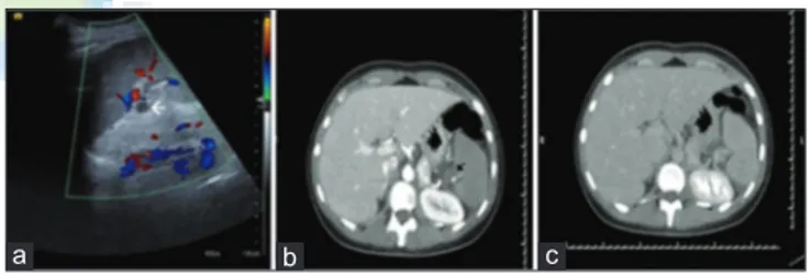
The patient had been consulted to radiology department. On abdominal ultrasound (US), the spleen was found in its normal anatomic position and Doppler US examination revealed that there is no blood flow in splenic vein [Figure 1a].
A contrast-enhanced computed tomography (CT) scan was also performed. Portal-phase CT images showed hypoperfused spleen, patent splenic artery, and contrast in proximal splenic vein but no contrast filling in distal part. There was no thrombus in splenic vein [Figure 1b]. Delayed-phase CT images revealed
Partial Splenic Torsion in situ: Revealed by Ultrasound and
Computed Tomography
Mert Bayramoglu1, K. Murat Haberal1, Gokhan Kahraman1*, Tevfik Avci2
1Department of Radiology, Faculty of Medicine, Baskent University, Ankara, Turkey, 2Department of General Surgery, Faculty of Medicine, Baskent University, Ankara, Turkey
We present a case of acute abdominal pain due to partial torsion of spleen located in its own normal anatomical position in a 20-year-old woman, diagnosed by ultrasound and confirmed on computed tomography and treated laparoscopically.
Keywords: Abdominal pain, splenopexy, partial splenic torsion
Access this article online
Quick Response Code:
Website: www.jmuonline.org DOI:
10.4103/JMU.JMU_25_18
Address for correspondence: Dr. Gokhan Kahraman,
Department of Radiology, Faculty of Medicine, Baskent University, Ankara, Turkey. E‑mail: [email protected]
How to cite this article: Bayramoglu M, Haberal KM, Kahraman G,
Avci T. Partial splenic torsion in situ: Revealed by ultrasound and computed tomography. J Med Ultrasound 2018;26:107-8.
Abstract
Case Report
This is an open access journal, and articles are distributed under the terms of the Creative Commons Attribution-NonCommercial-ShareAlike 4.0 License, which allows others to remix, tweak, and build upon the work non-commercially, as long as appropriate credit is given and the new creations are licensed under the identical terms.
For reprints contact: [email protected] Received: 27-11-2017 Accepted: 19-01-2018 Available Online: 12-06-2018
Figure 1: (a) Preoperative Doppler ultrasound image showing flow in splenic
artery and absence of flow in splenic vein (white arrow). (b) Preoperative axial portal‑phase computed tomography image. Spleen is relatively hypodense to liver. There is no contrast filling in splenic vein (black arrow). (c) Preoperative axial late‑phase computed tomography image. Spleen becomes isodense to liver
a b c
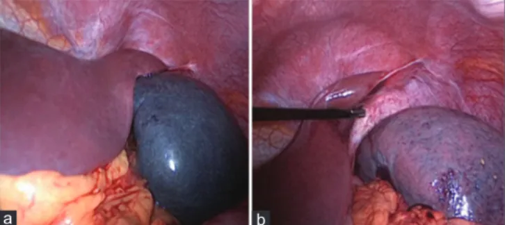
a patent splenic vein [Figure 1c]. These data showed that the partial torsion of spleen caused the splenic vein obstruction. The patient underwent laparoscopic operation and detorsion of spleen was performed. Because of the shortness of the pedicle of the spleen, splenopexy could not be performed [Figure 2]. Postoperative Doppler US examination confirmed patency of splenic vein. At the first postoperative follow-up after 1 month, the patient was evaluated as normal. The patient did not come to the next follow-up.
d
isCussionThe spleen is hold in its normal position by the gastrosplenic and splenorenal ligaments. While the former one connects the greater curvature of the stomach to the ventral aspect of the spleen, the latter attaches the spleen to the posterior abdominal wall. Inadequate development of these ligaments resulting in a long splenic mesentery and acquired factors such as abdominal wall laxity, the hormonal effects of pregnancy, and splenomegaly may increase splenic mobility.[2]
A wandering spleen or splenic ptosis is defined as a migration of the spleen from its anatomic location in anywhere in the abdomen or pelvis.[3]
Normally located spleen with ligamentous laxity as wandering spleen is usually asymptomatic. Torsion, a complication of wandering spleen, makes the situation symptomatic. Torsion of wandering spleen is a relatively rare clinical occurrence with about 500 cases reported and an incidence of < 0.2%.[4] It is fifteen times more common in females, usually detected between 20 and 40 years of age.[5]
Serious and constant abdominal pain suggests splenic torsion and secondary ischemia due to compromise in venous outflow, which produces congestion and impairment of arterial inflow. Pain appears as a result of both stretching in splenic capsule with rapid splenic enlargement and local peritonitis.[6] To the best of our knowledge, only one case with torsion of a spleen located in a normal anatomical position has been reported in the literature to date.[1] The patient in the referred case underwent an urgent laparotomy and splenectomy for 360° torsion of her spleen. Hence, the present case is unique because the 180º torsion occurred in normal anatomical position of spleen.
Bayramoglu, et al.: A case: Partial splenic torsion in situ
108 Journal of Medical Ultrasound ¦ Volume 26 ¦ Issue 2 ¦ April-June 2018 Ultrasonography and CT are key elements in diagnosing because physical symptoms and laboratory data are nonspecific. In the present case, no flow could be detected in the splenic vein [Figure 1a]. Splenic parenchyma was normal on gray scale imaging. Only arterial flow could be detected in the splenic hilus and parenchyma. We performed a contrast-enhanced CT scan to rule out splenic vein thrombosis [Figure 1b and c]. The presence of blood flow in proximal splenic vein segments could be explained with the tributary veins, namely inferior mesenteric vein or varices due to chronic venous congestion joining the main splenic vein proximal to the torsion.[7] Preservation of the spleen with laparoscopic splenopexy is highly recommended to avoid recurrence of torsion and complication of splenectomy.
C
onClusionSplenic torsion is relatively rare pathology and is usually found in wandering spleen cases. Partial splenic torsion occurring in normal anatomic position is more rare, and to diagnose of such torsion is more difficult even when it becomes symptomatic. A delay in diagnosis could lead to serious consequences, including splenectomy. The possibility of the splenic torsion in its normal anatomic position should come to mind in cases with sharp and persistent left upper abdominal pain, especially in cases without diagnosis, when there are not any other explanations. Laparoscopic splenopexy is an excellent option for organ-preserving treatment in splenic torsion. Doppler US is indispensable diagnostic method in such cases.
Declaration of patient consent
The authors certify that they have obtained all appropriate patient consent forms. In the form the patient(s) has/have given his/her/ their consent for his/her/their images and other clinical information to be reported in the journal. The patients understand that their names and initials will not be published and due efforts will be made to conceal their identity, but anonymity cannot be guaranteed. Financial support and sponsorship
Nil.
Conflicts of interest
There are no conflicts of interest.
r
EfErEnCEs1. Hussain M, Deshpande R, Bailey ST. Splenic torsion: A case report. Ann R Coll Surg Engl 2010;92:W51-2.
2. Raissaki M, Prassopoulos P, Daskalogiannaki M, Magkanas E, Gourtsoyiannis N. Acute abdomen due to torsion of wandering spleen: CT diagnosis. Eur Radiol 1998;8:1409-12.
3. Freeman JL, Jafri SZ, Roberts JL, Mezwa DG, Shirkhoda A. CT of congenital and acquired abnormalities of the spleen. Radiographics 1993;13:597-610. 4. Sharma A, Salerno G. A torted wandering spleen: A case report. J Med
Case Rep 2014;8:133.
5. Desai DC, Hebra A, Davidoff AM, Schnaufer L. Wandering spleen: A challenging diagnosis. South Med J 1997;90:439-43.
6. Sayeed S, Koniaris LG, Kovach SJ, Hirokawa T. Torsion of a wandering spleen. Surgery 2002;132:535-6.
7. Nemcek AA Jr., Miller FH, Fitzgerald SW. Acute torsion of a wandering spleen: Diagnosis by CT and duplex Doppler and color flow sonography. AJR Am J Roentgenol 1991;157:307-9.
Figure 2: (a) A laparoscopic appearance of partially torsioned spleen. The
spleen is significantly congested. (b) After laparoscopic detorsion, the color and appearance of the spleen show an improved circulation and reduced congestion
a b