VALUE OF AXIAL LOADING MAGNETIC RESONANCE IMAGING AND
COMPUTERIZED TOMOGRAPHY-MYELOGRAPHY IN DECISION MAKING IN
THE TREATMENT OF DEGENERATIVE LUMBAR SPINAL STENOSIS*
DEJENERAT‹F LOMBER SP‹NAL STENOZUN TEDAV‹S‹NDE KARAR VER‹RKEN
AKS‹YEL YÜKLENMEYLE B‹RL‹KTE YAPILAN MANYET‹K REZONANS
GÖRÜNTÜLEME VE B‹LG‹SAYARLI TOMOGRAF‹-M‹YELOGRAF‹N‹N DE⁄ER‹*
Mustafa fi‹RVANCI, Kürflat GAN‹YUSUFO⁄LU, Mona BHATIA, Ça¤atay ÖZTÜRK, Neslihan AKSU, Azmi HAMZAO⁄LU
SUMMARY:
Aim: The degenerative lumbar spinal stenosis
(LSS) is a dynamic phenomenon. Changes in posture and physical activities as standing and walking can aggravate symptoms. The aim of the study was to evaluate the results of post axial loading Computed Tomography Myelography (CTM) versus Magnetic Resonance Imaging (MRI) in patients undergoing surgery for multilevel degenerative lumbar spinal stenosis.
Methods: Thirty patients with multilevel degenerative LSS scheduled for elective surgery were enrolled in the study. Preoperatively, all patients underwent both MRI and CTM, in supine psoas relaxed position and post loading by axial compression in slight extension. Quantitative evaluation for LSS was conducted by two experienced radiologists. The parameters included dural sac cross sectional area, lateral recess and foraminal evaluations for stenosis, on CTM and MRI. Statistical analysis of the data was performed to evaluate relative advantages and additional information depicted by axial loaded CTM versus MRI.
Results: In 21 of 30 of patients, axially loaded
images demonstrated additional information, such as findings of spinal canal encroachment, deformation of the dural sac and nerve roots, reduction in cross sectional dural sac area, narrowing of the lateral recess, increased number and severity of affected stenotic sites particularly in borderline cases after axial compression. In 11 patients, borderlines for stenosis were passed in at least one level. CTM and MRI findings were comparable.
Conclusion: Axial loading mimics erect posture on
CTM and MRI, thus partially eliminating the diagnostic dilemma associated with the dynamic nature of LSS. The study demonstrated underestimation of extent and severity of LSS on preloading images. Post axial loaded CTM and MRI added valuable information to the preoperative assessment of patients of multilevel spinal stenosis by identifying additional levels and severity of stenosis, particularly in borderline cases.
Keywords: Lumbar spinal stenosis, myelography,
spinal CT · spinal MR
Level of Evidence: Comperative clinical study,
Level III
ÖZET:
Amaç: Dejeneratif lomber spinal stenoz
(LSS) dinamik bir olgudur. Postüral de¤ifliklikler ile ayakta durma ve yürüme gibi fiziksel aktiviteler semptomlar› ortaya ç›karabilir. Çal›flman›n amac›, çok seviyeli dejeneratif LSS nedeniyle ameliyat edilen olgularda aksiyel yüklenme sonras› manyetik rezonans görüntüleme (MRG) ile bilgisayarl› tomografi-miyelografi (BTM) nin sonuçlar›n›n karfl›laflt›r›lmas›d›r.
Yöntem: Elektif cerrahi için randevu verilen
çok seviyeli dejeneratif LSS olan 30 hasta bu çal›flmaya dahil edildi. Preoperatif olarak tüm hastalara supin pozisyonunda psoas kas› gevflekken ve hafif ekstansiyonda uygulanan aksiyel kompresyon sonras›nda MRG ve BTM çekilmifltir. Deneyimli iki radyolog taraf›ndan LSS aç›s›ndan kantitatif olarak de¤erlendirilmifltir. Parametreler, dural kese kesit alan›, lateral reses ve foraminal stenozun de¤erlendirmesini içermifltir. Verilerin istatistiksel de¤erlendirmesiyle aksiyel yüklenme ile yap›lan BTM ve MRG tetkiklerinin göreceli avantajlar› ortaya konmufltur.
Sonuçlar: 30 hastan›n 21’inde aksiyel
yüklenme ile al›nan görüntüler spinal kanalda daralma, sinir kökleri ve duran›n deformasyonu, dural kesenin kesit aln›nda azalma, lateral reseste daralma, s›n›rdaki olgularda stenotik bölgelerin belirginleflmesi gibi ek bilgiler vermifltir. 11 hastada, en az bir seviyede stenoz gösterilmifltir. BTM ve MRG den elde edilen bulgular karfl›laflt›r›labilirdir.
Ç›kar›mlar: Aksiyel yüklenme, BTM ve
MRG de erect postürünü taklit eder. Böylece, LSS nin dinamik do¤as›yla ilintili olan tan›sal sorun k›smen afl›lm›fl olur. Çal›flma, yüklenme öncesi görüntülerle LSS nin a¤›rl›¤› ve yayg›nl›¤›n›n hafife al›nd›¤›n› göstermifltir. Çok seviyeli LSS olan hastalar›n preoperative de¤erlendirilmesinde aksiyel yüklenme sonras› çekilen MRG ve BTM den elde edilen bilgilerle özellikle s›n›rdaki olgularda, ek stenotik seviyeler ve stenozun fliddeti daha iyi saptanabilmektedir.
Anahtar Kelimeler: Lomber spinal stenoz,
miyelografi, spinal BT, spinal MR
Kan›t Düzeyi: Karfl›laflt›rmal› klinik çal›flma,
INTRODUCTION:
Degenerative Lumbar Spinal Stenosis (LSS) is a frequently encountered condition causing low back pain, radiculopathy, neurogenic claudication, and functional disability among middle-aged and elderly populations (10,20,32). Degenerative LSS is defined as a narrowing of the spinal canal, the lateral nerve root canals, or the intervertebral neural foramina, due to progressive hypertrophy of surrounding osteocartilaginous and/or ligamentous elements(2, 10).
It is well known that LSS is a dynamic phenomenon. Changes in posture as well as extension and physical activities, such as standing and walking, can aggravate symptoms (27,31). Conversely, forward flexion of the spine, squatting, and lying supine with slight hip flexion typically relieves the symptoms (21,23,30-31). Myelographic examination including flexion and extension of the lumbar spine in a standing position has long been used as a diagnostic tool in patients suspected of encroachment into the spinal canal (1,5,30-31). Since the advent of computed tomography (CT) and magnetic resonance imaging (MRI), these non-invasive techniques have replaced myelography in most cases, mainly because of their superior capabilities for analyzing intraspinal soft tissues (4,9,29,32). Plain CT underestimates root impingement documented by conventional myelography due to its limitations, such as inability to identify intrathecal nerve root compression due to similar surrounding CSF densities. This limitation is overcome by intrathecal contast CT-myelography (CTM).
Traditionally, CT (with or without myelography) and MRI are performed with the patient in a supine, psoas-relaxed position (PRP) with consequent flexion of the lumbar
spine and widening of the spinal canal (21-22). Supine position reduces the compressive load of the spine approximately 75 % from upright standing load (18,39). Thus, instead of creating a symptom-provoking examination position, the standard positioning of the patient during CT and MRI examinations today probably has the opposite effect (39-40). Thus, being static imaging techniques, CT and MRI poorly assess the dynamic status of the lumbar spinal stenosis, thus making dynamic evaluation with myelography still the preferred examination for many surgeons in the preoperative assessment of LSS(5,16,31).
Since the maximal thecal compression occurs in the erect, extended position, it is essential that CT and MRI be performed subsequent to axial loading of the mildly extended spine to simulate the mechanical effects of the upright position (17,21,28-31). Recently, a device has been constructed that makes it possible to perform a CT scan and MRI of the lumbar spine with the subject in an axially loaded position (axial compression in extension (ACE))(3,17,21,28-31).
The aim of this study was to evaluate the results and compare the diagnostic value of post-axial loading CTM versus post-axial loading MRI in the radiologic assessment of patients undergoing surgery for multi-level degenerative lumbar spinal stenosis.
MATERIALS AND METHODS:
Thirty patients with multi-level degenerative LSS scheduled for elective surgery were enrolled in the study. There were 12 males and 18 females in the age range of 43 to 85 years. All patients underwent a preoperative MRI and CTM. Each patient was first examined in the conventional supine psoas
relaxed position with slight flexion in the hips and knees followed by examination in a supine, slightly extended position during axial compression (36,40).
Axial loading of the spine was performed using a non-magnetic compression device and a harness (7,34,40). The patient was lying supine with extended hips and knees. To prevent flexion of the spine during compression, a cushion was placed behind the lumbar spine. The feet were positioned against a footplate on the compression device. The harness worn by the patient was attached to the compression device using side straps, which were tightened for axial loading of the lumbar spine (10,13). The chosen load was approximately 50% of the subject’s body weight, with equal load distribution on both legs. The load was applied for at least five minutes, after which imaging was performed (3,18,25).
As recommended by other authors, patient selection for the axially loaded examination was done after serious evaluation. Osteoporotic or fractured spine or a spine with a skeletal malignancy represented contraindications for loading (39,40). There were no patients that were excluded from the study because of these criteria.
MRI was performed on all subjects in a 1.5-T system (Siemens Sonata, Erlangen, Germany) using standard protocols and sequences. CTM was performed in all patients after intrathecal injection of 13-14 cc of 180 mgI/cc Iohexol. To mix the contrast with the spinal fluid, patients were told to turn around and to rest prone for a few minutes. The disc levels from L1 to S1 were examined. The box for transverse slices was placed parallel to the disc examined.
Quantitative and qualitative evaluation for LSS was conducted by a radiologist with 13 years experience in spinal imaging. The parameters included dural sac cross sectional area (DS-CSA) and lateral recess evaluations post-loading for nerve root compression or stenosis after carefully inspecting and selecting the most comparable images on MRI and CTM during ACE. DS-CSA was measured on the transverse angled sections through the central part of the disc on MR and CTM images from the first lumbar through the first sacral lumbar vertebrae in all patients. The image selected was the one in which the area seemed to be the smallest on each disc level. The quantitative criteria used for central anatomical LSS were in accordance with experimental(27) and clinical studies (5,13). DS-CSA values below 100 mm2 indicate a relative central stenosis, whereas values below 75 mm2indicate an absolute central lumbar spinal stenosis. On DS-CSA measurements, the difference was considered (40)significant if the value differed by 5 % or more between MRI and CTM.
For evaluation of lumbar root compression in the lateral recess caused by lumbar degenerative changes on MRI and CTM, a grading system was devised with similar objective features of impingement for both imaging modalities (36). Each lateral recess was evaluated and nerve root compression graded separately using the following 0- to 3-point scale. Grades were defined as follows: Grade 0, no lateral recess narrowing or root compression; Grade 1, evidence of lateral recess narrowing, but no objective evidence of root compression (nerve root is visualized and not widened, flattened, or altered); Grade 2, more significant lateral recess narrowing (angular or trefoil) with the nerve root judged
to be flattened or widened, but with preservation of CSF around the root in the recess; Grade 3, severe nerve root compression within the lateral recess with obliteration of CSF from the recess (root may or may not be clearly visible, root may be seen coursing through the compressed lateral recess, root may be seen as medially displaced) (36).
Statistical analysis of the data was performed. DS-CSA was measured by post-axial loading CTM and MRI in 30 patients, and 150 intervertebral levels for each method, with a total of 300 levels. At 11 levels, DS-CSA could not be reliably measured by one of the methods. Thus, a total of 289 DS-CSA measurements were statistically analyzed by the chi-square test. An independent t-test was also applied to the data to compare the DS-CSA results of CTM and MRI. Sixteen and one lateral recess could not be reliably evaluated by CTM and MRI, respectively. Thus, a total of 703 lateral recesses were statistically analyzed by the chi-square test.
The current study was approved by the ethical committee of our institution and informed consent was obtained from all patients.
RESULTS:
Central stenosis was present at 77 intervertebral levels of the 150 evaluated. Both CTM and MRI demonstrated relative and absolute stenotic DS-CSA in 16 and 28 intervertebral levels, respectively. Seventy-three intervertebral levels demonstrated normal DS-CSA on both CTM and MRI. Thus in 117 of the 150 (78 %) intervertebral levels evaluated, CTM and MRI demonstrated equivalent findings.
Among the remaining 33 intervertebral levels, 18 patients had relative stenosis on MRI with normal DS-CSA on CTM, three patients had normal DS-CSA on MRI but relative stenosis on
CTM, 10 patients had absolute stenosis on MRI and relative stenosis on CTM, and two patients had relative stenosis on MRI and absolute stenosis on CTM.
Non-filling of the dural sac was observed at six intervertebral levels on CTM and it was deduced to represent absolute stenosis. In these cases, MRI was superior to CTM due to its superior soft tissue contrast. In one patient of severe stenosis due to excessive bony hypertrophy, evaluation of the dural sac margins on MRI was difficult while CTM with its superior bony and spatial resolution clearly demonstrated the true DS-CSA.
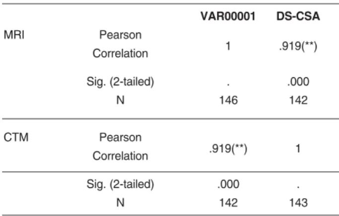
DS-CSA results, an independent t-test and correlation of the two imaging methods are summarized in Tables-1, 2, and 3. There is a statistically significant difference at the level of 0.05 between the spinal stenosis measurements of CTM and MRI by chi-square analysis. An independent sample t-test also showed similar results. The mean value of DS-CSA measured by CTM was 1.21, while it was 1.08 by MRI. This difference is statistically significant at the 0.05 level by independent t-test. Correlation of the two imaging methods regarding DS-CSA measurement was 0.92, and this correlation is statistically significant at 0.01 level.
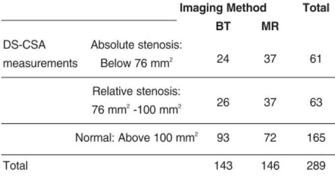
Table - 1. DS-CSA Measurements (Method: Cross tabulation).
Imaging Method Total
BT MR
DS-CSA Absolute stenosis:
measurements Below 76 mm2 24 37 61
Relative stenosis:
76 mm2-100 mm2 26 37 63
Normal: Above 100 mm2 93 72 165
Among the 30 patients, 360 lateral recesses were evaluated on ACE images by CTM and MRI. Of these 360 lateral recesses, 299 (83 %) sites demonstrated similar grade on both modalities while at 61 (17 %) sites there existed a discrepancy between CTM and MRI. Non-filling of the dural sac on CTM accounted for 16 patients where assessment was difficult, however, six of these lateral recesses demonstrated grade 3 stenosis on MRI, thus correlating well between CTM and MRI. In effect, 305 (85 %) lateral recesses thus had corroborative findings and grades of
stenosis. Non-filling of the dural sac on CTM overestimated the degree of stenosis in 10 patients where MRI demonstrated grade 0, grade 1 and grade 2 in four, four and two lateral recesses, respectively. Non-filled root sleeves on CTM overestimated the degree of stenosis due to its poorer soft tissue contrast than MRI.
CTM on account of its superior spatial resolution and thin section reconstructions better delineated the nerve roots in the lateral recesses. In 11 lateral recesses where MRI demonstrated grade 0, CTM demonstrated grade 1 and early nerve root deviation; in nine lateral recesses where MRI demonstrated grade 1, CTM demonstrated grade 2; and in five lateral recesses where MRI demonstrated grade 2, CTM demonstrated grade 3 stenosis. MRI overexaggerated the grade of nerve root compromise and lateral recess stenosis in 20 patients. While CTM demonstrated grade 0, MRI demonstrated grade 1 and 2 in 12 and one patient, respectively. In five lateral recesses, CTM demonstrated grade 1 while MRI demonstrated grade 2 and in two lateral recesses CTM demonstrated grade 2 while MRI demonstrated grade 3 stenosis. These
Table - 2. Independent Samples Test.
Levene's Test for Equality of
Variances t-test for Equality of Means
F Sig. t df Sig. Mean Std 95% Confidence (2-tailed) Difference Error Interval of the
Difference Difference Lower Upper DS-CSA Equal variances
Measurements assumed .416 .520 2.214 287 .028 .1287 .05811 .01428 .24303 Equal variances
not assumed 2.212 284.428 .028 .1287 .05815 .01418 .24312
Table - 3. Correlation of DS-CSA Measurements.
VAR00001 DS-CSA MRI Pearson Correlation 1 .919(**) Sig. (2-tailed) . .000 N 146 142 CTM Pearson Correlation .919(**) 1 Sig. (2-tailed) .000 . N 142 143
cases were explained by the superior spatial resolution of CT over MRI.
Lateral recess evaluation results and correlation of the two imaging methods are shown in Tables 4 and 5. No statistically significant differences (p>0.05) were observed between the imaging methods by chi-square analysis regarding lateral recess evaluation. Correlation of the two imaging methods regarding lateral recess evaluation is 0.84 and this correlation is statistically significant at 0.01 level.
DISCUSSION:
With increasing longevity of life and aging populations, the prevalence and associated clinical disability related to degenerative LSS is on the rise (33).
Spinal stenosis is a compressive process (6, 8). Combinations of discal bulging or disc-osteophyte complex usually coexist with facet and ligament hypertrophy and cause spinal canal narrowing. Besides the structural aspect, LSS has a dynamic component (22,24,28,31). Dynamic myelography has thus been the gold standard for the diagnosis of lumbar spinal stenosis for a while. The advent of non-invasive CT and MRI has shown advantages of improved spatial and soft tissue contrast definition in the assessment of the nature, level and severity of LSS and its impact on neural elements (16). However, the majority of CT and MRI are performed with the lumbar spine in a supine relaxed position, which results in unloading of the spine and enlargement of the canal and consequent underestimation of the lumbar spinal stenosis. Axial loading mimics erect posture on CTM and MRI, thus partially eliminating the diagnostic dilemma associated with the dynamic nature of LSS. Willen (40) and Danielson et al. (7) developed the axially loaded CTM technique, and later the MRI technique conducted with the subject in a supine position (39).
The changes in the spinal canal from PRP to simulated upright standing (ACE) have been described (7,40). According to these studies, there is a considerable risk of failing to detect an essential narrowing of the spinal canal if the examination is performed only in the unloaded traditional position. The specificity of the encroachment diagnosis was shown to increase considerably when the
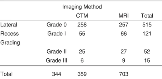
Table - 4. Lateral Recess Evaluation (Method: Cross tabulation). Imaging Method CTM MRI Total Lateral Grade 0 258 257 515 Recess Grade I 55 66 121 Grading Grade II 25 27 52 Grade III 6 9 15 Total 344 359 703
Table - 5. Correlation of Lateral Recess Evaluation.
Lateral Recess VAR00001 Grading
Lateral Recess
Evaluation Pearson Correlation 1 .841(**) by CTM
Sig. (2-tailed) . .000
N 344 343
Lateral Recess Pearson Correlation .841(**) 1 Evaluation
by MRI
Sig. (2-tailed) .000 .
N 343 359
patient was subjected to an axial load. In a study of healthy individuals, Kimura and Hargens (7) showed that the load comprising 50 % of a subject’s body weight applied by a compression device can morphologically simulate the lumbar spine in the upright position. Thus, in our study, all CTM and MRI examinations were conducted in the ACE position by an axial load of 50 % of the patient’s body weight.
The importance of spinal loading and posture during CT and MRI examinations has been reported in experimental and clinical studies by several authors (7,15,19,22,26,29,34,37-40). Penning and Wilmink (22)showed that the dural sac narrowed concentrically in the spinal canal during extension, and widened with relief of involved nerve roots in flexion. The structural changes like constriction of the dural sac and compression of the nerve roots caused by disc bulge or protrusion, thickened ligamentum flavum, and/or changed configuration of the dorsal fat pad during axial compression in slight extension are evident (39-40).
Authors have deemed MRI as the diagnostic study of choice due to its non-invasive nature, multiplanar capabilities, and the lack of ionizing radiation making it particularly desirable for patient and physician (12,14,35). In making the diagnosis of spinal stenosis, traditional non-loading CTM and MRI have been reported to be equally accurate (85.3 %) and sensitive (87.2 %) (4). In addition, in our study MRI did compare favorably with CTM. At 78% of intervertebral levels evaluated for central stenosis and 85 % of lateral recesses evaluated, post-loading CTM and MRI showed corroborative results. Although a statistically significant difference was found between the DS-CSA measurements of the two imaging methods, the correlation was
high. Moreover, in the intervertebral levels at which DS-CSA measurement discrepancies were demonstrated, detailed evaluation of the DS-CSA measurements showed that the figures were close to borderline between relative and normal or relative and absolute stenosis. Although discrepancy was noted in the grading of stenosis at these intervertebral levels, however, the actual figures did not demonstrate a significant difference, i.e., the difference was less than 5 % of the actual figure, thus making CTM and MRI comparable for grading of central stenosis. The most prominent discrepancies between the methods were caused by the differences at non-stenosed levels.
In our study, MRI, with its superior soft tissue contrast definition, was superior in cases where there was non-filling of the central dural sac or lateral recesses on CTM. This was observed in six out of the 150 intervertebral levels evaluated for central stenosis and in 16 lateral recesses of the 360 evaluated.
Other authors have seen a distinct advantage of CTM over MRI in one-third of cases for degenerative disease of the lumbar spine providing additional useful information. This included definitions of the extent of large disc herniations, demonstration of focal neural compression by small herniations, and clarifying abnormalities of the facets, including synovial cysts. These results should help to refine the indications for CTM (11). In our study, of the 45 discrepant lateral recesses evaluated, and in cases of non-filling of the nerve root sleeves, CTM overestimated the degree of stenosis due to poorer soft tissue contrast compared to MRI. However, CTM was found to be superior in the spatial resolution and more accurately delineated and
assessed lateral recess compromise than MRI in mild grades of nerve root deviation and compression (36).
In conclusion, although both modalities are extremely comparable, post-loading MRI has the distinct advantage of being non-invasive with no radiation effects and better soft tissue contrast, while post-loading CTM with its superior spatial resolution retains a place in the evaluation of nerve root compromise of the lateral recesses in the evaluation of degenerative LSS. As a preoperative investigation, postaxial loading MRI may be better choice than the invasive CTM. The next part of the current study will be an attempt to compare conventional dynamic myelography versus postaxial loading MRI using the data of the same patient group, in order to seek an answer to the question "Is it possible to totally abandon invasive techniques in this patient group?"
REFERENCES:
1. Amundsen T, Weber H, Lilleas F, Nordal HJ, Abdelnoor M, Magnaes B. Lumbar spinal stenosis: Clinical and radiological features. Spine 1995; 20: 1178–1186.
2. Arnoldi CC, Brodsky AE, Cauchoix J, Crock HV, Dommisse GF, Edgar MA, Gargano FP, Jacobson RE, Kirkaldy-Willis WH, Kurihara A, Langenskiold A, Macnab I, McIvor GW, Newman PH, Paine KW, Russin LA, Sheldon J, Tile M, Urist MR, Wilson WE, Wiltse LL. Lumbar spinal stenosis and nerve root entrapment syndromes. Definition and classification. Clin Orthop Relat Res 1976; 115: 4-5.
3. Danielson B, Willen J. Axially Loaded Magnetic Resonance Image of the Lumbar Spine in Asymptomatic Individuals. Spine 2001; 26: 2601-2606.
4. Bischoff RJ, Rodriguez RP, Gupta K, Righi A, Dalton JE, Whitecloud TS. A comparison of computed tomography-myelography, magnetic resonance imaging, and myelography in the diagnosis of herniated nucleus pulposus and spinal stenosis. J Spinal Disord 1993; 6: 289-295. 5. Bolender NF, Schonstrom NS, Spengler DM. Role
of computed tomography and myelography in the
diagnosis of central spinal stenosis. J Bone Joint Surg 1985; 67-A: 240–246.
6. Ciric I, Mikhael MA, Tarkington JA, Vick NA. The lateral recess syndrome: a variant of spinal stenosis. J Neurosurg 1980; 53: 433-443.
7. Danielson BI, Willen J, Gaulitz A, Niklason T, Hansson TH. Axial loading of the spine during CT and MR in patients with suspected spinal stenosis. Acta Radiol 1998; 39: 604-611.
8. Epstein JA, Epstein NE. Lumbar spondylosis and spinal stenosis. In: Wilkins RH, Rengachary SS (eds). Neurosurgery. McGraw Hill, New York, 1996; pp: 3831 –3840.
9. Moon ES, Kim HS, Park JO, Shin DE, Ha JW, Shim DJ, Kwak YH, Lee KI.. Comparison of the Predictive Value of Myelography, Computed Tomography and MRI on the Treadmill Test in Lumbar Spinal Stenosis. Yonsei Med J 2005; 46: 806-811.
10. Fritz JM, Delitto A, Welch WC, Erhard RE. Lumbar spinal stenosis: a review of current concepts in evaluation, management, and outcome measurements. Arch Phys Med Rehabil 1998; 79: 700-708.
11. Goldberg AL, Soo MS, Deeb ZL, Rothfus WE. Degenerative disease of the lumbar spine. Role of CT-myelography in the MR era. Clin Imaging 1991; 15: 47-55.
12. Goscinski I, Ulatowski S, Urbanik A. Comparison of the clinical usefulness of magnetic resonance (MR), computer tomography (CT) and radiculography (R) in diagnosing lumbar discopathy. Przegl Lek 2001; 58: 885-888.
13. Hamanishi C, Matukura N, Fujita M, Tomihara M, Tanaka S. Cross-sectional area of the stenotic lumbar dural tube measured from the transverse views of magnetic resonance imaging. J Spinal Disord 1994; 7: 388-393.
14. Janssen ME, Bertrand SL, Joe C, Levine MI. Lumbar herniated disk disease: comparison of MRI, myelography, and post-myelographic CT scan with surgical findings. Orthopedics 1994; 17: 121-127. 15. Kimura S, Hargens A. Lumbar spinal disc height
and curvature responds to an axial load using a MRI-compatible compression device. Spine 2001; 26: 2596–2600.
16. Morvan G. Imaging of lumbar stenosis. J Radiol 2002; 83: 1165-1175; discussion 1177-1179. 17. Muhle C, Wiskirchen J, Weinert D, Falliner A,
Wesner F, Brinkmann G, Heller M. Biomechanical aspects of the subarachnoid space and cervical
cord in healthy individuals examined with kinematic magnetic resonance imaging. Spine 1998; 23: 556–567.
18. Nachemson A, Elfstrom G. Intravital dynamic pressure measurements in lumbar discs. A study of common movements, maneuvers and exercises. Scand J Rehabil Med 1970; Suppl 1: 1-40.
19. Nowicki BH, Haughton VM, Schmidt TA, Lim TH, An HS, Riley LH 3rd, Yu L, Hong JW. Occult lumbar lateral spinal stenosis in neural foramina subjected to physiologic loading. Am J Neuroradiol 1996; 17: 1605-1614.
20. Panagiotis ZE, Athanasios K, Panagiotis D, Minos T, Charis M, Elias L. Functional outcome of surgical treatment for multilevel lumbar spinal stenosis. Acta Orthop 2006; 77: 670-676.
21. Penning L, Wilmink JT. Biomechanics of lumbosacral dural sac. A study of flexion-extension myelography. Spine 1981; 6: 398-408.
22. Penning L, Wilmink JT. Posture-dependent bilateral compression of L4 and L5 nerve roots in facet hypertrophy: a dynamic CT-myelographic study. Spine 1987; 12: 488–500.
23. Porter RW, Ward D. Cauda equina dysfunction: The significance of two-level pathology. Spine 1992; 17: 9-15 .
24. Panjabi MM, Takata K, Goel VK. Kinematics of the lumbar intervertebral foeramen. Spine 1983; 8: 348-357.
25 Sato K, Kikuchi S, Yonesawa T. In vivo intradiscal pressure measurements in healthy individuals and in patients with ongoing back problems. Spine 1999; 24: 2468–2474.
26. Schmid MR, Stucki G, Duewell S, Wildermuth S, Romanowski B, Hodler J. Changes in cross-sectional measurements of the spinal canal and intervertebral foramina as a function of body position: in vivo studies on an open-configuration MR system. AJR 1999; 172: 1095-1102.
27. Schonstrom N, Hansson T. Pressure changes following constriction of the cauda equina: An experimental study in situ. Spine 1988; 4: 385-388. 28. Schonstrom N, Lindahl S, Willen J, Hansson T. Dynamic changes in the dimensions of the lumbar spinal canal: an experimental study in vitro. J Orthop Res 1989; 7: 115-121.
29. Schonstrom N, Willen J. Imaging lumbar spinal stenosis. Radiol Clin North Am 2001; 39: 31–53. 30. Schumacher M. Die Belastungsmyelographie.
Fortschr Röntgenstr 1986; 145: 642-648.
31. Sortland O, Magnaes B, Hauge T. Functional myelography with metrizamide in the diagnosis of lumbar spinal stenosis. Acta Radiol Suppl 1977; 355: 42-54.
32 Spivak JM. Degenerative lumbar spinal stenosis. J Bone Joint Surg 1998; 80-A: 1053-1066.
33. Szpalski M, Gunzburg R. Lumbar spinal stenosis: Clinical features and new trends in surgical treatment. Geriatric Times 2004; 5 (4).
34. Tallroth K, Lindgren KA, Willen J. Axial Loading of the Lumbar Spine in CT Scanning: A Valuable Complement in the Diagnosis of Central Spinal Stenosis. Poster at the Nordic Orthopaedic Federation, Tampere, Finland, 2000.
35. Thornbury JR, Fryback DG, Turski PA, Javid MJ, McDonald JV, Beinlich BR, Gentry LR, Sackett JF, Dasbach EJ, Martin PA. Disk-caused nerve compression in patients with acute low-back pain: diagnosis with MRI, CT myelography and plain CT. Radiology 1993; 186: 731-738.
36. Bartynski WS, Luke Lin. Lumbar Root Compression in the Lateral Recess: MR Imaging, Conventional Myelography, and CT Myelography Comparison with Surgical Confirmation. AJNR 2003; 24: 348-360.
37. Weishaupt D, Schmid MR, Zanetti M, Boos N, Romanowski B, Kissling RO, Dvorak J, Hodler J. Positional MR imaging of the lumbar spine: does it demonstrate nerve root compromise not visible at conventional MR imaging? Radiology 2000; 215: 247-253.
38. Wildermuth S, Zanetti M, Duewell S, Schmid MR, Romanowski B, Benini A, Böni T, Hodler J. Lumbar spine: quantitative assessment of positional (upright flexion and extension) MR imaging and myelography. Radiology 1998; 207: 391-398. 39. Willen J, Danielson B. The diagnostic effect from
axial loading of the lumbar spine during computed tomography and magnetic resonance imaging in patients with degenerative disorders. Spine 2001; 26: 2607-1214.
40. Willen J, Danielson B, Gaulitz A, Niklason T, Schonstrom N, Hansson T. Dynamic Effects on the Lumbar Spinal Canal: Axially Loaded CT-Myelography and MRI in Patients With Sciatica and/or Neurogenic Claudication (Clinical Studies -Diagnosis). Spine 1997; 22: 2968-2976.