Contents lists available atScienceDirect
Journal of Drug Delivery Science and Technology
journal homepage:www.elsevier.com/locate/jddstPreparation and evaluation of QbD based fusidic acid loaded in situ gel
formulations for burn wound treatment
Ne
şe Buket Aksu
a, Vildan Yozgatl
ı
b, Mehmet Evren Okur
c,
Şule Ayla
d, Ay
şegül Yoltaş
e,
Neslihan Üstünda
ğ Okur
f,∗aAltınbas University, School of Pharmacy, Department of Pharmaceutical Technology, Bakırköy, Istanbul, Turkey bEge University, Faculty of Pharmacy, Department of Pharmaceutical Technology, Bornova, Izmir, Turkey cUniversity of Health Sciences, Faculty of Pharmacy, Department of Pharmacology, Üsküdar, Istanbul, Turkey dIstanbul Medipol University, School of Medicine, Department of Histology and Embryology, Beykoz, Istanbul, Turkey
eEge University, Faculty of Science, Department of Biology, Fundamental and Industrial Microbiology Division, Bornova, Izmir, Turkey fUniversity of Health Sciences, Faculty of Pharmacy, Department of Pharmaceutical Technology, Üsküdar, Istanbul, Turkey
A R T I C L E I N F O
Keywords: Fusidic acid
Thermo-sensitive in situ gel QBD Burn treatment Topical Rat Microbiological study A B S T R A C T
The purpose of this research was to prepare and evaluate the potential use of in situ gel formulations for dermal delivery of fusidic acid for burn wound treatment. Temperature sensitive in situ gels were successfully developed by the cold technique using poloxamer 188, poloxamer 407, poloxamer 338. Finally, the concentration of fusidic acid in formulations was 2% (w/w). The developed formulations were optimized using quality by design (QbD) approach. The prepared formulations were evaluated for clarity, sol-gel transition temperature, gelling capacity, pH, viscosity and drug content. The gelation temperatures of all the fusidic acid loaded formulations were within the range of 30–34 °C. Furthermore, sterility, antibacterial activity, stability, in vitro fusidic acid release, ex vivo permeation, and penetration study of these formulations were also examined. The wound healing feature was appraised by determining the wound contraction and by a histopathological survey. Based on the observed antimicrobial and wound healing effects, the formulations containing fusidic acid could be employed as an alternative to commercial cream. This novel formulation can be employed for making burn wound healing process more efficient.
1. Introduction
A wound can be defined as a defect in the skin, arising from thermal or physical damage or as a result of the existence of a medical or physiological situation. Other acute wounds comprise burns and che-mical damages, which come from various sources such as cheche-micals, electricity, radiation, and thermal. The temperature of the source and the exposure time efficacy the degree of a thermal burn [1,2]. Strong burn damages are the most traumatic damages affecting almost all or-gans and leading to significant morbidity [3,4]. In accordance with the World Health Organization, 180,000 deaths per year are concerned with burn damages [5].
The skin covers the whole body and serves as a line of defence against the external invasion of microorganisms and other peripheral stresses such as heat, entry of chemicals and toxins, as well as dehy-dration [6]. The stratum corneum is only between 20 and 25μm thick but nevertheless ensures a very effective barrier towards penetration
and the impermeability is a considerable problem in the delivery of medicines both to and through the skin [7]. Dermal drug delivery has the advantage that high concentrations of drugs can be localized at the site of action, reducing the systemic drug levels and therefore also de-creasing the systemic side effects [8].
The stiffness of gels is caused by gelling agents, which belong mainly to polymers. These polymers build up a three-dimensional network. Lately, an excellent overview not only of polymeric gels but also of other innovative gels was presented [7]. The gels can prove to be a beneficial carrier for the localized drug action on the skin. A gel is defined as a semisolid formulation, which exhibits an external solvent phase, and is immobilized within the spaces available of a three-di-mensional network structure. Compared to creams and ointments, gels, because of their high water content, permit a greater dissolution of drugs and facilitate migration of the drug through the carrier. More-over, gels can hydrate the skin by retaining a significant amount of transepidermal water and facilitate drug transport [6]. In situ activated
https://doi.org/10.1016/j.jddst.2019.04.015
Received 13 December 2018; Received in revised form 21 March 2019; Accepted 13 April 2019
∗Corresponding author. University of Health Sciences, Faculty of Pharmacy, Department of Pharmaceutical Technology, Uskudar, 34668, Istanbul, Turkey. E-mail address:[email protected](N. Üstündağ Okur).
Available online 17 April 2019
1773-2247/ © 2019 Elsevier B.V. All rights reserved.
gel-forming systems can be described as viscous liquids which by changing physiological conditions (pH, temperature, ionic strength, UV) can shift to a gel phase [9,10]. For the production of in situ gels several both natural and synthetic polymers have been applied [11]. Poloxamers are triblock copolymers between polyethylene oxide-poly-propylene oxide-polyethylene oxide (PEO-PPO-PEO) which have the ability to form optical clear gels in aqueous media. Their high water content which leads to the swellable hydrogels have a positive effect on drugs absorption [12].
Fusidic acid (FA), an antibiotic produced from the Fusidium cocci-neum fungus, belongs to the class of steroids but has no corticosteroid (anti-inflammatory or immunosuppressive properties) effects [13–15]. FA comes in a variety of formulations for oral, intravenous and topical use. It is a biopharmaceutics classification system (BCS) class II drug whose bioavailability is rate-limited by its dissolution [16]. After oral administration of 500 mg Cmax values range from 14.5 to 3.3 mg/l and an elimination half-life of 8.9–11.0 h [17]. Nowadays, topical fusidic acid is prescribed by doctors for the therapy of skin infections. The most frequent indications are boils, anthrax or carbuncle, erythema, cellu-litis, follicucellu-litis, acne, paronychia, hidradenitis, and wound infections due to burns and impetigo [14].
QbD was described as a systematic drug development approach that starts with predefined objectives and emphasizes the relevance of un-derstanding the product and process based on science and quality risk management and the most important element in use it to assist for-mulation and process design is to pre-define the desired final product quality profile [18–21]. According to the definition of ICH Q8 (R2), the Quality Target Product Profile is a prospective summary of the quality characteristics to be established taking into account the safety and ef-ficacy of a drug product to ensure the desired quality in the finished product, and the route of administration, dosage form, carrier system, affecting the pharmacokinetic properties such as stability and release of drugs for the intended end product. Critical Quality Attributes (CQAs) are defined in ICH Q8 (R) as physical, chemical, biological or micro-biological characteristics or characteristics that should be in a suitable limit, range or distribution to ensure the desired product quality [21,22]. In order to determine the parameters affecting the quality, risk evaluation methods are used, then the lower and upper limits of the formulation parameters are determined and a Design Space is formed which can be defined as a multidimensional combination which in-cludes the interaction of input variables with pre-formulation studies and literature data. In addition to enhanced product quality stability problems and manufacturing problems using the QbD approach (long and complex preparation methods, difficulty in scale-up, low reprodu-cibility) [23] disadvantages can be overcome.
The aim of this work was to develop novel FA loaded in situ gel formulations and to suggest a better formulation for FA for dermal delivery by using the QbD approach. For this aim, the physicochemical characterization, in vitro release, stability, ex vivo penetration/per-meation, optimization of the formulation with evaluating experimental data by modeling programs and in vivo of these formulations were evaluated.
2. Materials and methods
2.1. Materials
Poloxamer 407 (P407), poloxamer 188 (P188) and poloxamer 338 (P338) were the kind gift from BASF, Turkey. FA was a kind gift from Berkoİlaç, Turkey. High pressure liquid chromatography (HPLC) grade Acetonitrile was purchased from Sigma, Germany. Ultrapure water was obtained from Direct-Q®Water Purification System, Germany. Dialysis membrane (Spectra/por 4, diameter 16 mm, the molecular weight of 12–14 kDa) was purchased from Spectrum. For microbiological studies, Fluid Thioglycollate Medium (CM0173) and Soy Bean Casein Medium (CM0129) was purchased from Oxoid, Thermo-Fisher Scientific Inc.
Mueller-Hinton II Agar (70191) was purchased from Sigma-Aldrich Co. Type cultures of bacteria were purchased from ATCC, USA.
2.2. Preformulation studies
2.2.1. Preparation of in situ gels
The polymeric solutions were prepared by dispersing the required quantity of P407, P188 and P338 in water using a magnetic stirrer until the poloxamers completely dissolve. Aqueous solutions were stirred for about two hours by using magnetic stirrer. In situ gels were selected according to pH values and gelling temperatures of formulations (Table 1).
2.2.2. Determination of sol-gel temperature (Tsol-gel)
20 g of the cold solution was put into a beaker and placed in a temperature-controlled stirrer. A thermometer was immersed in the solution. The cold sample was heated at the rate of 2 ± 0.5 °C/min with the continuous with mixing at 200 rpm. The temperature at which the magnetic bar stopped moving due to gelation was reported as the gelation temperature [11]. Optimum poloxamer ratios were determined and selected with sol-gel temperature as 32–34 °C which is the skin surface temperature. The experiments were repeated four times.
2.2.3. QbD approach
An artificial intelligence software, INForm V.4 ANN from Intelligensys Ltd., used to examine the experimental results generated in the studies and to establish the relations between input and output variables. The software program includes ANOVA statistics to validate the predictability of trained models, the nonlinear coefficient of de-termination R2was computed against the validation data set. A higher train set r-squared value proves that more models have captured var-iation in the data; a value greater than 70%, supported by an f-ratio higher than 4, is considered appropriate [24].
In our study, the mixture of poloxamers in different amounts (CQAs) was considered as the inputs, and ideal gelling temperature and pH (CQAs) were the outputs. 19 formulations were used for model training. The criterion for judging the models,fitness type was selected as Mean Square Error (MSE). To validate the predictability of trained models, the nonlinear coefficient of determination was computed against the validation data set.
INForm ANN study conditions are,
Model type: Neural Network Number of hidden layers (HL): 1 Table 1
In Situ gel preformulation studies (for QbD).
Formulation code P188 (%w/w) P407 (%w/w) P338 (%w/w) F-1 5 20 – F-2 10 20 – F-3 15 20 – F-4 18 20 – F-5 20 20 – F-6 5 18 – F-7 10 18 – F-8 15 18 – F-9 18 18 – F-10 20 18 – F-11 5 15 – F-12 10 15 – F-13 15 15 – F-14 18 15 – F-15 20 15 – F-16 20 18 1 F-17 20 20 1 F-18 18 10 1 F-19 18 15 1
Current hidden layer (CHL): 1 Number of nodes (NN): 3
Transfer function: Asymmetric Sigmoid Output transfer function: Linear Training parameters
Target epochs 1000 Target MS error 0.0001 Random seed 10000
Back-propagation type: RPROP Back-propagation parameters: Momentum 0.8
Learning rate 0,7
After the actuation wasfinished, ANN suggested a set of conditions at which the optimum levels for the quality attributes could succeed. The optimization was performed in this study using the INForm V.4 ANN. After the model was trained, inputs were optimized with target values based on pharmacopeial and in-house specifications. Then, to find the formulation with the closest match to the optimized formula-tion, the best match property of the program was used.
Optimization parameters are:
Number of Populations: 1 Population Size: 100 Number of Iterations: 100 Mutation SD: 0.1 Random Seed: 1
2.3. Preparation and characterization of FA loaded in situ gels
The prepared optimum in situ gels was characterized such as clarity, gelling capacity, pH, drug content, spreadability, and viscosity. The experiments were repeated four times.
2.3.1. Clarity of formulations
The clarity of formulations was detected by visual inspection under a black and white background, and it was graded as follows: turbid, +; clear, ++; and very clear (glassy), +++ [11].
2.3.2. Gelling capacity
The gelling capacity of the gel was detected by dropping a drop of the gel in a beaker at 32–34 °C and it was visually observed for gelling time. It was graded as follows: +; gel after few minutes dissolves ra-pidly, ++; immediate gelation remains for few mins, +++; im-mediate gelation remains for nearly an hour [11].
2.3.3. Determination of pH
The pH of the gel formulation was detected using a pH meter (Mettler Toledo, Switzerland). Determinations were carried out four times.
2.3.4. Drug content
For drug loading experiment, the 2% FA loaded formulations (FA-IS1-4) were dissolved in methanol followed by HPLC estimation of the aliquot to determine drug concentration. The test was repeated four times.
2.3.4.1. HPLC studies. The FA samples were analyzed using an HPLC system (Agilent 1100). C18 column (5μm, 150 × 4.6 mm) was used. The mobile phase was prepared by mixing acetonitrile and ultrapure water (80:20) (v/v) with the aflow rate of 1.7 mL/min. The flow rate was 1.7 mL/min and the temperature of the column was maintained at 25 ± 1 °C. The volume of injection was adjusted 20μl. The column was equilibrated for at least 30 min. The run time was set at 10 min with the system operating at 25 ± 1 °C. The method validation was based on linearity, precision, selectivity, specificity, accuracy, stability,
precision, limit of detection and quantitation [25].
2.3.5. Spreadability of FA loaded in situ gels
To determine spreadability of FA loaded in situ gels, 0.1 g of FA loaded in situ gels were moved to a glass plate center (10 × 10 cm), which the plate's temperature 32 °C and this plate was compressed under another same size plate. Thus, the gel was spread out between the plates. After one minute, the weight was removed and the diameter of the spread area (cm) was detected. The measurement was performed in triplicate.
2.3.6. Rheological studies
The viscosity of gels was carried out using a Brookfield viscometer LVDV-E model with a cylindrical spindle. The in situ gels were placed in the tube. The gels were analyzed with 50 rpm at 4 ± 0.5 °C by a cir-culating bath.
2.4. Stability of FA loaded in situ gels
In physical stability studies, FA-IS1 formulation was stored at 4 ± 1 °C in the refrigerator and 25 ± 1 °C (relative humidity 60%) for 3 months in the stability cabinets. After storage for 3 months; visual appearance, clarity, pH, gelling capacity, gelling temperature, viscosity, spreadability of the gel and FA content were investigated. The experi-ments were repeated three times.
2.5. In vitro drug release studies
Dialysis membrane method was used for in vitro drug release studies for FA loaded in situ gels. In this method, physical separation of the dosage forms is achieved by usage of a dialysis membrane which allows for ease of sampling at periodic intervals. In vitro release experiment of formulations was performed in ethanol and pH 7.4 phosphate buffered saline (ratio of 30:70) at 50 rpm. Ethanol and pH 7.4 phosphate buf-fered saline (ratio of 30:70) to ensure sink condition was used as a medium in the study. The temperature was adjusted at 34 ± 0.5 °C to imitate skin temperature. 5 g of formulations were separated from the medium by means of dialysis membrane (Spectra/por, MW of 12–14 kDa). 1 mL of sample was withdrawn at a predetermined time interval of 0.5, 1, 2, 3, 4, 5, 6, 7, 8, 10, 12 and 24. hour and the same volume of fresh medium were replaced. The samples were analyzed with HPLC for the drug content. The experiment was repeated three times.
2.6. Microbiological studies of in situ gel
2.6.1. Sterility studies
In situ gel formulations in the presence or absence of FA were pre-pared at Laminar airflow Cabinet (Haier HR40-IIA2). To check the sterility of the prepared formulations sterility control testing were performed. Sterility testing of the in situ gel formulations with or without FA was carried out under aseptic conditions according to the international pharmacopoeia. For anaerobic bacteria, fluid thiogly-collate medium was used. For fungi and aerobic bacteria, soya-bean casein digest medium was used. 1 mL of formulation solution was added to each medium and incubated in BINDER GmbH incubator at 35 °C for bacteria and 25 °C for fungi for 14 days. No growth of the micro-organisms occurred.
To check the suitability of the used mediums for the sterility testing promotion test was performed. For growth promotion test of aerobes, anaerobes and fungi, fluid thioglycollate media (using the separate portion of media for each microorganism) were inoculated with 100 CFU of Staphylococcus aureus ATCC 6538, Clostridium sporogenes ATCC 19404 and Candida albicans ATCC 10231. Media were incubated at 35 °C for 48 h. Both microorganisms showed visible growth.
2.6.2. Disk diffusion testing
The antibacterial effect of the in situ gels both with FA and without FA and the commercial product (Fucidin, Abdiİbrahim) were tested on S. aureus (ATCC 6538), Bacillus cereus (ATCC 7064), Pseudomonas aer-uginosa (ATCC 27853), Escherichia coli (ATCC 8739), using both disk diffusion method according to the Clinical and Laboratory Standards Institute guidelines (CLSI standard M02-A). The gram positive Staphylococci are known to reason skin gathering and worldwide they are the most commonly identified agent managing from skin disorders [26]. Gram negative P. aeruginosa is also known to be an extensive and rising reason for skin infections that can end in septicemia [27].
Bacteria were incubated on Nutrient Agar at 37 ± 1 °C for 18 h. Active cultures were aseptically suspended in 0.85% saline solution and were arranged to give an inoculum with an equivalent 0.5 McFarland cell density. 100μL of each sample were effused onto Mueller-Hinton II Agar (Sigma-Aldrich 70191) and dried, and 10 mm diameter plates drilled with cork-borer werefilled with 100 μL formulations. Then the wells were then incubated at 37 ± 1 °C for 24–48 h and the zone dia-meters of formulation for each organism were detected by a digital ruler. Experiments were performed four times.
2.7. Ex vivo studies
2.7.1. Preparation of rat abdominal skin
Rats were sacrificed and the hair of skin was trimmed. The ab-dominal area was washed, stored at 4 ± 1 °C and then used for ex-periments.
2.7.2. Penetration and permeation of FA loaded in situ gel
For ex vivo permeation experiment of FA in situ gel, 0.78 cm2 dif-fusion area Franz diffusion cells were used. The skin was mounted in the cells. The receptor cell wasfilled with 10 mL phosphate buffered saline/ethanol (80/20) for sink condition, and the cell was then covered with Parafilm®. The temperature was adjusted at 37 ± 0.5 °C. The re-ceptor cell was sampled (0.1 mL) at a pre-determined time. The samples were assayed at 285 nm by HPLC method as described earlier. The cumulative FA amount of permeated through the skin was plotted as a function of time. For penetration study, the skin was washed after 24 h. To detect the amount of FA deposited in the skin, the skin was shredded. The small pieces skins were pooled in a tube with ethanol, vortexed for 5 min. The obtained sample was homogenized for 5 min at 24.000 rpm and centrifuged. The penetrated drug was assayed by HPLC. Experiments were performed four times.
2.8. In vivo studies
2.8.1. Animals
Male Wistar albino rats weighing 250 ± 20 g were purchased from the Istanbul Medipol University Animal Center (Istanbul, Turkey). The in vivo experimental protocol was approved by the Ethical-Scientific Committee of Istanbul Medipol University (approval number: 38828770–604.01.01-E.33032). All animal experiments comply with ARRIVE guidelines and were carried out in accordance with the UK
animals and associated guidelines such as EU Directive 2010/63/EU. Rats were housed in a room adjusted at 22 ± 1 °C with an alternating 12 h light/dark cycle. Animals had free access to a pellet diet and water ad libitum.
2.8.2. Experimental groups and excisional wound model
28 animals were divided into 4 groups (n = 7) each as follows;
1: Control group (CG) (Untreated group) 2: Blank in situ gel group (BG) (Vehicle group) 3: FA (2%) in situ gel group (FG)
4: Fucidin®(2% FA) cream group (RG) (Standard treatment group)
All rats were anesthetized with ketamine-xylazine (10mg/kg-80 mg/kg) and the back of each animal was depilated and washed with the solution of povidone-iodine. The 2nd degree burn wounds (two full-thickness burn wounds per rat) were induced on a surgical area by using temperature controlled modified heating instrument. The heated template (1 cm diameter) was applied perpendicularly to the designated area with gravitational pressure and temperature of 100 °C for 15s [28]. The standard drug and the formulations were topically applied once a day for 10 days.
2.8.3. Burned wound area measurement
A digital camera (Canon Japan) was used to take photographs of each burned wound site at days 0, 3, 5, 7, and 10 to measure wound contraction. The wound photographs were taken at the same angle. To calculate wound surface areas an image software (Image J.2.0, USA) was used [29]. The wound closure (%) was computed as follows: % Wound contraction= (Current Wound Area/Wound Area at the be-ginning) × 100
2.8.4. Histology
The rats were sacrificed and the wound tissues were taken on day 10. The skins werefixed in 10% formalin, for one day and then em-bedded in paraffin, sever to 5 μm-thick portions, and dyed with Hematoxylin & Eosine for scoring under light microscopy. The histo-logic score system ranged between 1 and 4. Burn healing for each formulation was performed with the scoring system for epidermal and dermal regeneration described by Galeano et al. (2006) [30]. Scoring of histological changes in wound healing is shown inTable 2.
2.8.5. Statistical analysis
GraphPad Prism 7.0 (CA, USA) was used for all statistical analyses and graphs. The results were expressed as means ± standard error of the mean (SEM). Significant differences between groups were de-termined by one-way ANOVA using a Kruskal–Wallis test. Values for p < 0.05 were considered statistically significant.
Table 2
Scoring of histological changes in wound healing.
Score Angiogenesis Granulation Tissue Reepithelialization
4 More than 7 vessels per site Complete tissue organization in≥80% of the tissue Complete epidermal remodelling in≥80% of the tissue
3 Slight edema, congestion and 5–6 vessel per site
Thick granulation layer and wellformed collagen matrix in ≥60% of the tissue
Moderate epithelial proliferation in≥60% of the tissue
2 Moderate edema, congestion and 3–4 vessel per site
Moderate remodelling in≥40% of the tissue Incomplete epidermal organization in≥40% of the tissue
1 Edema, congestion, hemorrhage and 1–2 vessel per site
3. Results and discussion
3.1. Preparation in situ gels
During the wound healing process, the microbes sequestration at the lesion site further represents a major problem. Because of the imbalance between the pathological factors and integrity of immune defense me-chanism, colonization of both gram-positive and gram-negative bac-teria increases on the lesion surface [31]. For wound infections treat-ment topical application has always been noted favorably due to the high concentration of antimicrobial at the infection area, easy appli-cation, better adherence and decreased risk of systemic toxicity [32]. Nevertheless, the classic topical carriers such as creams and ointment fail to stay adhered to the lesion area. This leads to non-specific delivery of antibiotic at the target infection area [33]. In situ gels give a soothing effect and reduce pain, rehydrate the wound bed, facilitate autolytic debridement,fill in dead space, and can be used when the infection is present [34]. When these in situ gels are used for wound treatment applications, the Tsol-gel is recognized as one of the most important parameters for in situ gel-forming systems, because they must satisfy two basic requirements: i) be free-flowing liquids at room temperature to allow spraying onto the injured tissue; ii) easily form in situ gels at skin temperature [35]. For this reason, in our study temperature sen-sitive in situ gels were developed and evaluated for burn healing.
Temperature sensitive in situ gels were successfully prepared by a cold method using P407, P188, P338 (Table 1). Cold method is one of the preferred methods as it provides a clear solution for in situ gel as well as it does not form lumps of the polymer as it is reported and observed by the hot process. Preliminary studies were carried out using different concentrations of polymers evaluated for their gelling tem-perature to identify the compositions suitable for use in the in situ gel formulations for dermal drug delivery. Poloxamer 407 (ethylene oxide and propylene oxide blocks) has excellent thermo-sensitive gelling properties, which is of the most interest in optimizing drug formulation. Poloxamer 407 formulations led to improved solubilization of poorly soluble drugs and prolonged release profile for many applications (e.g., ophthalmic, oral, rectal, topical, nasal and injectable preparations) but did not clearly show any proper advantages when used alone [36]. Poloxamer 407 and Poloxamer 188 are used for several pharmaceutical products due to their non-toxicity, safety and suitability as controlled-release agents [37]. The ethanol was used to enhance the solubility of the drug. Ethanol concentration was taken as 5% to reduce any irrita-tion or damage to the burned skin. The gelling temperature and pH data of prepared formulations without FA are presented inTable 3.
3.1.1. Optimization with ANN modeling software
Models were derived based on the ANOVA test results of the pro-gram were tested and r2values for outputs are accordingly: for Gelling Temperature r2is 99.1, for pH r2is 99.5. According to modeling, re-lations between independent input variables (poloxamer ratios) and dependent output variables (pH and gelling temperature) are demon-strated inFig. 1. Optimized formulation of in situ gels given by program and which formulation is similar to the optimized formulation that is presented inTable 4.
3.1.2. Preparation and characterization of FA loaded in situ gels After QbD studies on conducted of blank in situ gels, four formula-tions were chosen for upload FA. The components of dermal in situ gels presence of FA are shown inTable 5. Physicochemical characterization of in situ gels is an important issue to be considered in the formulation stage, especially when the formulations intended for dermal applica-tion. The physicochemical parameters of in situ gels with FA are seen in Table 6. All developed in situ gels were assessed for clarity by visual observation against a black and white background, demonstrating that the clarity of all the gels was sufficient. The gelation temperature of prepared in situ gel formulations with FA was changed as about
31.367 ± 0.666 °C to 31.967 ± 0.208 °C. This indicates that the for-mulations can be converting to gel when they installed the skin surface. Between 32 and 34 ± 2 °C, the solutions are converted into gels with high viscosity. The gelling capacity data of prepared formulations with FA are presented inTable 6. It was established that the formulations had immediate gelation existing for 1.33 s. When the in situ solutions were installed the 32–34 ± 2 °C skin surface temperature, the solutions were converted to gel form after only a few seconds. The pH of the developed in situ gels with FA ranged between 5.777 ± 0.07 and 6.053 ± 0.1. Moreover, spreading diameter of the developed for-mulations demonstrated that is similar for all forfor-mulations. The drug entrapment was high in all tests ranging from 91 to 97%.
3.2. Stability studies of FA loaded in situ gel
The stability experiments were performed at 4 ± 1 °C in the re-frigerator and 25 ± 1 °C (relative humidity 60%) in the stability ca-binets for a period of 3 months. The samples were analyzed periodically every month, and it was found that there are no changes in visual ap-pearance, clarity, gelling temperature, gelation capacity, viscosity and spreadability for 3 months. Drug content was analyzed with HPLC using a validated method. No indication of aggregation/precipitation or pH change was observed over a period of 90 days. This fact revealed that the formulations were stable after such period. According to the sta-bility studies, FA-IS1 formulation can be stored at 4 and 25 °C (Table 7).
3.3. In vitro drug release study of FA loaded in situ gels
The prepared FA loaded in situ gel formulations FA-IS1– FA-IS4 were evaluated for their in vitro release, using pH 7.4 as release media. In vitro drug release results of FA loaded in situ gels are shown inFig. 2. Firstly, it can be said that FA presents an improved release rate since FA-IS1– FA-IS4 formulations exhibit that MUP was released at least in 95–99%. In addition, a progressive release rate is shown in the case of FA-IS1– FA-IS4 in situ gels which also present a low burst effect. 3.4. Microbiological studies of FA loaded in situ gels
Wound infection is a big problem in burn therapy and is the most prevalent reason of mortality [38–40]. Pathogens have evolved over time in line with antibiotic use [4]. With limited new antibiotics dis-coveries increasing the efficacy of currently available antimicrobials is remarkable to enhance morbidity in burn sufferer [41].
Table 3
Gelling temperature and pH results of prepared in situ gels for preformulation studies.
Formulation code Gelling temperature pH
F1 31.200 ± 0.100 7.270 ± 0.059 F2 33.867 ± 0.764 7.401 ± 0.034 F3 31.133 ± 1.002 7.521 ± 0.066 F4 32.833 ± 0.379 7.669 ± 0.046 F5 34.633 ± 0.802 7.693 ± 0.031 F6 39.333 ± 0.451 7.244 ± 0.019 F7 41.267 ± 0.924 7.288 ± 0.066 F8 44.800 ± 0.300 7.437 ± 0.013 F9 41.000 ± 0.265 7.508 ± 0.061 F10 40.900 ± 0.458 7.609 ± 0.009 F11 52.333 ± 0.569 7.201 ± 0.040 F12 57.667 ± 1.060 7.266 ± 0.052 F13 54.300 ± 0.600 7.403 ± 0.028 F14 50.867 ± 0.862 7.449 ± 0.052 F15 47.600 ± 0.520 7.501 ± 0.053 F16 25.967 ± 0.208 7.690 ± 0.010 F17 31.667 ± 0.586 7.600 ± 0.010 F18 36.200 ± 0.200 7.590 ± 0.184 F19 38.600 ± 0.200 7.547 ± 0.025
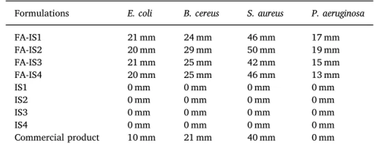
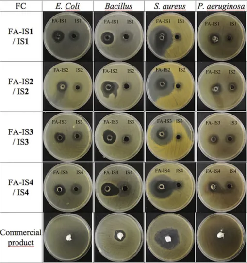
The results of the sterility studies of the in situ gel formulations with FA, no growth any of the microorganisms inoculated occurred after the incubation period. All of the prepared in situ gels showed an anti-bacterial effect on selected microorganisms. Blank gels not containing FA did not show any inhibition zones. Both preparations showed greater inhibition zones for all of the bacteria compared to the com-mercial product (Table 8, Fig. 3). According to our results, while P. aeruginosa and E. coli showed the narrowest antibacterial spectrum, the largest zone was observed in S. aureus which is a well known bacterium that causes infections on soft tissues. Of all our preparations, FI-2 seems
to be most effective on all bacteria except E. coli.
The treatment of topical infections has always been a difficult pro-position because of the lack of efficacy of existing anti-infectives, longer period of treatment and yet incomplete recovery [42]. Bacterias indu-cing burn infection can be classified into 2 categories, gram positive and negative. Gram negative bacteria induce most burn wound infections, with similar incidence, prevalence and pathogens regardless of geo-graphy or institution [43,44]. Burn area infection by Klebsiella pneu-monia, P. aeruginosa, E. coli, and S. aureus are independent predictors of mortality [38]. S. aureus is the essential reason of gram-positive burn Fig. 1. Graphical demonstration of relations between independent input variables and dependent output variables.
Table 4
QbD results of in situ gels.
Formulation code P188 (%w/w) P407 (%w/w) P338 (%w/w) Gelling temperature pH Best match formula
O-1 5 17 1 26.205 7.406 F6
infections all over the world [40] and a widespread reason of septi-cemia. It is one of the most predominant pathogens found in burn wounds [45]. Since 1962, FA has been widely used in the systemic and topical therapy of staphylococcal infections. It is also effective against anaerobic Gram-positive strains and shows activity against Neisseria spp., Bordetella pertussis, and Moraxella catarrhalis, but has no activity against other aerobic Gram-negative species [14].
3.5. Ex vivo studies
According to in vitro and characterization study results, FA-IS1 formulation was chosen for further ex vivo penetration and permeation studies. For ex vivo studies, pH 7.4 buffer was used as the medium. After 24 h of the contact period, the amount of FA that permeated through
the skin from FA-IS1 was determined as 0.932 ± 0.116% in the re-ceptor compartment. The penetration ratio to the skin tissue for FA-IS1 was recorded as 0.834 ± 0.230%. Topical therapy perspective plays a critical role in the management of skin therapy such as infections, burn or wound healing. In such cases, topical antibiotics may be the choice of preference with respect to systemic options, since they enhance the effective doses at the site of infection while minimizing the systemic side effects of the drugs [46]. When drug reaches the dermal layer, it becomes available for systemic absorption. As drug skin permeation is determined not only by the partition coefficient (logP of FA 4.97), but also by other physico-chemical properties as water solubility, molecular size and diffusivity; very low solubility of FA in water (0.00521 mg/mL) might justify its poor capability of skin permeation.
Under disease clinical situations such as burn, dermatitis and Table 5
FA loaded ideal in situ gel formulations components.
Formulation code P407 (g) P188 (g) P338 (g) FA (g) Ethanol (g) Water (g) (q.s)
FA-IS1 20 5 – 2 5 100
FA-IS2 20 15 – 2 5 100
FA-IS3 16 18 1 2 5 100
FA-IS4 16 20 1 2 5 100
Table 6
Characterization results of FA loaded in situ gel formulations.
Formulation code Gelling temperature (oC) pH Gelling capacity (Sec) Clarity Viscosity (cP) Spreadability (cm) Drug content (%)
FA-IS1 31.367± 0.666 5.777± 0.07 1.067± 0.115 +++ 147.033± 0.252 1.550 ± 0.050 92.867 ± 2.26 FA-IS2 31.967 ± 0.208 5.907 ± 0.01 1.333± 0.153 +++ 149.033 ± 0.252 1.383 ± 0.104 97.300 ± 3.39 FA-IS3 32.467 ± 0.200 6.053 ± 0.10 1.100± 0.100 ++ 103.267 ± 0.751 1.733 ± 0.058 96.667 ± 1.64 FA-IS4 31.800 ± 0.600 6.027 ± 0.06 0.927± 0.110 ++ 175.333 ± 0.058 1.733 ± 0.153 91.400 ± 5.49
Table 7
Stability studies results of formulation FA-IS1.
Parameters t = 0 t = 3 4 and 25 °C 4 °C 25 °C Gelling temperature (oC) 31.367± 0.666 32.10 ± 0.624 31.300 ± 0.529 pH 5.777± 0.07 6.23 ± 0.026 6.107 ± 0.015 Gelling capacity (Sec) 1067± 0,115 1.06 ± 0.010 1.080 ± 0.010 Clarity +++ +++ +++ Viscosity (cP) 147.033± 0.252 133.333 ± 2.887 158.333 ± 2.887 Spreadability (cm) 1.550 ± 0.05 1.15 ± 0.010 1.4 ± 0.100 Drug content (%) 92.867 ± 2.26 92.141 ± 0.641 92.168 ± 2.675
Fig. 2. In vitro drug release results of FA-IS1-4 (n:3, ± STD). Table 8
Microbiological studies results of in situ gels with FA (FA-IS1-FA-IS4), without FA (IS1-IS4) and commercial product of FA.
Formulations E. coli B. cereus S. aureus P. aeruginosa
FA-IS1 21 mm 24 mm 46 mm 17 mm FA-IS2 20 mm 29 mm 50 mm 19 mm FA-IS3 21 mm 25 mm 42 mm 15 mm FA-IS4 20 mm 25 mm 46 mm 13 mm IS1 0 mm 0 mm 0 mm 0 mm IS2 0 mm 0 mm 0 mm 0 mm IS3 0 mm 0 mm 0 mm 0 mm IS4 0 mm 0 mm 0 mm 0 mm Commercial product 10 mm 21 mm 40 mm 0 mm
psoriasis, the skin especially SC is disrupted. The obtained results confirmed the key role of the vehicle in determining the API's dermal or transdermal fate for topically administered formulations. In this study, in situ gels were used for treatment. Topical gel formulations might be considered as suitable delivery systems for skin since they are less greasy and their removal from the skin surface is very simple. Higher viscosity of topical gel formulations may also result in attenuated pe-netration and permeation. This attenuation is very important for APIs having irritation potency at high concentrations. Especially when such kinds of molecules are to be needed to be administered to skin for a long period of time, topical gel formulations may provide not only atte-nuated irritation but also reduced systemic exposure. In our experi-mental studies, higher viscosity of topical gel formulations maintained low permeation and penetration of FA in addition to controlled in vitro release tendency. According to the penetration and permeation results, the present data demonstrated that sufficient active FA is retained on the skin. Considering the overall results, developed formulation may be used for topical application and the prepared in situ gel could be safely applied for topical delivery of FA. The antimicrobial activity of FA is specifically active for eradicating skin pathogens like Methicillin-re-sistant S. aureus and other wound infections. However, the currently available topical formulations of FA fail to penetrate across the wound eschar leading to sub-optimal therapeutic efficacy [47]. Thus, the de-veloped FA loaded in situ gels offer a safer and promising solution in wound healing effect. Ex vivo studies do not exactly reflect the true permeation since normalfirm skin was used in the experiments. For this reason, in vivo experiments were also performed on 2nd degree burned rat skin. Second-degree burns affect the epidermis and the dermis.
Thus, the amount of penetrated drug can be increased by using 2nd degree burned skin for ex vivo studies.
3.6. In vivo studies
3.6.1. Macroscopic burn healing
Male Wistar albino rats were used for burn experimental model. Rats and humans share physiological and pathological characteristics in many organ systems that have been already well established in the literature. Similar to humans, the rat skin is also composed of the major layers of epidermis and dermis [48]. This experimental model was es-tablished to standardize thermal burn injuries in order to obtain injuries with the same size and depth degree. The choice of Wistar rats due to these animals shows ease of handling, accommodation and resistance to surgical aggressions, and infectious processes, with low mortality [49,50].
The in situ gel was applied to wound area in the rat dorsum for 10 days to estimate the wound healing effect of the FA containing in situ gel for the acceleration of burn healing. The daily behaviors (food in-take, activity, e.g.) of rats were observed as normal. The determination of the wound closure rate is beneficial for scoring the progress of healing [29].
Both minor burn and major burn damages initiate the wound healing process which consists of several highly integrated and over-lapping phases: inflammation, cell recruitment, matrix deposition, epithelialization and tissue remodelling. Moreover local wound repair, severe large burns also stimulate a persistent pathophysiological stress response and a systemic hypermetabolic-catabolic condition [4]. Fig. 3. Zone inhibition diameters of in situ gels with FA, without FA and commercial product of FA.
The macroscopic images on days 0, 3, 5, 7, and 10 of burn wounds experimentally induced in rats are presented in Fig. 4. The wounds were initially characterized by a white eschar with an affected epi-dermis and epi-dermis and a hyperaemic zone in the periphery. During the first day, the wound area became fully hyperaemic due to red blood cell extravasation and a post-traumatic inflammatory process. All rats sur-vived throughout the postoperative period. Crust formations were ob-served on the skin after several days. It was found out that FG and BG treatments did not get irritated the skin.
During thefirst 48 h, the severity and extent of local tissue injury result in inflammatory mediators being released into the systemic cir-culation. These induce specific haemodynamic alternations, including increased capillary permeability (small protein molecules leak out of the circulation leading to edema and significant fluid loss. Burn edema is a result of circulating inflammatory mediators including histamine, prostaglandins, leukotrienes and kinins that result in increased capillary permeability), peripheral and splanchnic vasoconstriction, and myo-cardial depression [1,51,52].
The wound contraction ratio was assessed as the percentage redu-cing in wound size on days 3, 5, 7, and 10 after the burning procedure. As it is illustrated inFig. 5, the treated group with FG and RG displayed a significant burn healing progression on the day 3 (P < 0.01), 5 (P < 0.01), 7 (P < 0.01), and 10 (P < 0.01) compared to control group. The % of wound area ranged from 90.109 ± 0.654% to 65.492 ± 0.722% in the period from 3 to 10 days in the control group. The % of wound area in rats treated with BG ranged from 90.011 ± 1.620% to 60.786 ± 1.897% in the period from 3 to 10 days. Besides that the % of wound area in rats treated with RG ranged from 77.298 ± 2.109% to 46.714 ± 2.733% as well as FG ranged from 68.020 ± 1.578% to 43.776 ± 2.967% in the period from 3 to 10 days. RG and FG recovered quickly and the wound area rapidly shrank to half by the seventh day as compared to the untreated group.
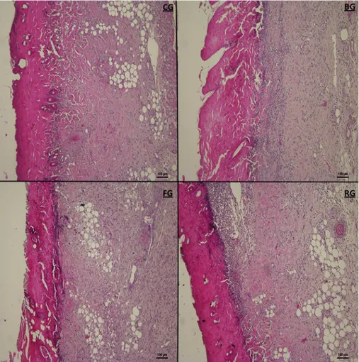
3.6.2. Histology of wound healing
The microscopic photos were taken during the histological ex-amination of the wound tissues on the 10th day are given inFig. 6. Histopathological examination of wound tissues on the 10th day is il-lustrated inFig. 7.
Angiogenesis (neovascularization) is a complicated phase including membrane disintegration, endothelial cell reproduction and migration, and the forming of the new basement membrane from endothelial cells leading to the production of new capillaries from existent blood vessels [53]. In addition to this, enhancement of the epidermal and dermal regeneration is one of the main purposes of the wound healing. Fig. 4. Effects of FA in situ gel on wound's evolution. Macroscopic examples of wound healing with control (CG), blank in situ gel (BG), FA (2%) in situ gel (FG), and Fucidin®(RG) groups after excision on days 0, 3, 5, 7, and 10.
Fig. 5. The rate of wound healing in animal groups on days 0–10. Healing percentage of scar tissue surface area in each group. Each data point represents the mean ± SEM.
Moreover, granulation tissue thickness is a critical parameter to ob-jectively evaluate of wound treatment [29,54].
As stated in the obtained outcomes, it was determined that FG and RG had a significant effect compared to the control group (Figs. 6and 7). According to the histological evaluation results, more blood vessel
formation was detected on FG (P < 0.01) and RG (P < 0.05) in comparison to the untreated group. It has been also revealed that there were significantly higher percentages of dermal and epidermal re-generation with the FG (P < 0.05) and RG (P < 0.05) in comparison to the untreated group. Moreover, it was determined that thicker Fig. 6. Histopathological view of scarred tissues of the control (CG), blank in situ gel (BG), FA (2%) in situ gel (FG) and Fucidin®groups (RG) on the 10th day after the burning process (Hematoxylin and eosin (H&E) (original magnification×10). The scale bars represent 100 μm for the figure.
Fig. 7. Microscopic examination of granulation tissue thickness, angiogenesis and epidermal-dermal regeneration on control (CG), blank in situ gel (BG), FA (2%) in situ gel (FG) and Fucidin®groups (RG) by histological wound healing scores among. Statistically significant as compared to control; P < 0.05(∗), P < 0.01(∗∗). Values are presented as the mean ± SEM.
granulation tissue was formed in the FG (P < 0.05) compared to the untreated group after treatment (Figs. 6and7).
4. Conclusion
In this study, the potential of FA loaded in situ gels as drug carriers for dermal delivery was evaluated. Preparing in situ gels which were optimized with the pre-studies and artificial intelligence program. The optimum formulations provided by the ANN software have not been described so far in the literature and cannot be provided by experi-mental data. These formulations were examined experiexperi-mentally and optimized successfully. It was ruled out that in situ gels of FA can be successfully prepared with the cold method. The clarity, pH, gelation time and drug content of all formulations were found to be satisfactory. In addition, the gel was found to be stable for 3 months. Clearly, these results indicate the suitability of the formulation with significant im-provement of the in vitro release, ex vivo permeation and thus possibly improved in vivo efficacy. In conclusion, the present study can open up a window for dermal application of in situ gels loaded with FA, they would be a better alternative to conventional dermal creams in burn healing. In addition, ANN programs have big advantageous for Research and Development, especially for Industry. Because they have the ability to provide detailed results and prevent unnecessary studies which cause a loss of time and money. Consequently; the emphasis of this study is to demonstrate the practical gain of the QbD approach in pharmaceutical drug development.
Declaration of interests
The authors declare no conflict of interest. Acknowledgements
The authors would like to acknowledge to Berkoİlaç for providing fusidic acid and we also thank the BASF for providing the poloxamers.
References
[1] A.A. Udy, J.A. Roberts, J. Lipman, S. Blot, The effects of major burn related pa-thophysiological changes on the pharmacokinetics and pharmacodynamics of drug use: an appraisal utilizing antibiotics, Adv. Drug Deliv. Rev. 123 (2018) 65–74,
https://doi.org/10.1016/j.addr.2017.09.019.
[2] J.S. Boateng, K.H. Matthews, H.N.E. Stevens, G.M. Eccleston, Wound healing dressings and drug delivery Systems : a review, J. Pharm. Sci. 97 (2008) 2892–2923,https://doi.org/10.1002/jps.21210.
[3] K. Thakur, G. Sharma, B. Singh, A. Jain, R. Tyagi, S. Chhibber, O.P. Katare, Cationic-bilayered nanoemulsion of fusidic acid: an investigation on eradication of methicillin-resistant Staphylococcus aureus 33591 infection in burn wound, Nanomedicine 13 (2018) 825–847,https://doi.org/10.2217/nnm-2017-0227. [4] Y. Wang, J. Beekman, J. Hew, S. Jackson, A.C. Issler-Fisher, R. Parungao,
S.S. Lajevardi, Z. Li, P.K.M. Maitz, Burn injury: challenges and advances in burn wound healing, infection, pain and scarring, Adv. Drug Deliv. Rev. 123 (2018) 3–17,https://doi.org/10.1016/j.addr.2017.09.018.
[5] R.F. Pereira, C.C. Barrias, P.L. Granja, P.J. Bartolo, Advanced biofabrication stra-tegies for skin regeneration and repair, Nanomedicine 8 (2013) 603–621,https:// doi.org/10.2217/nnm.13.50.
[6] K. Rehman, M.H. Zulfakar, Recent Advances in Gel Technologies for Topical and Transdermal Drug Delivery, 9045, (2013), pp. 1–8,https://doi.org/10.3109/ 03639045.2013.828219.
[7] C. Valenta, B.G. Auner, The Use of Polymers for Dermal and Transdermal Delivery vol. 58, (2004), pp. 279–289,https://doi.org/10.1016/j.ejpb.2004.02.017. [8] P.L. Honeywell-nguyen, J.A. Bouwstra, Vesicles as a Tool for Transdermal and
Dermal Delivery vol. 2, (2005), pp. 67–74,https://doi.org/10.1016/j.ddtec.2005. 05.003.
[9] G. Sharma, S. Kamboj, K. Thakur, P. Negi, K. Raza, O.P. Katare, Delivery of ther-moresponsive-tailored mixed Micellar nanogel of lidocaine and prilocaine with improved dermatokinetic profile and therapeutic efficacy in topical anaesthesia, AAPS PharmSciTech 18 (2017) 790–802, https://doi.org/10.1208/s12249-016-0561-8.
[10] M.P. Venkatesh, P.K. Liladhar, T.M.P. Kumar, H.G. Shivakumar, In situ gels based drug delivery systems, Curr. Drug Ther. 6 (2011) 213–222,https://doi.org/10. 2174/157488511796392004.
[11] N. Üstündağ Okur, A. Yoltaş, V. Yozgatli, O. Uygulama, V. Yüklü, I. Situ, J. Formülasyonlarının, G. Ve Karakterizasyonu, Development and characterization
of voriconazole loaded in situ gel formulations for ophthalmic application, Turk J Pharm Sci 13 (2016) 311–317,https://doi.org/10.4274/tjps.2016.05.
[12] H. Qi, W. Chen, C. Huang, L. Li, C. Chen, W. Li, C. Wu, Development of a poloxamer analogs/carbopol-based in situ gelling and mucoadhesive ophthalmic delivery system for puerarin, Int. J. Pharm. 337 (2007) 178–187,https://doi.org/10.1016/j. ijpharm.2006.12.038.
[13] K. Thakur, G. Sharma, B. Singh, S. Chhibber, A.B. Patil, O.P. Katare, Chitosan-tai-lored lipidic nanoconstructs of Fusidic acid as promising vehicle for wound infec-tions: an explorative study, Int. J. Biol. Macromol. 115 (2018) 1012–1025,https:// doi.org/10.1016/j.ijbiomac.2018.04.092.
[14] B. Ramachandra, A critical review of properties of modafinil and analytical, bioa-nalytical methods for its determination, Crit. Rev. Anal. Chem. 46 (2016) 482–489,
https://doi.org/10.1080/10408347.2016.1153948.
[15] S. Wadhwa, B. Singh, G. Sharma, K. Raza, O.P. Katare, Liposomal fusidic acid as a potential delivery system: a new paradigm in the treatment of chronic plaque psoriasis, Drug Deliv. 23 (2016) 1204–1213,https://doi.org/10.3109/10717544. 2015.1110845.
[16] C.M. Hosey, R. Chan, L.Z. Benet, BDDCS predictions, self-correcting aspects of BDDCS assignments, BDDCS assignment corrections, and classification for more than 175 additional drugs, AAPS J. 18 (2016) 251–260,https://doi.org/10.1208/ s12248-015-9845-2.
[17] J. Turnidge, Fusidic acid pharmacology, pharmacokinetics and pharmacodynamics, Int. J. Antimicrob. Agents 12 (Suppl 2) (1999) S23–S34http://www.ncbi.nlm.nih. gov/pubmed/10528784, Accessed date: 16 March 2019.
[18] P. Negi, B. Singh, G. Sharma, S. Beg, O.P. Katare, Biocompatible lidocaine and prilocaine loaded-nanoemulsion system for enhanced percutaneous absorption: QbD-based optimisation, dermatokinetics and in vivo evaluation, J. Microencapsul. 32 (2015) 419–431,https://doi.org/10.3109/02652048.2015.1046513. [19] P. Negi, B. Singh, G. Sharma, S. Beg, K. Raza, O.P. Katare, Phospholipid
micro-emulsion-based hydrogel for enhanced topical delivery of lidocaine and prilocaine: QbD-based development and evaluation, Drug Deliv. 23 (2016) 941–957,https:// doi.org/10.3109/10717544.2014.923067.
[20] X. Xu, M.A. Khan, D.J. Burgess, A quality by design (QbD) case study on liposomes containing hydrophilic API: II. Screening of critical variables, and establishment of design space at laboratory scale, Int. J. Pharm. 423 (2012) 543–553,https://doi. org/10.1016/j.ijpharm.2011.11.036.
[21] ICH, Pharmaceutical Development Q8 (R2), ICH, 2009 doi:EMEA/CHMP/167068/ 2004.
[22] B. Mesut, Y. Özsoy, B. Aksu, The Place of Drug Product Critical Quality Parameters in Quality by Design, QBD, 2015.
[23] B. Sylvester, A. Porfire, D.-M. Muntean, L. Vlase, L. Lupuţ, E. Licarete, A. Sesarman, M.C. Alupei, M. Banciu, M. Achim, I. Tomuţă, Optimization of prednisolone-loaded long-circulating liposomes via application of Quality by Design (QbD) approach, J. Liposome Res. 28 (2018) 49–61,https://doi.org/10.1080/08982104.2016. 1254242.
[24] B. Aksu, G. Yegen, S. Purisa, E. Cevher, Y. Ozsoy, Optimisation of ondansetron orally disintegrating tablets using artificial neural networks, Trop. J. Pharmaceut. Res. 13 (2014) 1374–1383,https://doi.org/10.4314/tjpr.v13i9.1.
[25] N. Üstündağ Okur, E.Ş. Çağlar, V. Yozgatli, Development and validation of an Hplc method for voriconazole active substance in bulk and its pharmaceutical formula-tion, Marmara Pharm. J. 20 (2016) 79,https://doi.org/10.12991/mpj. 20162076793.
[26] S.L. Kaplan, K.G. Hulten, B.E. Gonzalez, W.A. Hammerman, L. Lamberth, J. Versalovic, E.O. Mason, Three-year surveillance of community-acquired Staphylococcus aureus infections in children, Clin. Infect. Dis. 40 (2005) 1785–1791,https://doi.org/10.1086/430312.
[27] F. Siebenhaar, W. Syska, K. Weller, M. Magerl, T. Zuberbier, M. Metz, M. Maurer, Control of Pseudomonas aeruginosa skin infections in mice is mast cell-dependent, Am. J. Pathol. 170 (2007) 1910,https://doi.org/10.2353/ajpath.2007.0607706. [28] A. Oryan, M. Jalili, A. Kamali, B. Nikahval, The concurrent use of probiotic
mi-croorganism and collagen hydrogel/scaffold enhances burn wound healing: an in vivo evaluation, Burns 44 (2018) 1775–1786,https://doi.org/10.1016/J.BURNS. 2018.05.016.
[29] M.E. Okur,Ş. Ayla, D. Çiçek Polat, M.Y. Günal, A. Yoltaş, Ö. Biçeroğlu, Novel in-sight into wound healing properties of methanol extract of Capparis ovata Desf. var. palaestina Zohary fruits, J. Pharm. Pharmacol. 70 (2018) 1401–1413,https://doi. org/10.1111/jphp.12977.
[30] M. Galeano, D. Altavilla, A. Bitto, L. Minutoli, M. Calò, P. Lo Cascio, F. Polito, G. Giugliano, G. Squadrito, C. Mioni, D. Giuliani, F.S. Venuti, F. Squadrito, Recombinant human erythropoietin improves angiogenesis and wound healing in experimental burn wounds, Crit. Care Med. 34 (2006) 1139–1146,https://doi.org/ 10.1097/01.CCM.0000206468.18653.EC.
[31] R. Serra, R. Grande, L. Butrico, A. Rossi, U.F. Settimio, B. Caroleo, B. Amato, L. Gallelli, S. de Franciscis, Chronic wound infections: the role of Pseudomonas aeruginosa and Staphylococcus aureus, Expert Rev. Anti Infect. Ther. 13 (2015) 605–613,https://doi.org/10.1586/14787210.2015.1023291.
[32] B.A. Lipsky, C. Hoey, Topical antimicrobial therapy for treating chronic wounds, Clin. Infect. Dis. 49 (2009) 1541–1549,https://doi.org/10.1086/644732. [33] K. Thakur, G. Sharma, B. Singh, S. Chhibber, A.B. Patil, O.P. Katare,
Chitosan-tai-lored lipidic nanoconstructs of Fusidic acid as promising vehicle for wound infec-tions: an explorative study, Int. J. Biol. Macromol. 115 (2018) 1012–1025,https:// doi.org/10.1016/j.ijbiomac.2018.04.092.
[34] V. Thakkar, V. Korat, L. Baldaniya, M. Gohel, T. Gandhi, N. Patel, Development and characterization of novel hydrogel containing antimicrobial drug for treatment of burns, Int. J. Pharm. Investig. 6 (2016) 158,https://doi.org/10.4103/2230-973X. 187343.
[35] G.G. Pereira, F.A. Dimer, S.S. Guterres, C.P. Kechinski, J.E. Granada, N.S.M. Cardozo, Formulation and characterization of poloxamer 407®: thermo-reversible gel containing polymeric microparticles and hyaluronic acid, Quim. Nova 36 (2013) 1121–1125,https://doi.org/10.1590/S0100-40422013000800008. [36] G. Dumortier, J.L. Grossiord, F. Agnely, J.C. Chaumeil, A review of poloxamer 407
pharmaceutical and pharmacological characteristics, Pharm. Res. (N. Y.) 23 (2006) 2709–2728,https://doi.org/10.1007/s11095-006-9104-4.
[37] S.D. Singh-Joy, V.C. McLain, Safety assessment of poloxamers 101, 105, 108, 122, 123, 124, 181, 182, 183, 184, 185, 188, 212, 215, 217, 231, 234, 235, 237, 238, 282, 284, 288, 331, 333, 334, 335, 338, 401, 402, 403, and 407, poloxamer 105 benzoate, and poloxamer 182 dibenzoate as use, Int. J. Toxicol. 27 (2008) 93–128,
https://doi.org/10.1080/10915810802244595.
[38] L.C. D'Avignon, B.K. Hogan, C.K. Murray, F.L. Loo, D.R. Hospenthal, L.C. Cancio, S.H. Kim, E.M. Renz, D. Barillo, J.B. Holcomb, C.E. Wade, S.E. Wolf, Contribution of bacterial and viral infections to attributable mortality in patients with severe burns: an autopsy series, Burns 36 (2010) 773–779,https://doi.org/10.1016/J.BURNS. 2009.11.007.
[39] N. Merchant, K. Smith, M.G. Jeschke, An ounce of prevention saves tons of lives: infection in burns, Surg. Infect. 16 (2015) 380–387,https://doi.org/10.1089/sur. 2013.135.
[40] W. Norbury, D.N. Herndon, J. Tanksley, M.G. Jeschke, C.C. Finnerty, Infection in burns, Surg. Infect. 17 (2016) 250–255,https://doi.org/10.1089/sur.2013.134. [41] M.S. Kinch, E. Patridge, M. Plummer, D. Hoyer, An analysis of FDA-approved drugs
for infectious disease: antibacterial agents, Drug Discov. Today 19 (2014) 1283–1287,https://doi.org/10.1016/J.DRUDIS.2014.07.005.
[42] K. Thakur, G. Sharma, B. Singh, S. Chhibber, O.P. Katare, Current state of nano-medicines in the treatment of topical infectious disorders, Recent Pat. Anti-Infect. Drug Discov. 13 (2018) 127–150,https://doi.org/10.2174/
1574891X13666180529103804.
[43] E.A. Azzopardi, E. Azzopardi, L. Camilleri, J. Villapalos, D.E. Boyce, P. Dziewulski, W.A. Dickson, I.S. Whitaker, Gram negative wound infection in hospitalised adult burn patients-systematic review and metanalysis-, PLoS One 9 (2014) e95042, ,
https://doi.org/10.1371/journal.pone.0095042.
[44] A.C. Issler-Fisher, R.M. Fakin, O.M. Fisher, G. McKew, R. Gazzola, A.-K. Rauch,
T. Gottlieb, P. Haertsch, M. Guggenheim, P. Giovanoli, P.K.M. Maitz,
Microbiologicalfindings in burn patients treated in a general versus a designated intensive care unit: effect on length of stay, Burns 42 (2016) 1805–1818,https:// doi.org/10.1016/J.BURNS.2016.06.019.
[45] T. Chhibber, S. Wadhwa, P. Chadha, G. Sharma, O.P. Katare, Phospholipid struc-tured microemulsion as effective carrier system with potential in methicillin sen-sitive Staphylococcus aureus (MSSA) involved burn wound infection, J. Drug Target. 23 (2015) 943–952,https://doi.org/10.3109/1061186X.2015.1048518. [46] D. Bonamonte, A. Belloni Fortina, L. Neri, A. Patrizi, Fusidic acid in skin infections
and infected atopic eczema, G. Ital. Dermatol. Venereol. 149 (2014) 453–459
http://www.ncbi.nlm.nih.gov/pubmed/25068235.
[47] H. Schöfer, L. Simonsen, Fusidic acid in dermatology: an updated review, Eur. J. Dermatol. 20 (2010) 6–15,https://doi.org/10.1684/EJD.2010.0833.
[48] A. Abdullahi, S. Amini-Nik, M.G. Jeschke, Animal models in burn research, Cell. Mol. Life Sci. 71 (2014) 3241–3255,https://doi.org/10.1007/s00018-014-1612-5. [49] R.A. Santos Heredero FX, C. Hamann, J.M. Obispo Martin, C.M. S. C, Experimental
burn models, ann burn, Fire Disast 9 (1996) 96–97.
[50] D. dos, S. Tavares Pereira, M.H.M. Lima-Ribeiro, N.T. de Pontes-Filho, A.M. dos, A. Carneiro-Leão, M.T. dos, S. Correia, Development of animal model for studying deep second-degree thermal burns, J. Biomed. Biotechnol. (2012) 1–7,https://doi. org/10.1155/2012/4608412012.
[51] K. Ganrot, S. Jacobsson, U. Rothman, Transcapillary passage of plasma proteins in experimental burns, Acta Physiol. Scand. 91 (1974) 497–501,https://doi.org/10. 1111/j.1748-1716.1974.tb05705.x.
[52] H. Douglas, J. Dunne, J. Rawlins, Management of burns, Surg. (United Kingdom) 36 (2018) 435–440,https://doi.org/10.1016/j.mpsur.2018.05.004.
[53] J.Y. Park, J.H. Kwak, K.S. Kang, E.B. Jung, D.S. Lee, S. Lee, Y. Jung, K.H. Kim, G.S. Hwang, H.L. Lee, N. Yamabe, S.N. Kim, Wound healing effects of deoxy-shikonin isolated from Jawoongo: in vitro and in vivo studies, J. Ethnopharmacol. 199 (2017) 128–137,https://doi.org/10.1016/j.jep.2016.10.031.
[54] S. Ayla, M.E. Okur, M.Y. Günal, E.M. Özdemir, D. Çiçek Polat, A. Yoltaş, Ö. Biçeroğlu, S. Karahüseyinoğlu, Wound healing effects of methanol extract of Laurocerasus officinalis roem, Biotech. Histochem. 00 (2018) 1–9,https://doi.org/ 10.1080/10520295.2018.1539242.