Relationship between Calcium Stone Disease and Metabolic Syndrome
Emre Can Polat,1* Levent Ozcan,2 Suleyman Sami Cakir,3 Murat Dursun,3 Alper Otunctemur,3 Emin Ozbek3
Purpose: We aimed to investigate relationship between metabolic syndrome and calcium-oxalate stone formation.
Materials and Methods: Between January 2008 and February 2015 we retrospectively investigated biochemical parameters and anthropometric characteristics (height, weight, and waist circumference) of 198 patients who had calcium-oxalate stones and we also randomly selected 200 participants who had no history of urolithiasis as the controls.
Results: The presence of obesity increased the risk of calcium stones in both men (P = .003, OR = 2.92) and wom-en (P = .03, OR = 2.18). Diabetes was significantly correlated to the risk of calcium stones (P = .04, OR = 1.94). However, when calculated separately for men and women, diabetic men had a higher risk of calcium-oxalate stone disease (P = .04, OR = 2.59), but diabetic women did not (P > .05). Hypertension also significantly increased the risk of calcium stones when compared with normotensive individuals (P = .0001, OR = 3.03).
Conclusion: The risk for the development of calcium-oxalate stone disease is most significantly associated with the patient’s body mass index and the presence of hypertension.
Keywords: metabolic syndrome; epidemiology; outcome assessment; prevalence; risk assessment; urolithiasis; etiology.
INTRODUCTION
M
etabolic syndrome (MS), the simultaneousoc-currence of hyperglycemia, hyperlipidemia, hy-pertension, and visceral obesity, is a chronic disease associated with high mortality. In addition, this condi-tion substantially increases the risk of developing
cardi-ovascular diseases and type 2 diabetes.(1) In the United
States, the prevalence of MS is 24% in men and 23.4% in women, increasing at ages 60-69 years to 43.5% in
both sexes.(2) Through out the years, numerous
defi-nitions of MS have been proposed by various organi-zations. The National Cholesterol Education Program Adult Treatment Panel III (NCEP/ATP III) definition is the one most used today because it incorporates the key concepts of MS, relies on commonly used laboratory studies available to most physicians, and is less
restric-tive than the other classifications.(3) The prevalence of
urolithiasis ranges from 2 to 20% throughout the world based on the geographic and socioeconomic character-istics of the different populations. The worldwide prev-alence of the disease appears to have increased in the last quarter of the twentieth century for both men and women. The identification of common, modifiable risk
factors for kidney stones may result in new approaches
to the treatment and prevention of urolithiasis.(4)
Much like MS and obesity, the prevalence of nephro-lithiasis in the United States and other countries is in-creasing. There is evidence that these parallel changes
might be linked.(5) Studies have shown that MS and its
components (obesity/increased waist circumference [WC], hypertension, and etc.) are associated with
in-creased rates of nephrolithiasis.(6-8) Although the exact
pathophysiologic mechanisms underlying the associa-tion between MS and nephrolithiasis are unclear, MS has been associated with changes in urinary constitu-ents, including lower urinary pH, decreased citrate ex-cretion, and increased uric acid and calcium exex-cretion, leading to increased risks of uric acid and calcium stone
formation.(9-12)
Since 80% of kidney stones consist of calcium oxalate
(CaOx),(13,14) studies exploring the relationship between
urinary risk factors for calcium stone formation and features of the MS are critical. Therefore in this study, we aimed to investigate the relationship between CaOx stone disease and MS components.
1 Department of Urology, Istanbul Medipol University, Istanbul, Turkey.
2 Department of Urology, Derince Training and Research Hospital, Kocaeli, Turkey. 3 Department of Urology, Okmeydani Training and Research Hospital, Istanbul, Turkey.
*Correspondence: Department of Urology, Istanbul Medipol University, Istanbul, Turkey. Tel: +90 532 7149604. E-mail: [email protected].
MATERIALS AND METHODS
We retrospectively investigated biochemical parame-ters and anthropometric characteristics (height, weight, and WC) of 198 patients who had CaOx stones and we also randomly selected 200 participants who had no history of urolithiasis as the controls between January 2008 and February 2015.
Patients who had a surgery for urolithiasis (open neph-rolithotomy, percutaneous nephrolithotomy and ureter-orenoscopy) and whose stones were stones with a CaOx content ˃ 70% and a calcium apatite content ˂ 5% with Fourier transform infrared spectroscopy and
high-res-olution X-ray diffraction(15) were enrolled in the study.
Patients were excluded from the study if they had prima-ry hyperparathyroidism, chronic diarrheal syndromes, intestinal malabsorption, complete distal renal tubular acidosis, primary hyperoxaluria, recurrent or active uri-nary tract infection, history of kidney transplantation, ongoing 5-α reductase inhibitor therapy, liver disease, primary gout, any debilitating chronic illness, or a cal-culated creatinine clearance of ≤ 50 mL/minute. Weight, WC, and blood pressure were measured after an overnight fasting, and a blood sample was drawn. Fast-ing blood glucose, serum total cholesterol, high-density lipoprotein (HDL) cholesterol, and triglycerides were measured using enzymatic methods with an autoanalyz-er. MS was defined according to the criteria established in 2005 by the NCEP/ATP III. For the criteria for MS, abdominal obesity was defined as WC ≥ 102 cm in men
and ≥ 89 cm in women, according to the NCEP/ATP III obesity criteria.
MS was diagnosed in those who satisfied at least 3 of the following 5 criteria: WC ≥ 102 cm in men, ≥ 89 cm in women, triglyceride concentration >150 mg/dL or undergoing treatment for hypertriglyceridemia, HDL cholesterol concentration < 40 mg/dL in men and ˂ 50 mg/dL in women or undergoing treatment for low HDL-cholesterol level, blood pressure > 130/85 mmHg or undergoing treatment for hypertension, and fasting blood glucose level > 100 mg/dL or undergoing
treat-ment for hyperglycemia.(3)
Statistical Analysis
Analyses were performed using Chi-square tests. Odds ratios (OR) were calculated. Statistical determinations were within the 95% confidence interval (CI). All P values were two-tailed, and P < .05 was considered statistically significant. The data were analyzed with Statistical Package for the Social Science (SPSS Inc, Chicago, Illinois, USA) version 16.
RESULTS
Baseline demographic characteristics of the 398 partic-ipants are shown in Table 1. In the study population, 198 were patients with CaOx stone disease, aged 36-58 years and 200 were patients without urolithiasis aged 31-64 years.
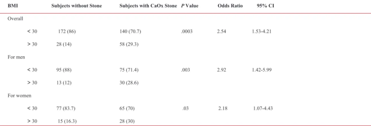
Body Mass Index (BMI) was significantly correlated with the risk of CaOx stone disease. Overall,
partic-ipants with a BMI > 30 kg/m2 increased their risk of
calcium stones by 2.54-fold when compared with
par-ticipants with a BMI < 30 kg/m2 (Table 2). The
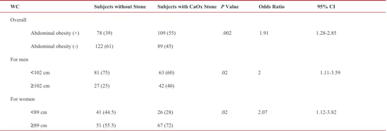
pres-ence of obesity increased the risk of calcium stones in both men (P = .003, OR = 2.92) and women (P = .03, OR = 2.18). Increased WC also increased the risk of CaOx stone disease (P =.002, OR = 1.91). When WC
Table 1. Demographic characteristics of the participants.
Variables CaOx Stone (+) Stone (-) P Value Gender, male/female; n (%) 105/93 (53/47) 108/92 (54/46) ˃ .05 Age (years) 43.3 ± 9.3 42.7 ± 9.7 ˃ .05
Abbreviation: CaOx, calcium oxalate.
Abbreviations: BMI, Body Mass Index; CI, confidence interval; CaOx, calcium oxalate. * Data are presented as no (%).
BMI Subjects without Stone Subjects with CaOx Stone P Value Odds Ratio 95% CI
Overall ˂ 30 172 (86) 140 (70.7) .0003 2.54 1.53-4.21 ˃ 30 28 (14) 58 (29.3) For men ˂ 30 95 (88) 75 (71.4) .003 2.92 1.42-5.99 ˃ 30 13 (12) 30 (28.6) For women ˂ 30 77 (83.7) 65 (70) .03 2.18 1.07-4.43 ˃ 30 15 (16.3) 28 (30)
was calculated separately for men and women, both ab-dominally obese men (WC ≥ 102 cm) and abab-dominally obese women (WC ≥ 89 cm) had higher risk of CaOx stone disease respectively (P = .02, OR = 2; P = .02, OR = 2.07) (Table 3).
Of the participants, 50 (12.5%) had been diagnosed with diabetes mellitus and 99 (24.8%) with hyperten-sion. Diabetes was significantly related to the risk of calcium stones (P = .04, OR = 1.94). However, when calculated separately for men and women, diabetic men had a higher risk of CaOx stone disease (P = .04, OR = 2.59), but diabetic women did not (P > .05) (Table
4). Hypertension also significantly increased the risk of
calcium stones when compared with normotensive indi-viduals (P = .0001, OR = 3.03) (Table 5). Multi-variant analysis revealed that only hypertension and obesity had significant impacts on the development of CaOx stone disease (P = .001 and P = .02, respectively). The calculated OR was 2.32 (95% confidence interval [CI]: 1.32–3.51) for hypertension and 1.43 (CI: 1.21–2.42)
for obesity.
DISCUSSION
Our study was a retrospective analysis designed to explore the relationship between MS factors and the CaOx stone disease. We found that, both obesity and hypertension were independently and significantly associated with CaOx stone disease. Insulin resistant individuals often have an abnormal distribution of fat that is predominantly characterized by upper body fat. (16) Upper body obesity may result in insulin resistance in otherwise normal weight individuals, so we analyzed our data for BMI and WC as separate entities. Our re-sults showed that the number of individuals with a WC of over 100 cm (n = 106, 26.6%) was greater than the
number of individuals with a BMI of over 30 kg/m2(n
= 86, 21.6%).
Uric acid and infectious stones are both linked with
in-creased body weight as well as insulin resistance.(17,18)
Although the association between body weight and cal-cium nephrolithiasis has not been clearly established,
Table 3. The association between waist circumference and CaOx stone disease.*
WC Subjects without Stone Subjects with CaOx Stone P Value Odds Ratio 95% CI
Overall Abdominal obesity (+) 78 (39) 109 (55) .002 1.91 1.28-2.85 Abdominal obesity (-) 122 (61) 89 (45) For men ˂102 cm 81 (75) 63 (60) .02 2 1.11-3.59 ≥102 cm 27 (25) 42 (40) For women ˂89 cm 41 (44.5) 26 (28) .02 2.07 1.12-3.82 ≥89 cm 51 (55.5) 67 (72)
Abbreviations: WC, waist circumference; CI, confidence interval; CaOx, calcium oxalate. * Data are presented as no (%).
Abbreviations: DM, diabetes mellitus; CI, confidence interval; CaOx, calcium oxalate. * Data are presented as no (%).
DM Subjects without Stone Subjects with CaOx Stone P Value Odds Ratio 95% CI
Overall DM (+) 18 (9) 32 (16.2) .04 1.94 1.05 - 3.6 DM (-) 182 (91) 166 (83.8) For men DM (+) 7 (6.5) 16 (15.2) .04 2.59 1.02 - 6.59 DM (-) 101 (93.5) 89 (84.8) For women DM (+) 11 (12) 16 (17.2) - DM (-) 81 (88) 77 (82.8)
Sarica and colleagues showed that an increased body size increased the excretion of urinary stone-forming
risk factors such as oxalate, calcium, and citrate.(19)
Parvin and colleagues realized that oxalate play the most important role as a urinary stone risk factor in idi-opathic calcium stone disease followed by calcium and uric acid and that the adjusted values for body weight is a stronger and more determinant factor in calcium
stone formation than their concentration.(20) Siener and
colleagues(21) evaluated the relationship between BMI
and 24 h urine parameters in a population of idiopathic CaOx stone formers and found that an increased BMI was strongly associated with an increase in the excre-tion of stone promoters but not inhibitors. In our study, the OR was 2.92 for men and 2.18 for women with BMI
≥ 30 kg/m2 versus BMI ˂ 30 kg/m2. As with BMI, WC
also showed a significant correlation with the risk of CaOx stone disease. The calculated OR was 2 for men and 2.07 for women with WC of 102 and 89 cm, respec-tively. Although our study showed a strong association between body weight (BMI and WC) and CaOx stone disease, one of the limitations of our research is the lack of the metabolic evaluation of the individuals.
Taylor and colleagues(22) showed a higher prevalence of
a past history of kidney stones and a higher incidence of stone episodes among diabetic patients than among non-diabetic patients. This association was independent from age and BMI. The crosssectional study conducted by Meydan and colleagues compared the prevalence of kidney stone disease between diabetic and age-matched non-diabetic subjects. Diabetic individuals had a sig-nificantly higher prevalence of nephrolithiasis (21%
among 321 vs. 8% among 115).(23) Lieske and
col-leagues,(24) in a case–control community-based study,
compared 3,561 stone formers with 3,561 age and gen-der-matched control subjects to show the relationship
between urolithiasis and diabetes. Their results showed that a higher proportion of stone formers were diabetic and that stone formers had a 22% increased risk of be-ing diabetic. The frequency of diabetes was much high-er in patients with uric acid nephrolithiasis. Our results showed that diabetes was significantly related to the risk of calcium stones (P = .04, OR = 1.94). However, when calculated separately for men and women, dia-betic men had a higher risk of CaOx stone disease (P = .04, OR = 2.59), but diabetic women did not (P > .05). Differences in racial/ethnic variables, age distribution, and study populations may have affected the results of analyses. Therefore, additional studies are needed to de-termine whether diabetes is an independent risk factor for the formation of calcium stones.
To date, several epidemiologic studies have analyzed the association between hypertension and nephrolithia-sis. In cross sectional studies, it has been reported that nephrolithiasis is more frequent in hypertensive patients than in those who are normotensive, but the pathologic link between hypertension and stone disease remains to
be clarified.(21-24) In addition, some prospective studies
reported the risk of stones in hypertensive patients.(22,25)
Animal studies have consistently shown hypercalciuria and metabolic acidosis in hypertensive rodent models.
(26) The research of Eisner and colleagues(27) has
con-firmed that there is an increased excretion of calcium in hypertensive patients. Another possible mechanism may be the hypocitraturia, which occurs secondary to acidosis in hypertensive patients. In our study, we found a significant correlation between hypertension and CaOx stone disease with an OR of 3.
Our study has several inherent limitations. Due to the retrospective nature of the study, 24-hour urine data, urine pH and metabolic evaluation were either unavail-able or unobtainunavail-able for an unacceptunavail-able number of
pa-Table 5. Correlation of CaOx stone disease with hypertension.*
HT Subjects without Stone Subjects with CaOx Stone P Value Odds Ratio 95% CI
Overall HT (+) 30 (15) 69 (34.8) .0001 3.03 1.86-4.92 HT (-) 170 (85) 129 (65.2) For men HT (+) 13 (12) 32 (30.5) .002 3.2 1.57-6.53 HT (-) 95 (88) 73 (69.5) For women HT (+) 17 (18.5) 37 (39.8) .002 2.91 1.49-5.7 HT (-) 75 (81.5) 56 (60.2)
Abbreviations: HT, hypertension; CI, confidence interval; CaOx, calcium oxalate. * Data are presented as no (%).
tients. Also due to the lack of number of the patients, we could not make age related statistical analysis. We be-lieve that since 80% of kidney stones consist of CaOx, studies exploring the relationship between urinary risk factors for calcium stone formation and features of the MS with metabolic evaluation will be more critical on this topic.
CONCLUSIONS
Patients who have MS components are at a higher risk for developing CaOx stone formation. Among the com-ponents, the risk for the development of CaOx stone disease is most significantly associated with the pa-tient’s BMI and the presence of hypertension with the lack of the patient’s metabolic evaluation.
CONFLICT OF INTEREST
None declared.
REFERENCES
1. Eckel RH, Grundy SM, Zimmet PZ. The
metabolic syndrome. Lancet. 2005;365:1415-28.
2. Ford ES, Giles WH, Dietz WH. Prevalence
of the metabolic syndrome among US adults: findings from the Third National Health and Nutrition Examination Survey. JAMA. 2002;287:356-9.
3. Huang PL. A comprehensive definition
for metabolic syndrome. Dis Model Mech. 2009;2:231-7.
4. Binbay M, Yuruk E, Akman T, et al. Updated
epidemiologic study of urolithiasis in Turkey II: role of metabolic syndrome components on urolithiasis. Urol Res. 2012;40:247-52.
5. Stamatelou KK, Francis ME, Jones CA,
Nyberg LM, Curhan GC.Time trends in reported prevalence of kidney stones in the United States: 1976-1994. Kidney Int. 2003;63:1817-23.
6. Taylor EN, Stampfer MJ, Curhan GC. Obesity,
weight gain, and the risk of kidney stones. JAMA. 2005;293:455-62.
7. Taylor EN, Stampfer MJ, Curhan GC. Diabetes
mellitus and the risk of nephrolithiasis. Kidney Int. 2005;68:1230-5.
8. Curhan GC, Willett WC, Rimm EB, Speizer
FE, Stampfer MJ. Body size and risk of kidney stones. J Am Soc Nephrol. 1998;9:1645-52.
9. Jeong IG, Hwang SS, Kim HK, Ahn H, Kim
CS. The association of metabolic syndrome and its components with serum prostate-specific antigen levels in a Korean-screened population. Cancer Epidemiol Biomarkers Prev. 2010;19:371-80.
10. Sakhaee K, Maalouf NM. Metabolic syndrome
and uric acid nephrolithiasis. Semin Nephrol. 2008;28:174-80.
11. Sakhaee K. Recent advances in the
pathophysiology of nephrolithiasis. Kidney Int. 2009;75:585-95.
12. Iba A, Kohjimoto Y, Mori T, et al. Insulin resistance increases the risk of urinary stone formation in a rat model of metabolic syndrome. BJU Int. 2010;106:1550-4.
13. Pak CY, Sakhaee K, Peterson RD, Poindexter
JR, Frawley WH. Biochemical profile of idiopathic uric acid nephrolithiasis. Kidney Int. 2001;60:757-61.
14. Pak CY, Sakhaee K, Moe O, et al. Biochemical profile of stone forming patients with diabetes mellitus. Urology. 2003;61:523-7.
15. Amato M, Lusini ML, Nelli F. Epidemiology
of nephrolithiasis today. Urol Int. 2004;72 Suppl 1:1-5.
16. Grundy SM, Cleeman JI, Daniels SR,
et al. Diagnosis and management of the metabolic syndrome: an American Heart Association/National Heart, Lung and Blood Institute scientific statement. Circulation. 2005:112:2735-52.
17. Curhan GC, Willett WC, Rimm EB, Speizer
FE, Stampfer MJ. Body size and risk of kidney stones. J Am Soc Nephrol.1998;9:1645-52.
18. Taylor EN, Stampfer MJ, Curhan GC. Obesity, weight gain and the risk of kidney stones. JAMA. 2005;293:455-62.
19. Sarıca K, Altay B, Erturhan S. Effect of being overweight on stone forming risk factors. Urology. 2008;71:771-4.
20. Parvin M, Shakhssalim N, Basiri A, et al. The most important metabolic risk factors in recurrent urinary stone formers. Urol J. 2011;8:99-106.
21. Siener R, Glatz S, Nicolay C, Hesse A. The role of overweight and obesity in calcium oxalate stone formation. Obes Res. 2004;12:106-13.
22. Taylor EN, Stampfer MJ, Curhan GC. Diabetes
mellitus and the risk of nephrolithiasis. Kidney Int. 2005;68:1230-5.
23. Meydan N, Barutca S, Caliskan S, Camsari
T. Urinary stone disease in diabetes mellitus. Scand J Urol Nephrol. 2003;37:64-70.
24. Lieske JC, de la Vega LS, Gettman MT, et al.
Diabetes mellitus and the risk of urinary tract stones: a population-based case-control study. Am J Kidney Dis. 2006;48:897-904.
25. Daudon M, Traxer O, Conort P, Lacour B,
Jungers P. Type 2 diabetes increases the risk for uric acid stones. J Am Soc Nephrol. 2006;17:2026-33.
26. Batlle DC1, Sharma AM, Alsheikha MW,
Sobrero M, Saleh A, Gutterman C. Renal acid excretion and intracellular pH in salt-sensitive genetic hypertension. J Clin Invest. 1993;91:2178-84.
27. Eisner BH, Porten SP, Bechis SK, Stoller ML.
Hypertension is associated with increased urinary calcium excretion in patients with urol ithiasis J Urol. 2010;183:576-9.