See discussions, stats, and author profiles for this publication at: https://www.researchgate.net/publication/258445238
Evaluation of articular eminence pneumatization using cone beam
computed tomography
Article in Minerva stomatologica · October 2013 Source: PubMed CITATIONS 3 READS 212 6 authors, including:
Some of the authors of this publication are also working on these related projects: Imaging of the Temporomandibular JointView project
FWO-TUBITAK Bilateral projectView project Cagri Delilbasi
Istanbul Medipol University School of Dentistry, School of Medicine
67 PUBLICATIONS 550 CITATIONS SEE PROFILE Kaan Orhan Ankara University 264 PUBLICATIONS 1,844 CITATIONS SEE PROFILE Murat Icen
Zonguldak Bülent Ecevit University, Faculty of Dentistry
12 PUBLICATIONS 57 CITATIONS SEE PROFILE
Secil Aksoy
Near East University
38 PUBLICATIONS 280 CITATIONS SEE PROFILE
surgery and spread of infections in this re-gion. Practitioners who are dealing with TMJ surgery and pathology should have detailed information about this anatomical variation since it may cause serious compli-cations.
Key words: Temporomandibular joint -
Radiog-raphy, panoramic - Cone-beam computed tomog-raphy.
P
neumatization refers to the development of air-filled cavities in bone. In addition to the major paranasal sinuses, accessory air cells may arise in numerous locations in the skull, including the temporal, either singly or in cluster.1-5 The phrase of pneumatizedarticular eminence (PAT) was introduced by Tyndall and Matteson to describe accessory air cells which occur in the root of the zy-gomatic arch and in the articular eminence of the temporal bone that are similar to air 1Department of Oral and Maxillofacial Surgery
Istanbul Medipol University School of Dentistry Istanbul, Turkey
2Ankara University
Department of Dentomaxillofacial Radiology Faculty of Dentistry, Ankara, Turkey
3Department of Dentomaxillofacial Radiology
Faculty of Dentistry Bulent Ecevit University, Zongulelak, Turkey
4Teknodent Imaging Center, Istanbul, Turkey 6Department of Biostatistics Ankara University
Faculty of Medicine Ankara, Turkey
ORIGINAL ARTICLES
MINERVA STOMATOL 2013;62:349-54C. DELILBASI 1, K. ORHAN 2, M. ICEN 3, S. AKSOY 3, S. HORASAN 5,S. KENAN KOSE 6
Evaluation of articular eminence pneumatization
using cone beam computed tomography
Aim. Knowledge about the anatomy of the temporomandibular joint (TMJ) and its sur-rounding structures is mandatory for success in surgical interventions and for understand-ing the pathway of spread of infections in this region. This study aims to investigate the anatomy and morphology of pneumatized articular eminence (PAT) using cone beam computed tomography (CBCT) imaging in a group of patients.
Methods. A retrospective study on 825 pa-tients (377 males, 448 females) aged between 18 and 91 years was performed using sagit-tal and coronal CBCT images. PAT was de-fined as non-expansile, nondestructive cyst-like radiolucency in the zygomatic process of the temporal bone, which appears similar to the mastoid air cells. It was classified in two groups as unilocular and multilocular. Statis-tical comparison of gender, age and localiza-tion was performed using chi-square test and correspondence analysis. P<0.05 was consid-ered as significant.
Results. Twenty-eight pneumatized articular eminence were found in 21 patients, repre-senting a prevalence of 2.54%. Patients with PAT had a mean age of 28.9 years with a range of 21-78 years. Ten cases (47.6%) curred in females and 11 cases (52.4%) oc-curred in males. The results of statistical tests showed no significant differences among the groups with respect to gender, age, and lo-calization (P>0.05).
Conclusion. Pneumatization of the articu-lar eminence is a challenging entity for TMJ
Corresponding author: C. Delilbasi, Istanbul Medipol University School of Dentistry, Department of Oral and Maxillofacial Surgery, Ataturk Bulvari No:27, 34083 Fatih, Istanbul, Turkey. E-mail: [email protected]
MINER
VA MEDICA
COPYRIGHT®
y inter national cop yr ight la ws .No additional reproduction is author
iz ed. It is per mitted f or persona l use to do wnload and sa v
e only one file and pr
int only one cop
y of this Ar ticle . It is not per mitted to mak e additional copies adically or systematically , either pr
inted or electronic) of the Ar
ticle f or an y pur pose . It is not per mitted to dis tr ib
ute the electronic cop
y of the ar
ticle through online inter
net and/or intr
anet file shar
ing systems , electronic mailing or an y other y allo w access to the Ar ticle .
The use of all or an
y par
t of the Ar
ticle f
or an
y Commercial Use is not per
mitted.
The creation of der
iv
ativ
e w
or
ks from the Ar
ticle is not per
mitted.
The production of repr
ints f
or personal or commercial use is
mitted. It is not per mitted to remo v e , co v er , o v er la y, obscure , b loc k, or change an y cop yr
ight notices or ter
ms of use wh
ich the Pub
lisher ma y post on the Ar ticle . It is not per mitted to fr ame or use fr
aming techniques to enclose an
y tr ademar k, logo , ietar y inf or
mation of the Pub
lisher
DELILBASI CBCT EVALUATION OF PAT
cells in the mastoid process and ethmoid bone.6 Studies about the mastoid air cell
system and temporal bone pneumatization have been discussed and the distribution of temporal bone pneumatization has previ-ously been described in the literature.1, 2, 6-8
Pneumatization in the articular eminence can be challenging to the surgeon during the surgery, since serious complications can be encountered.
Air cells in the articular eminence of the temporal bone can be seen on panoramic radiographs and most of the studies about the subject have been conducted using pan-oramic radiographs.1, 2, 6-8 The use of cone
beam computed tomography (CBCT) for identifying PAT is very limited in the litera-ture.9 Hence, it was considered worthwhile
to determine the prevalence and character-istics of PAT in a group of Turkish patients in order to make a contribution to studies about pneumatization of the temporal bone.
Materials and methods
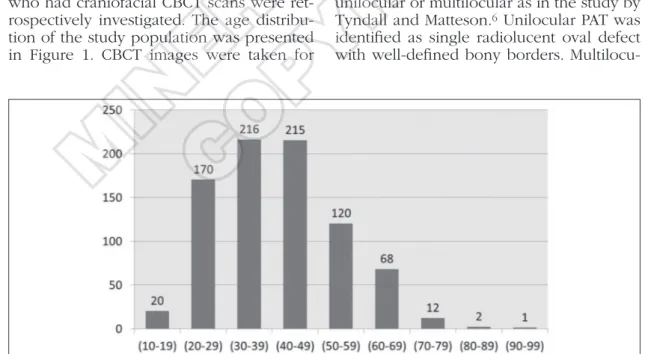
Eight-hundred and twenty-five patients (377 men and 448 women) ranging in age from 18 to 91 years (mean: 37.82 years) who had craniofacial CBCT scans were ret-rospectively investigated. The age distribu-tion of the study populadistribu-tion was presented in Figure 1. CBCT images were taken for
various purposes such as preimplant imag-ing, paranasal sinus examinations, tempo-romandibular joint (TMJ) examinations or orthodontic purposes. CBCT scans were made with Newtom 3G (Quantitative Radi-ology s.r.l., Verona, Italy). The imaging pro-tocol used a 9-inch field of view to include the mandibular and maxillary anatomy. The axial slice thickness was 0.3 mm, and the voxels were isotropic. Cases in which the zygomatic arch was not adequately seen for technical reasons and the cases, that had maxillofacial fracture history or having maxillofacial anomalies, were not included. The study protocol was approved by the lo-cal Ethilo-cal Committee.
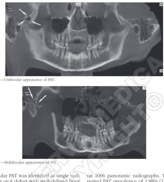
Blind fashion to the clinical status of the patient, an oral and maxillofacial radiolo-gist examined the radiographs. The age and sex were recorded for all patients and for the cases of PAT; gender, age, localization and radiographic appearance were noted as well. Diagnosis of PAT on the radiographs was done only if unequivocal pneumatiza-tion of the articular eminence could be seen or if the defect was located in the articular eminence posterior to the zygomaticotem-poral suture as a well-defined uni- or mul-tilocular radiolucency. PAT was classified as unilocular or multilocular as in the study by Tyndall and Matteson.6 Unilocular PAT was
identified as single radiolucent oval defect with well-defined bony borders.
Multilocu-Figure 1.—Age distribution of 825 patients by decade.
MINER
VA MEDICA
COPYRIGHT®
y inter national cop yr ight la ws .No additional reproduction is author
iz ed. It is per mitted f or persona l use to do wnload and sa v
e only one file and pr
int only one cop
y of this Ar ticle . It is not per mitted to mak e additional copies adically or systematically , either pr
inted or electronic) of the Ar
ticle f or an y pur pose . It is not per mitted to dis tr ib
ute the electronic cop
y of the ar
ticle through online inter
net and/or intr
anet file shar
ing systems , electronic mailing or an y other y allo w access to the Ar ticle .
The use of all or an
y par
t of the Ar
ticle f
or an
y Commercial Use is not per
mitted.
The creation of der
iv
ativ
e w
or
ks from the Ar
ticle is not per
mitted.
The production of repr
ints f
or personal or commercial use is
mitted. It is not per mitted to remo v e , co v er , o v er la y, obscure , b loc k, or change an y cop yr
ight notices or ter
ms of use wh
ich the Pub
lisher ma y post on the Ar ticle . It is not per mitted to fr ame or use fr
aming techniques to enclose an
y tr ademar k, logo , ietar y inf or
mation of the Pub
lisher
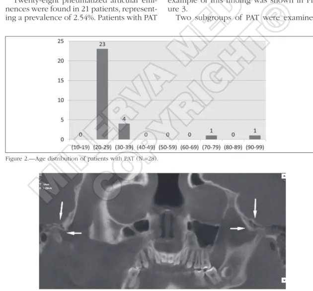
had a mean age of 28.9 (SD 3.13) years with a range of 21-78 years. Ten cases (47.6%) occurred in females and 11 cases (52.4%) occurred in males. All cases were located in the zygomatic process of the temporal bone, did not extend anteriorly beyond the zygomaticotemporal suture, and lacked ex-pansile or destructive characteristics. The mean age of males was 32.7 (SD 2.54) years with a range of 21-78 years while mean age of females was 24.6 (SD 3.9) years with a range of 21-25 years (Figure 2). A total of 28 PATs were detected; unilateral PAT was found in 14 cases (66.6%), with 6 cases on the right and 8 cases on the left. Bilateral PAT was found in 7 cases (33.4%) and an example of this finding was shown in Fig-ure 3.
Two subgroups of PAT were examined; lar PAT was identified as numerous
radiolu-cent small cavities.
Statistical analysis
Statistical analyses were performed us-ing the SPSS 15.0 program (SPSS Inc, Chi-cago, IL, USA) for WINDOWS. Comparison of gender, age and localization in patients with PAT was performed using chi-square test and correspondence analysis. P<0.05 was considered significant.
Results
Twenty-eight pneumatized articular emi-nences were found in 21 patients, represent-ing a prevalence of 2.54%. Patients with PAT
Figure 3.—Bilateral appearance of PAT.
Figure 2.—Age distribution of patients with PAT (N.=28).
MINER
VA MEDICA
COPYRIGHT®
y inter national cop yr ight la ws .No additional reproduction is author
iz ed. It is per mitted f or persona l use to do wnload and sa v
e only one file and pr
int only one cop
y of this Ar ticle . It is not per mitted to mak e additional copies adically or systematically , either pr
inted or electronic) of the Ar
ticle f or an y pur pose . It is not per mitted to dis tr ib
ute the electronic cop
y of the ar
ticle through online inter
net and/or intr
anet file shar
ing systems , electronic mailing or an y allo w access to the Ar ticle .
The use of all or an
y par
t of the Ar
ticle f
or an
y Commercial Use is not per
mitted.
The creation of der
iv
ativ
e w
or
ks from the Ar
ticle is not per
mitted.
The production of repr
ints f
or personal or commercial use is
mitted. It is not per mitted to remo v e , co v er , o v er la y, obscure , b loc k, or change an y cop yr
ight notices or ter
ms of use wh
ich the Pub
lisher ma y post on the Ar ticle . It is not per mitted to fr ame or use fr
aming techniques to enclose an
ietar
y inf
or
mation of the Pub
lisher
DELILBASI CBCT EVALUATION OF PAT
on 1006 panoramic radiographs. They re-ported PAT prevalence of 1.88% (19 cases) of which 63.1% (12 cases) was in females and 36.9% in males (7 cases). Bilateral PAT was found in 36.9% of cases (7 cases).2
Ya-vuz et al. investigated the prevalence of PAT in 8107 panoramic radiographs. The preva-lence was found to be 1.03% (83 cases). Forty-one of the cases were females and 42 were males. PATs were located on the right side in 60 cases and on the left side in 50 cases. A total of 110 PATs were observed of which 44 were unilocular and 66 were multilocular. Fifty-six cases had unilateral and 27 cases had bilateral PATs.8 Miloglu et al. carried out a study using CBCT in 514
cases. PAT was found in 8% of cases (41 cases) of whom 61% was female (25 cases) and 39% was male (16 cases). Age range unilocular PAT was identified as single
radi-olucent oval defect with well-defined bony borders (Figure 4). Seven of the PATs were unilocular type. Multilocular PAT was identi-fied as numerous radiolucent small cavities, which resemble mastoid air cells (Figure 5). Twenty-one of the PATs were multilocular type. The Chi-square tests showed no statis-tical significant differences with respect to gender, localization and age (P>0.05).
Discussion
The first study on the prevalence of PAT was reported by Tyndall and Matteson in 1985 by using panoramic radiographs. They reported a prevalence of 2.6% (28 cases) in 1061 cases.6 Orhan et al. conducted a study Figure 4.—Unilocular appearance of PAT.
Figure 5.—Multilocular appearance of PAT.
MINER
VA MEDICA
COPYRIGHT®
y inter national cop yr ight la ws .No additional reproduction is author
iz ed. It is per mitted f or persona l use to do wnload and sa v
e only one file and pr
int only one cop
y of this Ar ticle . It is not per mitted to mak e additional copies adically or systematically , either pr
inted or electronic) of the Ar
ticle f or an y pur pose . It is not per mitted to dis tr ib
ute the electronic cop
y of the ar
ticle through online inter
net and/or intr
anet file shar
ing systems , electronic mailing or an y other y allo w access to the Ar ticle .
The use of all or an
y par
t of the Ar
ticle f
or an
y Commercial Use is not per
mitted.
The creation of der
iv
ativ
e w
or
ks from the Ar
ticle is not per
mitted.
The production of repr
ints f
or personal or commercial use is
mitted. It is not per mitted to remo v e , co v er , o v er la y, obscure , b loc k, or change an y cop yr
ight notices or ter
ms of use wh
ich the Pub
lisher ma y post on the Ar ticle . It is not per mitted to fr ame or use fr
aming techniques to enclose an
y tr ademar k, logo , ietar y inf or
mation of the Pub
lisher
fications make CBCT a growing imaging technique to evaluate osseous components of TMJ.11-13
It is not necessary to perform a treatment for PAT, but it can be an obstacle when eminoplasty or eminectomy is planned. Al-though small pneumatizations may not be contraindication for surgical interventions, Lindenmuth and Clark warned the surgeons about knowing the details of PAT.3 PAT is
important as it is on the pathway of spread of pathological lesions and infections, frac-tures may occur in the TMJ region owing to minimal resistance due to pneumatiza-tion. During TMJ surgery, perforation can occur because of firm dissection or force-ful flap retraction. Besides, while inserting fossa-eminence prosthesis between condyle and skull base, special care is needed to avoid screw perforation. During the opera-tion, special care must be taken while using osteotoms and burs to avoid unintentional penetration into the defect.8, 14, 15 In order
to be safe, the anesthesiologist should per-form Valsalva manoeuvre on the patient to control dural tear. The defect must be observed for the presence of cerebrospinal fluid leakage.4, 15
Temporal air spaces play an important role in suppuration of various pathological processes. Tumors of the mastoid process and ear may extend into the TMJ, whereas otitis media or otomastoiditis may involve the TMJ and may result in ankylosis.1, 2, 15, 16
Conclusions
In this study, it is figured out that pneu-matization of accessory air cells can be accurately detected using CBCT. Practi-tioners who are interested in TMJ surgery and pathology should have detailed infor-mation about anatomical variations in the region. This will prevent misdiagnose and operative complications thus will enhance the result of treatment. Furthermore, ad-vanced radiological techniques such as CBCT are useful to support panoramic ra-diographs to detect anatomical variations in the bone.
of patients with PAT was 15-62 years with a mean of 30.6 years. Bilateral PAT was de-tected in 24.4% cases (10 cases) and unilat-eral PAT was detected in 75.6% cases (31 cases). Detected PATs were unilocular in 41.5 cases (17 cases) and multilocular in 58.5% cases (24 cases). The authors men-tioned that prevalence of PAT is higher than suspected; therefore, panoramic ra-diographs should be supported with CBCT before surgical interventions in the region.9
Recently, Laderia et al. investigated pneu-matization of the glenoid fossa (PGF) and articular eminence (PAT) with CBCT in 658 patients. The prevalence of PGF and PAT was 38.3% and 21.3%, respectively. Consid-ering the TMJ, PAT was found in 15.5% of the patients and PGF was found in 30.2% of the patients. They concluded that preva-lence of PAT and PGF is higher than previ-ously reported.10
We found a PAT prevalence of 2.54% which is higher than most of the studies using panoramic radiographs but lower than that of Miloglu et al.9 and Laderia et al.10 This study contributes to a few
stud-ies about CBCT and anatomical variations of the TMJ and emphasizes the use of CBCT for detecting such entities.
Panoramic radiograph is a useful tech-nique to diagnose the PAT, since posterior aspect of the zygomatic arc is usually dis-placed. Computed tomography (CT) can be used for evaluating bone surface and air spaces in the skull. CT is not subject to superimposition, and it is superior to plain radiographs for evaluating temporal air spaces. Also less superficially located (i.e., medial portion of the articular eminence) air cells, adjacent to TMJ may only be vis-ible on CT.3 CBCT provides high-resolution
multiplanar images and delivers substan-tially lower radiation dose, compared with multislice CT. CBCT is useful for TMJ evalu-ation without superimposition and distor-tion for bone morphology analysis. The diagnostic efficacy of CBCT is as good as conventional CT and better than panoramic radiography. More accurate images and a lower radiation dose enable CBCT to be commonly preferred modality. These
quali-MINER
VA MEDICA
COPYRIGHT®
y inter national cop yr ight la ws .No additional reproduction is author
iz ed. It is per mitted f or persona l use to do wnload and sa v
e only one file and pr
int only one cop
y of this Ar ticle . It is not per mitted to mak e additional copies adically or systematically , either pr
inted or electronic) of the Ar
ticle f or an y pur pose . It is not per mitted to dis tr ib
ute the electronic cop
y of the ar
ticle through online inter
net and/or intr
anet file shar
ing systems , electronic mailing or an y allo w access to the Ar ticle .
The use of all or an
y par
t of the Ar
ticle f
or an
y Commercial Use is not per
mitted.
The creation of der
iv
ativ
e w
or
ks from the Ar
ticle is not per
mitted.
The production of repr
ints f
or personal or commercial use is
mitted. It is not per mitted to remo v e , co v er , o v er la y, obscure , b loc k, or change an y cop yr
ight notices or ter
ms of use wh
ich the Pub
lisher ma y post on the Ar ticle . It is not per mitted to fr ame or use fr
aming techniques to enclose an
ietar
y inf
or
mation of the Pub
lisher
354 MINERVA STOMATOLOGICA October 2013
DELILBASI CBCT EVALUATION OF PAT
2. Orhan K, Delilbasi C, Cebeci I, Paksoy C. Prevalence and variations of pneumatized articular eminence: a study from Turkey. Oral Surg Oral Med Oral Pathol Oral Radiol Endod 2005;99:349-54.
3. Lindenmuth JE, Clark MS. Pneumatization of the ar-ticular eminence. Cranio 1986;4:86-7.
4. Hofmann T, Friedrich RE, Wedl JS, Schmelzle R. Pneu-matization of the zygomatic arch on pantomography. Mund Kiefer Gesichtschir 2001;5:173-9.
5. Ganc DT, Jyung RW. Temporal bone pneumatiza-tion in otosclerosis. Otolaryngol Head Neck Surg 2008;139:850-3.
6. Tyndall DA, Matteson SR. Radiographic appearance and population distribution of the pneumatized ar-ticular eminence of the temporal bone. J Oral Maxil-lofac Surg 1985;43:493-7.
7. Orhan K, Oz U, Orhan AI, Ulker AE, Delilbasi C, Ak-cam O. Investigation of pneumatized articular emi-nence in orthodontic malocclusions. Orthod Cranio-fac Res 2010;13:56-60.
8. Yavuz MS, Aras MH, Güngör H, Büyükkurt MC. Prev-alence of the pneumatized articular eminence in the temporal bone. J Craniomaxillofac Surg 2009;37:137-9.
9. Miloglu O, Yilmaz AB, Yildirim E, Akgul HM. Pneu-matization of the articular eminence on cone beam computed tomography: prevalence, characteristics and a review of the literature. Dentomaxillofac Radiol 2011;40:110-4.
10. Ladeira DB, Barbosa GL, Nascimento MC, Cruz AD, Freitas DQ, Almeida SM. Prevalence and characteris-tics of pneumatization of the temporal bone evalu-ated by cone beam computed tomography. Int J Oral Maxillofac Surg 2013;42:771-5.
11. Barghan S, Merrill R, Tetradis S. Cone beam comput-ed tomography imaging in the evaluation of the tem-poromandibular joint. J Calif Dent Assoc 2010;38:33-9.
12. Barghan S, Tetradis S, Mallya S. Application of cone beam computed tomography for assessment of the temporomandibular joints. Aust Dent J 2012;57(Suppl 1):109-18.
13. Hill CA, Richtsmeier JT. A quantitative method for the evaluation of three-dimensional structure of temporal bone pneumatization. J Hum Evol 2008;55:682-90. 14. Kaugars GE, Mercuri LG, Laskin DM. Pneumatization
of the articular eminence of the temporal bone: prev-alence, development, and surgical treatment. J Am Dent Assoc 1986;113:55-7.
15. Kulikowski BM, Schow SR, Kraut RA. Surgical man-agement of a pneumatized articular eminence of the temporal bone. J Oral Maxillofac Surg 1982;40:311-3. 16. de Rezende Barbosa GL, Nascimento MD, Ladeira
DB, Bomtorim VV, da Cruz AD, Almeida SM. Accu-racy of digital panoramic radiography in the diag-nosis of temporal bone pneumatization: A study in vivo using cone-beam-computed tomography. J Cra-niomaxillofac Surg 2013 [In press].
Conflicts of interest.—The authors certify that there is no conflict of interest with any financial organization regarding the material discussed in the manuscript.
Received on February 21, 2013.
Accepted for publication on October 17, 2013. Riassunto
Valutazione della pneumatizzazione dell’eminenza articolare tramite tomografia computerizzata a fa-scio conico
Obiettivo. La conoscenza dell’anatomia
dell’arti-colazione temporo-mandibolare (ATM) e delle strut-ture circostanti è indispensabile per il successo degli interventi chirurgici e per la comprensione del per-corso di diffusione delle infezioni in questa regione. Questo studio si propone di indagare l’anatomia e la morfologia dell’eminenza articolare pneumatizza-ta (PAT) tramite tomografia computerizzapneumatizza-ta a fascio conico (CBCT) in un gruppo di pazienti.
Metodi. Uno studio retrospettivo su 825 pazienti
(377 maschi, 448 femmine) di età compresa tra i 18 ei 91 anni è stato eseguito utilizzando immagini CBCT sagittali e coronali. La PAT è stata definita come radiotrasparenzasimil-cistica non espansiva e non distruttiva nel processo zigomatico dell’osso temporale, che appare simile alle cellule mastoi-dee. Èstata classificata in due gruppi come uni-loculare e multiuni-loculare. Il confronto statistico di genere, età e localizzazione è stato effettuato uti-lizzando il test del chi-quadrato e l’analisi delle corrispondenze. P <0,05 è stato considerato signi-ficativo.
Risultati. Ventotto casi di eminenza articolare
pneumatizzata sono stati riscontrati in 21 pazienti, che rappresentano una prevalenza del 2,54%. I pa-zienti con PAT avevano un’età media di 28,9 anni, con un range di 21-78 anni. Dieci casi (47,6%) si sono verificati nelle femmine e 11 casi (52,4%) nei maschi. I risultati dei test statistici non hanno mo-strato differenze significative tra i gruppi per quanto riguarda sesso, età e localizzazione (P> 0,05).
Conclusione. La pneumatizzazione
dell’eminen-za articolare è una materia difficile per la chirurgia dell’ATM e la diffusione delle infezioni in questa regione. I professionisti che si occupano di chirur-gia e patolochirur-gia ATM dovrebbero avere informazioni dettagliate su questa variante anatomica in quanto può causare gravi complicazioni.
Parole chiave: Articolazione temporo-mandibolare
- Radiografia panoramica - Tomografia Computeriz-zata a fascio conico.
References
1. Orhan K, Delilbasi C, Orhan AI. Radiographic evaluation of pneumatized articular eminence in a group of Turkish children. Dentomaxillofac Radiol 2006;35:365-70.
MINER
VA MEDICA
COPYRIGHT®
This document is protected b
y inter national cop yr ight la ws .
No additional reproduction is author
iz ed. It is per mitted f or persona l use to do wnload and sa v
e only one file and pr
int only one cop
y of this Ar ticle . It is not per mitted to mak e additional copies (either spor adically or systematically , either pr
inted or electronic) of the Ar
ticle f or an y pur pose . It is not per mitted to dis tr ib
ute the electronic cop
y of the ar
ticle through online inter
net and/or intr
anet file shar
ing systems , electronic mailing or an y other means which ma y allo w access to the Ar ticle .
The use of all or an
y par
t of the Ar
ticle f
or an
y Commercial Use is not per
mitted.
The creation of der
iv
ativ
e w
or
ks from the Ar
ticle is not per
mitted.
The production of repr
ints f
or personal or commercial use is
not per mitted. It is not per mitted to remo v e , co v er , o v er la y, obscure , b loc k, or change an y cop yr
ight notices or ter
ms of use wh
ich the Pub
lisher ma y post on the Ar ticle . It is not per mitted to fr ame or use fr
aming techniques to enclose an
y tr ademar k, logo , or other propr ietar y inf or
mation of the Pub
lisher