Introduction
Functional orthodontic appliances aim to correct malfunctions, dentofacial problems, crowding, and anterior and posterior overjet. In recent years, interest to the functional orthopaedic treatment modalities has gained a great increase.
Fränkel developed four different functional orthopaedic appliances on the basis of functional orthopaedics principles, and they are used worldwide today. These are orthopaedic exercise devises that aid in maturation, training, and ‘reprogramming’ of the orofacial neuromusculature (Fränkel, 1970).
Fränkel (1970) emphasized the importance of soft tissue environment, maintaining that aberrant postural behaviour of the orofacial musculature played a primary role in the development of skeletal and dentoalveolar deformities. The following sentences belong to Rolf Fränkel and his daughter Christine Fränkel (1989): ‘Function regulator (FR) appliance is capable of producing maxillary expansion in the alveolar basal area even after the permanent premolars have erupted. We believe that the pull of the projecting buccal shields of the FR on the soft tissues at the sulci is transferred to the periosteum which results in deposition of new bone on the buccal aspect of the alveoli with subsequent remodelling of the outer alveolar walls.’
It has been stated by Fränkel (1970), Fränkel and Fränkel (1989), and McNamara (2002) that early treatment with FR-3 appliance offers some opportunities including normal displacement of teeth and facial bones and space gaining in dental arches. Most of the studies (Loh and Kerr, 1985; Ülgen and Firatli, 1994; Baik et al., 2004; Kalavritinos et al., 2005; Levin et al. 2008) on this subject evaluated the
Effects of the functional regulator III on transversal changes:
a postero-anterior cephalometric and model study
Nihat Kilic*, Mevlüt Celikoglu* and Hüsamettin Oktay**
Departments of Orthodontics, Faculties of Dentistry, *Atatürk University, Erzurum and **Istanbul Medipol University, Istanbul, Turkey
Correspondence to: Dr Husamettin Oktay, İstanbul Medipol Üniversitesi Diş Hekimliği Fakültesi, Unkapanı, Atatürk Bulvarı No. 27, Fatih-İstanbul 34083, Turkey. Email: [email protected]
SUMMARY Studies assessing the transversal treatment changes caused by the functional regulator III (FR-3) are
limited in number. This clinical study was planned to analyse the transversal effects of the FR-3 appliance therapy. The treatment group consisted of 17 patients (8 males and 9 females) with Class III malocclusion, who were treated with the FR-3 appliance. The control group consisted of 17 subjects (7 males and 10 females) with a normal occlusion. Mean ages of the subjects were 10.73 and 10.66 years in the treatment and control groups, respectively. Postero-anterior radiographs and stone casts were obtained before (T1) and after (T2) treatment/observation.
The results of the Student’s t-test comparing initial values showed that maxillary dentoalveolar and skeletal widths are significantly larger in the control group than those in the treatment group. At the end of the treatment, significant transverse increments occurred only at the dentoalveolar level of the maxilla. The transversal changes in the mandible were not statistically significant. Buccal shields of FR-3 did not stimulate the growth of maxillary apical base but caused an enhanced and supplementary widening of maxillary dental and alveolar structures.
sagittal and vertical effects of the FR-3 appliance. Only a limited number of studies investigated the transversal dentoalveolar changes and demonstrated favourable maxillary dentoalveolar effects produced by FR-3 (Firatli and Ulgen, 1996; Miethke et al., 2003; Kalavritinos et al., 2005). Firatli and Ulgen (1996) found that the FR-3 appliance produced a significant dental and alveolar widening in maxilla, but it did not affect mandibular apical base. According to our knowledge, however, no study has conducted to evaluate if the FR-3 appliance stimulate the growth of maxillary apical base. Thus, as claimed by Fränkel (1970), the argument that the FR-3 appliance is capable of stimulating skeletal growth of maxillary base in transverse direction remained unanswered.
The present study was planned to evaluate the transversal dentoalveolar and skeletal changes in subjects with Class III, who were treated with the FR-3 appliance.
Materials and methods
This study was approved by the local ethics committee. Informed consent was obtained from the parents of the children.
Seventeen patients (8 males and 9 females) treated with the original FR-3 appliance and 17 subjects (7 males and 10 females) with a normal occlusion were included to the present study. The chronological ages of the subjects in both groups are shown in Table 1. The normal occlusion group was chosen from the longitudinal archive of the Department of Orthodontics at Atatürk University. Selection of the control subjects was based on normal growth and development, balanced skeletal profile, Class I occlusion,
cephalostat (Siemens Nanodor 2; Siemens AG, Munich, Germany). In order to determine the skeletal changes, three linear measurements were made on the PA films. These are shown in Figure 1.
Cephalometric measurements:
1. Nasal cavity width (NL–NR): The distance between the left and right most lateral points of nasal cavity.
2. Maxillary skeletal width (MxL–MxR): The distance between the left and right deepest points of lateral maxillary contours.
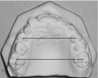
3. Mandibular skeletal width (AgL–AgR): The distance between the left and right deepest points of antegonial notches. On the stone casts, six linear measurements were done to assess dentoalveolar changes (Figures 2–4). When deciduous first molars have not yet been replaced by their counterparts, the distance between the first premolars was not measured, and this caused different subject numbers in Tables 2 and 3. Orthodontic model measurements:
Maxilla
4. Upper inter-molar width: The distance between central fossae of the right and left first maxillary molars. 5. Upper inter-premolar width: The distance between
central fossae of the right and left first maxillary premolars. 6. Maxillary first molar alveolar width: The distance
between the alveolar regions of maxillary first molars (5 mm above the most apical points of gingival margin). 7. Maxillary first premolar alveolar width: The distance
between the alveolar regions of maxillary first premolars or deciduous first molars (5 mm above the most apical points of gingival margin).
Mandible
8. Lower inter-molar width: The distance between the medio-buccal cusp tips of the right and left first molar teeth. 9. Lower inter-premolar width: The distance between the
buccal cusp tips of the right and left first premolar teeth. Dental arch widths on the stone casts were measured using the reference points of the first premolar and molar teeth in both jaws, which are the projection of each other. In the subjects with normal occlusion, the upper and lower dental arch widths are the same if the dental reference points are used. Table 1 Initial chronological ages and observation periods of the groups and their comparisons. NS, not significant.
Parameters Gender
Study group Control group
Test
N Mean Standard deviation N Mean Standard deviation
Chronological age (years) Male 8 11.06 0.75 7 10.75 0.47 NS Female 9 10.44 1.32 10 10.60 0.45
Total 17 10.73 1.11 17 10.66 0.45
Observation period (months) 15.35 7.31 14.82 5.25 NS
Figure 1 Measurements used in the postero-anterior films: nasal cavity
width (NL–NR): The distance between the left and right most lateral points of nasal cavity. Maxillary skeletal width (MxL–MxR): The distance between the left and right deepest points of lateral maxillary contours. Mandibular skeletal width (AgL–AgR): the distance between the left and right deepest points of antegonial notches.
and absence of anterior and/or posterior crossbites. The patients in the treatment group had maxillary retrusion or a combination of maxillary retrusion and mandibular protrusion (skeletal Class III relationship with anterior crossbite and retrusive nasomaxillary area). The subjects with a history of orthodontic treatment were not included.
During the first 2 weeks, the patients wore their appliances 4–5 hours during the day for slow adaptation. If, after this time, the mucosa looked healthy, the patients were instructed to wear the appliance all day, but not at nights. After the patients adapted to wearing the FR-3 appliance during the day, they were asked to wear their appliances day and night. The appliance and treatment progress were checked at four weekly intervals. Active functional treatment was terminated when the negative overjet and concave facial profile were corrected.
Postero-anterior (PA) radiographs and stone casts were obtained from all the subjects at the beginning and after the treatment/control periods. The films were taken in a standard position by one operator using the same X-ray machine and
Statistical analysis
To determine the errors associated with digitizing and measurements, 15 PA films and stone casts were randomly selected. All procedures were repeated 2 weeks after by the same author. Intra-class correlation coefficients were performed to assess the reliability of the measurements as described by Houston (1983).
Comparisons between the groups were carried out by means of Student’s t-test. All statistical analyses were performed using the SPSS software package program (SPSS for Windows 98, version 10.0; SPSS Inc., Chicago, Illinois, USA).
Results
The reliability coefficients of the measurements were above 0.90 for all parameters. Initial chronological ages and observation periods of the groups and their comparisons are shown in Table 1. There was no statistically significant difference between the groups in relation to these parameters.
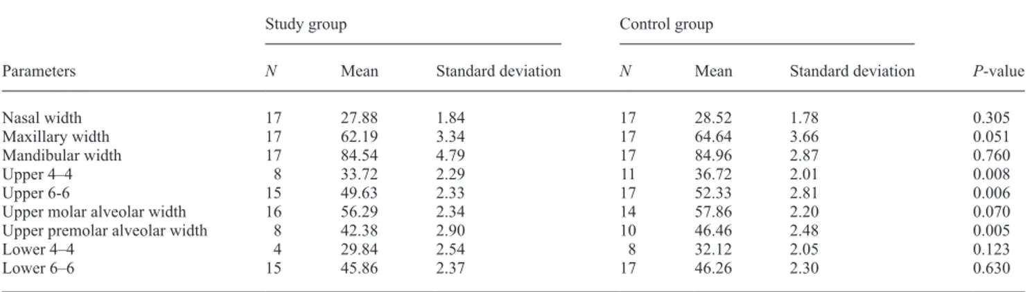
The results of Student’s t-test comparing initial values for the groups are shown in Table 2. Upper molar and inter-premolar widths and upper first inter-premolar alveolar width are significantly larger in the control group. There were no statistically significant differences in the other parameters although the P-values of maxillary width and upper molar alveolar width were close to significance level (0.051 and 0.070, respectively).
Changes occurring during the treatment and observation periods are shown in Table 3. The changes in upper inter-molar and inter-preinter-molar widths and upper first inter-molar and premolar alveolar widths showed statistically significant differences between the groups. In other words, these measurements increased more in the FR-3 group.
Discussion
One of the most confusing conditions in orthodontics is diagnosis and treatment of the subjects with a Class III malocclusion. Children with a Class III malocclusion may have an underdeveloped maxilla, an overdeveloped mandible, or a combination of both. Orthopaedic, functional, or orthodontic treatments or combination of these are among the treatment options of Class III cases. Orthodontists are among the health care professionals who can undertake functional orthopaedics.
Since ethical consideration did not allow postponing the treatment of Class III subjects for scientific purposes, the control group was formed by the subjects with an acceptable occlusion and a skeletal Class I relationship.
In the present study, the functional regulator III appliance was applied to the patients aged from 8 to 13 years, mean age 10.73 years. All the subjects were in prepubertal and in the mixed or early permanent dentition stages at the beginning of treatment. There is a consensus that treatment with FR-3 should be commenced during the prepubertal ages (Fränkel, 1970; Loh and Kerr, 1985; Ülgen and Firatli, 1994; Firatli and Ulgen, 1996; Baik et al., 2004; Levin et al., 2008).
Figure 4 Alveolar width assessment. Maxillary first molar alveolar width:
the distance between alveolar regions of maxillary first molars (measured from 5 mm above the most apical points of the gingival margins of first molars). Maxillary first premolar alveolar width: the distance between the alveolar regions of the maxillary first premolars (measured from 5 mm above the most apical points of the gingival margins of first premolars).
Figure 2 The measurements between upper molars and premolars. Upper
molar width: the distance between upper first molars. Upper inter-premolar width: the distance between upper first inter-premolars.
Figure 3 The measurements between lower molars and premolars.
Lower inter-molar width: the distance between lower first molars. Lower inter-premolar width: the distance between lower first premolars.
Comparisons of initial values between the groups showed that upper inter-molar and inter-premolar widths and upper first premolar alveolar width are significantly larger in the control group (P < 0.01). Upper first molar alveolar width and maxillary skeletal width are also larger in this group, but their P-values did not reach the significance level (Table 2). These findings indicated that the subjects in the treatment group had maxillary deficiency. These pretreatment findings are consistent with the findings of Miethke et al. (2003).
Numerous studies have been carried out to evaluate dentoskeletal and soft tissue changes in sagittal direction, and a great number of them supported the idea that the FR-3 appliance therapy produced a favourable growth effect on the maxilla and surrounding soft tissue structures (Fränkel, 1970; McNamara and Huge, 1985; Kohmura et al.,1986; Aytan et al., 1989; Isiksal and Seckin, 1990; Miethke et al., 2003; Falck and Zimmermann-Menzel, 2008; Levin et al., 2008; Kilic et al., 2010). However, the studies on transversal dentoalveolar changes are limited in number, and according to their findings, favourable dentoalveolar effects can be obtained with the FR-3 appliance therapy (Firatli and Ulgen, 1996; Miethke et al., 2003; Kalavritinos et al., 2005).
Fränkel (1970), Fränkel and Fränkel (1989), and McNamara (2002) explained that early treatment with FR-3 appliance offers some opportunities including normal displacement of teeth and facial bones and space gaining in dental arches.
Fränkel (1970) and Fränkel and Fränkel (1989) claimed that a force produced by buccal shields is sufficient to overcome the functional disorders associated with dentoskeletal maldevelopment and to produce skeletal expansion. Expanding the external soft tissue capsule encircling the maxillary structures by means of buccal shields of FR-3 appliance, which exert a considerable tension to periosteal attachments in the depth of the maxillary sulcus, induces load-specific responses in maxillary bony structures. However, the question as to whether these tension forces reached the upper structures of naso-maxillary complex and whether produced a skeletal expansion in maxilla and nasal cavity remained unanswered.
The findings of the present study showed that maxillary dental and alveolar widths increased significantly with the FR-3 appliance treatment. In other words, the FR-3 appliance therapy produced significant improvements in dentoalveolar regions of maxilla, but it failed to promote transversal skeletal maxillary and nasal growth. Dentoalveolar and skeletal widths of mandible showed similar increments in both groups. All these findings imply that FR-3 appliance is not effective on increasing the nasal cavity width and stimulating growth of maxillary apical base as well as restricting the growth of mandibular apical base.
A few reports are available regarding with the transversal changes induced by the FR-3 appliance therapy, and thus, it is difficult to make a valuable comparison between our findings Table 2 Results of the Student’s t-test comparing the initial values between the groups.
Study group Control group
Parameters N Mean Standard deviation N Mean Standard deviation P-value
Nasal width 17 27.88 1.84 17 28.52 1.78 0.305 Maxillary width 17 62.19 3.34 17 64.64 3.66 0.051 Mandibular width 17 84.54 4.79 17 84.96 2.87 0.760
Upper 4–4 8 33.72 2.29 11 36.72 2.01 0.008
Upper 6-6 15 49.63 2.33 17 52.33 2.81 0.006 Upper molar alveolar width 16 56.29 2.34 14 57.86 2.20 0.070 Upper premolar alveolar width 8 42.38 2.90 10 46.46 2.48 0.005
Lower 4–4 4 29.84 2.54 8 32.12 2.05 0.123
Lower 6–6 15 45.86 2.37 17 46.26 2.30 0.630
Table 3 Results of Student’s t-test comparing mean changes between groups.
Study group Control group
Parameters N Mean Standard deviation N Mean Standard deviation P-value
Nasal width 17 1.10 0.11 17 1.03 0.17 0.160 Maxillary width 17 1.39 0.77 17 1.18 0.65 0.380 Mandibular width 17 2.11 0.93 17 2.13 1.03 0.945
Upper 4–4 8 1.71 0.85 11 0.42 0.16 0.000
Upper 6-6 15 1.75 0.67 17 0.73 0.27 0.000
Upper molar alveolar width 16 1.66 0.45 14 0.79 0.46 0.000 Upper premolar alveolar width 8 1.79 0.58 10 0.66 0.27 0.000
Lower 4–4 4 0.57 0.25 8 0.41 0.15 0.186
Lower 6–6 15 0.84 0.43 17 0.66 0.24 0.133
and the findings of other clinicians. Fränkel (1970, 1974) and Fränkel and Fränkel (1989) declared that soft tissue matrix formed by cheeks, lips, and tongue has an important influence on the development of skeletal structures. He theorized that apical extensions of the shields into the buccal vestibulum cause a tension on buccinator muscle fibres and dentoalveolar periosteum, stimulating bone deposition. Our findings do not support that hypothesis since we found no significant differences between transversal skeletal maxillary and nasal growth in both
groups. Fränkel (1974) accepted the hyperactivity of the
muscles as a causative role in the development of a narrowed maxilla. However, electromyographic studies on this subject found no association between narrow maxillary arches and buccinator hyperactivity (Stavridi and Ahlgren, 1992).
The present study clearly revealed that buccal shields of FR-3 appliance leaded to an enhanced and supplementary widening of maxillary dental and alveolar structures when compared with those of control group. These findings are in accordance with the studies of Fränkel (1970, 1974), Fränkel and Fränkel (1989), Owen (1983), Firatli and Ulgen (1996), and Kalavritinos et al. (2005) who observed an increase at the maxillary dentoalveolar width. Owen (1983) suggested that function regulator allows a passive expansion of the dental arches. According to Fränkel (1970, 1974) and Fränkel and Fränkel (1989), this expansion is accomplished mainly through the action of the shields since the vestibular shields create a tension at the depth of vestibular fold in a lateral direction, which influence the permanent teeth to erupt in a more buccal position, leading to arch expansion. Vestibular shields of the appliance lead to a change in the equilibrium of tooth position by reducing strong buccal pressure, particularly during the growth periods. Changes in the equilibrium produced by both relieving buccal pressure and continuing tongue pressure against to maxillary arches might lead to passive dentoalveolar expansion of upper arch as observed in the present study. This passive dental and alveolar arch expansion may cause a chance for ‘decrowding’ of the
teeth in the individuals with crowding (Fränkel, 1970, 1974;
Fränkel and Fränkel, 1989). In addition, the function regulator acts as a training devise for correcting and regulating abnormal activities of circumoral and perioral muscles.
Another finding of the present study is that the FR-3 appliance treatment did not restrict the transversal mandibular growth, and this finding is in agreement with
the findings of Firatli and Ulgen (1996). An insignificant
transverse increase was observed in the mandible, and this issue can be attributed to combined effects of growth and appliance treatment. The increase in maxillary dentition may lead to an increase in mandibular arch dimensions due to the interdigitation forces from functional jaw movements.
Conclusions
Pretreatment cephalometric measurements showed that the Class III treatment group had reduced maxillary dentoalveolar and skeletal widths compared with the Class I control.
During the treatment period, there was enhanced and supplementary widening of maxillary dental and dentoalveolar structures in the FR-3 group compared with the control group, but there was no evidence of statistically significant increase in the maxillary skeletal width.
Mandibular inter-premolar and -molar widths increased approximately in the same amount in both groups, showing that the FR-3 appliance therapy did not restrict the transversal mandibular growth.
References
Aytan S, Yukay F, Ciger S, Enacar A, Aksoy A, Telli A E 1989 Fränkel III appliance. Turkish Journal of Orthodontics 2: 338–345
Baik H S, Jee S H, Lee K J, Oh T K 2004 Treatment effects of Fränkel functional regulator III in children with Class III malocclusions. American Journal of Orthodontics and Dentofacial Orthopedics 125: 294–301 Falck F, Zimmermann-Menzel K 2008 Cephalometric changes in the
treatment of Class III using the Fränkel appliance. Journal of Orofacial Orthopedics 69: 99–109
Firatli S, Ulgen M 1996 The effects of the FR-3 appliance on the transversal dimension. American Journal of Orthodontics and Dentofacial Orthopedics 110: 55–60
Fränkel R 1970 Maxillary retrusion in Class 3 and treatment with the function corrector III. Transactions of the European Orthodontic Society, pp. 249–259
Fränkel R 1974 Decrowding during eruption under the screening influence of vestibular shields. American Journal of Orthodontics 65: 372–406 Fränkel R, Fränkel C 1989 Orofacial orthopedics with the function
regulator. Karger, Basel
Houston W J 1983 The analysis of errors in orthodontic measurements. American Journal of Orthodontics 83: 382–390
Isiksal E, Seckin O 1990 [The functional regulators (FR III, FR IV)]. Turkish Journal of Orthodontics 3: 150–156
Kalavritinos M, Papadopoulos M A, Nasiopoulos A 2005 Dental arch and cephalometric changes following treatment for Class III malocclusion by means of the function regulator (FR-3) appliance. Journal of Orofacial Orthopedics 66: 135–147
Kilic N, Celikoglu M, Oktay H 2010 Effects of the functional regulator III on profile changes in subjects with maxillary deficiency. European Journal of Orthodontics May 7 [Epub ahead of print]
Kohmura T et al. 1986 Effects of therapy using the function regulator (FR III) on the cases with the reversed occlusion. Nippon Kyosei Shika Gakkai Zasshi 45: 693–711
Levin A S, McNamara J A Jr. Franchi L, Baccetti T, Fränkel C 2008 Short-term and long-term treatment outcomes with the FR-3 appliance of Fränkel. American Journal of Orthodontics and Dentofacial Orthopedics 134: 513–524 Loh M K, Kerr W J 1985 The Function Regulator III: effects and indications
for use. British Journal of Orthodontics 12: 153–157
McNamara J A 2002 In memoriam Rolf Fränkel, 1908-2001. American Journal of Orthodontics and Dentofacial Orthopedics 121: 238–239 McNamara J A, Huge S A 1985 Functional regulator (FR-3) of Fränkel.
American Journal of Orthodontics 88: 409–424
Miethke R R, Lindenau S, Dietrich K 2003 The effect of Fränkel’s function regulator type III on the apical base. European Journal of Orthodontics 25: 311–318
Owen A H 1983 Morphologic changes in the transverse dimension using the Fränkel appliance. American Journal of Orthodontics 83: 200–217 Stavridi R, Ahlgren J 1992 Muscle response to the oral-screen activator.
An EMG study of the masseter, buccinator, and mentalis muscles. European Journal of Orthodontics 14: 339–349
Ülgen M, Firatli S 1994 The effects of the Fränkel’s function regulator on the Class III malocclusion. American Journal of Orthodontics and Dentofacial Orthopedics 105: 561–567